Key Points
Morphological dissection of the progression of nuclear expulsion reveals complex F-actin rearrangements in primary erythroblasts.
Enucleation depends upon a novel, conserved, F-actin/myosin IIB/Tmod1 structure (the “enucleosome”) at the rear of the translocating nucleus.
Abstract
Biogenesis of mammalian red blood cells requires nuclear expulsion by orthochromatic erythoblasts late in terminal differentiation (enucleation), but the mechanism is largely unexplained. Here, we employed high-resolution confocal microscopy to analyze nuclear morphology and F-actin rearrangements during the initiation, progression, and completion of mouse and human erythroblast enucleation in vivo. Mouse erythroblast nuclei acquire a dumbbell-shaped morphology during enucleation, whereas human bone marrow erythroblast nuclei unexpectedly retain their spherical morphology. These morphological differences are linked to differential expression of Lamin isoforms, with primary mouse erythroblasts expressing only Lamin B and primary human erythroblasts only Lamin A/C. We did not consistently identify a continuous F-actin ring at the cell surface constriction in mouse erythroblasts, nor at the membrane protein-sorting boundary in human erythroblasts, which do not have a constriction, arguing against a contractile ring-based nuclear expulsion mechanism. However, both mouse and human erythroblasts contain an F-actin structure at the rear of the translocating nucleus, enriched in tropomodulin 1 (Tmod1) and nonmuscle myosin IIB. We investigated Tmod1 function in mouse and human erythroblasts both in vivo and in vitro and found that absence of Tmod1 leads to enucleation defects in mouse fetal liver erythroblasts, and in CD34+ hematopoietic stem and progenitor cells, with increased F-actin in the structure at the rear of the nucleus. This novel structure, the “enucleosome,” may mediate common cytoskeletal mechanisms underlying erythroblast enucleation, notwithstanding the morphological heterogeneity of enucleation across species.
Introduction
Mammalian erythroblast enucleation is a rapid cellular process (<10 minutes in cultured erythroblasts1,2) involving nuclear polarization to one side of the cell, membrane protein sorting to remove unwanted membrane proteins from the nascent reticulocyte, nuclear translocation and expulsion, followed finally by separation of the pyrenocyte (expelled nucleus) from the nascent reticulocyte.3-7 Many genes have been linked directly or indirectly to enucleation,8,9 from transcription factors (eg, KLF110) to signaling molecules (eg, Rac GTPases11-14), structural proteins (eg, mDia2,14 dematin15), molecular motors (eg, dynein16,17 and nonmuscle myosin IIB [NMIIB]2,11,12), and endocytic pathway components (eg, EPS15, clathrin18,19), converging on a model in which enucleation is proposed to be analogous to asymmetric cell division, with microtubules important for cell polarization and nuclear movement, and F-actin assembly into an actomyosin contractile ring driving nuclear expulsion. Local activation of phosphoinositide-3-kinase near the microtubule-organizing center regulates cell polarity and nuclear movement to the opposing side of the erythroblast.2 Nuclear polarization and movement also require degradation of dynein microtubule motors by Trim58, a ubiquitin ligase upregulated during terminal erythroid differentiation.16 F-actin assembly and actomyosin contractile activity are important for nuclear expulsion, based on impaired enucleation by cytochalasin D inhibition of actin assembly2,11,20 and by blebbistatin inhibition of myosin II in cultured mouse and human erythroblasts.2,12 However, the subcellular location of F-actin and NMIIB assembly and force production with respect to initiation, progression, and completion of the process of nuclear expulsion are not clearly understood.
Key regulators of F-actin assembly and stability in postmitotic, differentiated cells are tropomodulins (Tmods), which bind to tropomyosin and cap the pointed (slow-growing) ends of F-actin, regulating filament length and stability in contractile structures as well as in the membrane skeleton.21,22 Tmod1 is the sole member of the Tmod family present in mouse and human erythrocytes, where it caps the pointed ends of the short F-actins in the membrane skeleton.23 Loss of Tmod1 in mice results in a mild compensated hemolytic anemia with variable F-actin lengths and a defective spectrin-actin lattice.24 However, Tmod3 is expressed during erythroid differentiation25 and is upregulated in Tmod1−/− mouse erythrocytes, providing partial compensation and accounting for the mild anemia.24 By contrast, Tmod3−/− mice are embryonic lethal with anemia due to impaired fetal liver (FL) erythropoiesis with defective macrophage-erythroblast interactions and defective enucleation, but tropomodulin 1 (Tmod1) levels do not increase and Tmod1 does not compensate for loss of Tmod3.25
Here, we used high-resolution confocal fluorescence microscopy to study erythroblast morphology and F-actin rearrangements during the progression of mammalian enucleation in vivo, finding that some but not all of the structural rearrangements of F-actin and morphological events of enucleation are conserved between primary mouse and human erythroblasts. We found that primary mouse erythroblasts form a cell surface constriction (neck) through which the nucleus passes, acquiring a dumbbell-shaped morphology, while primary human bone marrow erythroblasts do not form a cell surface constriction, and nuclei retain their spherical morphology throughout the process of expulsion. Notably, during the initiation and progression of nuclear expulsion, F-actin fails to form a continuous contractile ring at the membrane sorting boundary between the emerging nucleus and the nascent reticulocyte in human or mouse erythroblasts. Instead, a different F-actin population (the “enucleosome”) forms in polarized erythroblasts, defined by a large F-actin spot or collection of smaller spots in the cytoplasm that follows the rear of the translocating nucleus. This F-actin population is associated with NMIIB and Tmod1, but not mDia2, Arp3, or Tmod3. Using in vivo and in vitro models, we found that Tmod1 plays an important role in enucleation of human and mouse erythroblasts by regulating F-actin in the enucleosome at the rear of the nucleus. Altogether, these observations bring novel insights into F-actin rearrangements during enucleation and indicate the need for a reexamination of the cytoskeleton contribution to the forces required for nuclear expulsion in this critical process.
Materials and methods
Animals
E13-E15.5 embryos from wild-type or Tmod1−/− mice on C57Bl6/J background were used to obtain FL erythroblasts. Bone marrow erythroblasts were from 2-month-old adult C57Bl6/J mice. Splenic erythroblasts were from adult C57Bl6/J mice phlebotomized on days 1, 2, and 3, harvesting spleens on day 4. All experiments were performed according to National Institutes of Health animal care guidelines, as approved and enforced by Institutional Animal Care and Use Committee at The Scripps Research Institute.
Erythroblast collection and fluorescence staining
Mouse FLs, bone marrow, or spleens were dissociated into phosphate-buffered saline by gentle trituration using a cutoff P200 tip, followed by 4 hours or overnight fixation in 4% paraformaldehyde in phosphate-buffered saline at room temperature, and immunostaining as described.25 All studies involving human samples were conducted in accordance with the Declaration of Helsinki and under institutional review board approval of the Northwell Health System.
In vitro differentiation of CD34+ cells and flow cytometry
CD34+ isolation from cord blood and differentiation toward the erythroid lineage were carried out as previously described.26
Confocal imaging and analysis
Images were acquired on a Bio-Rad Radiance 2100 or a Zeiss LSM 780 laser scanning confocal microscope with a 100×/1.4 numerical aperture objective, zoom 2 (LSM 780) or 3 (Radiance 2100). For Z-stacks, the step size was 0.3 µm, and sufficient Z-steps were collected to image the entire cell (usually between 17 and 25 steps). Images and 3-dimensional (3D) animations were processed and analyzed using Volocity 6.1.1 and Adobe Photoshop.
Statistics
Statistical analysis was performed using Prism 7.0 (GraphPad Software, Inc). Data are expressed as mean ± standard error of the mean. Significance was defined as P < .05.
Additional methods and details are described in supplemental Methods, available on the Blood Web site.
Results
F-actin rearranges as nuclei adopt a dumbbell morphology during mouse erythroblast enucleation in vivo
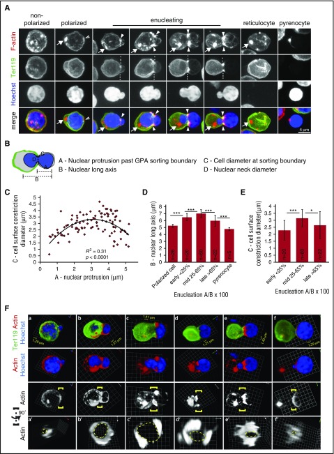
The actin cytoskeleton is critical for efficient erythroblast enucleation,11,12,14,20,25,27,28 but F-actin organization with respect to the morphological progression of enucleation is not understood. Examination of F-actin in mouse FL erythroblasts revealed that nonpolarized erythroblasts exhibit dispersed F-actin fibrils and foci around the periphery of the nucleus (Figure 1A), whereas polarized erythroblasts have a bright spot of F-actin in the cytoplasm on the side of the nucleus opposite its membrane point of contact (Figure 1A arrows). A small F-actin cap is also seen at the site of nuclear contact with the membrane (Figure 1A open arrowhead). Once a nuclear protrusion forms, a membrane protein-sorting boundary appears with low Ter119 signal surrounding the nuclear protrusion in comparison with the remainder of the cell membrane (Figure 1A dashed lines). At this sorting boundary, the enucleating erythroblasts develop a prominent cell surface constriction with bright F-actin foci connecting to small F-actin cables surrounding the protruding nuclear lobe, resembling an F-actin “basket” cradling the nuclear protrusion (Figure 1A closed arrowheads; supplemental movies 1-3). A bright spot of cytoplasmic F-actin remains at the rear of the nucleus, traveling with the nucleus throughout expulsion, approaching the F-actin foci at the cell constriction zone late in enucleation (Figure 1A arrows). The resulting Ter119-positive reticulocyte and Ter119-negative pyrenocyte both contain a bright F-actin spot once they have separated from one another (Figure 1A arrows). Similar F-actin rearrangements and morphological features are evident in erythroblasts prepared without cytospinning, or imaged with structured illumination microscopy (supplemental Figure 1). Mouse bone marrow erythroblasts and splenic erythroblasts from phlebotomized mice also show similar morphologies and patterns of F-actin during enucleation (supplemental Figure 2). Thus, F-actin rearrangements are coordinated with the striking morphological changes observed during erythroblast polarization and nuclear expulsion.
Figure 1.
Mouse FL erythroblast enucleation is characterized by dumbbell-shaped nuclear morphology and prominent F-actin foci at the cell surface constriction and at the rear of the translocating nucleus. (A) Extended-focus projections of confocal Z-stacks of mouse FL erythroblasts at various stages of enucleation immunostained for Ter119, phalloidin for F-actin, and Hoechst for nuclei. Arrows, bright F-actin spot in polarized cells and at the rear of the translocating nucleus opposite its membrane point of contact. Open arrowheads, F-actin cap at the site of nuclear contact with the membrane in a polarized cell. Closed arrowheads, F-actin foci at the cell surface constriction in enucleating cells, connecting to fine F-actin cables extending over the nuclear protrusion. Dashed lines, Ter119 membrane sorting boundary. Bar, 4 µm. (B) Schematic representation of quantitative image analysis of nuclear and cell surface constriction geometry in mouse FL enucleating erythroblasts. (C) Scatter plot of cell surface constriction diameter plotted as a function of nuclear protrusion for erythroblasts at all stages of nuclear expulsion. Parabolic regression was applied to the data (P < .0001). (D) Bar graph of nuclear long axis plotted as a function of nuclear protrusion for erythroblasts binned according to extent of enucleation. ***P < .001. (E) Bar graph of the cell surface constriction diameter size for erythroblasts binned according to extent of enucleation. *P < .05; ***P < .001. Images for quantification of FL erythroblasts were obtained from 133 confocal Z-stacks of cells obtained from 23 embryos (12 different litters). A total of 128 polarized or enucleating cells were analyzed. One hundred two enucleating cells with protruding nuclei were analyzed in panel C and separated into mid, early, and late enucleation stages for panels D-E. Numbers (n) of polarized or enucleating (mid, early, late) cells, or pyrenocytes, indicated on bars in panels D and E. (F) 3D reconstructions of confocal Z-stacks of (a-f) mouse FL erythroblasts at various stages of enucleation immunostained for Ter119, phalloidin for F-actin, and Hoechst for nuclei. Also shown is F-actin at the Ter119-sorting boundary, defined by yellow brackets, of (a′-f′) erythroblasts rotated 90° around the axis running perpendicular to the direction of nuclear expulsion, providing a view through the neck of the enucleating erythroblast. Dashed yellow lines demarcate location of the nuclear perimeter in the neck region. In erythroblasts beginning to enucleate or nearly completely enucleated with smaller constriction diameters, F-actin completely surrounds the nucleus beneath the cell surface constriction (b′, e′). In erythroblasts with larger constriction diameters, F-actin exhibits small gaps (c′) and/or large F-actin–free regions (d′). 3D grid dimensions are as indicated on each image for A-F.
F-actin at the cell surface constriction of mouse erythroblasts is associated with a prominent nuclear constriction, resulting in a dumbbell-shaped nuclear morphology (Figure 1A). This morphology is also observed in histological sections and transmission electron micrographs of mouse FLs (supplemental Figure 3).2,29-31 We performed morphometric analyses of cell and nuclear dimensions to obtain insights into the nature of the cell surface and nuclear constrictions. The diameter of the cell surface constriction can be approximated as a parabolic function of the extent of nuclear protrusion (Figure 1B-C), indicating that the opening through which the nucleus passes is initially small, then enlarges, and finally shrinks as the nucleus is expelled. The length of the nucleus along its long axis (ie, in the direction of expulsion) also increases and then decreases (Figure 1D), indicating that elongation and shortening of the nucleus occurs coincidently with widening and narrowing of the constriction, respectively (Figure 1E). Even at its greatest diameter, the maximum diameter of the opening is smaller than that of the round condensed nucleus in a polarized erythroblast, providing a necessity for nuclear deformation during expulsion (supplemental Table 1). Notably, regardless of the extent of nuclear expulsion, nuclear diameter occupies a constant fraction (∼75%) of the cell surface constriction diameter (supplemental Table 1), implying that the remaining ∼25% of the cell surface constriction diameter is occupied by cytoplasmic and/or cytoskeletal components, such as F-actin (Figure 1A). The biphasic variations in diameter of the constriction neck during nuclear expulsion are distinct from cytokinesis, where the membrane constriction site progressively decreases in diameter.32
F-actin forms an incomplete ring during mouse erythroblast enucleation in vivo and associates with NMIIB and Tmod1 at the rear of the nucleus
To further evaluate F-actin organization at the cell surface constriction, we visualized F-actin in enucleating erythroblasts using 3D reconstructions of confocal Z-stacks, reorienting the cells 90° to view the neck circumference (Figure 1F). In mouse FL erythroblasts early in the process of nuclear expulsion, characterized by a partially protruding nuclear lobe and small neck diameter, F-actin nearly surrounds the nucleus at the cell surface constriction, although the accumulation of F-actin is not uniform around the constriction (Figure 1Fa,a′). In erythroblasts more advanced in the process with a wider cell surface constriction, F-actin fails to completely surround the constriction, exhibiting small gaps and/or large F-actin–free regions (Figure 1Fc,c′-d,d′; supplemental Figure 4A). The discontinuous F-actin at the constriction site in enucleating FL erythroblasts contrasts with dividing FL cells in which a continuous F-actin ring can be observed at the constriction site during cytokinesis (supplemental Figure 4B). Only at the final stage of nuclear expulsion, when the nucleus is nearly completely expelled, and separation of the reticulocyte from the pyrenocyte is imminent, is a continuous F-actin ring evident at the constriction site (Figure 1Fe,e′-f,f′), as previously observed.11,20 By contrast, earlier stages of nuclear expulsion through the erythroblast neck do not involve a continuous F-actin contractile ring (Figure 1Fc,c′-d,d′).
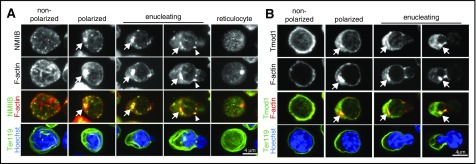
Bipolar myosin filaments comprising NMIIB are proposed to interact with F-actin to produce force for nuclear expulsion.12,33 Indeed, NMIIB colocalizes with the bright F-actin spot at the rear of the translocating nucleus in polarized and enucleating erythroblasts, and to a lesser extent with the F-actin spots in the neck region, but similar to F-actin, NMIIB appears in discrete foci and does not form a continuous ring (Figure 2A arrows and arrowheads). Moreover, mDia2, which nucleates F-actin assembly into the contractile ring in dividing cells,34 is not detected in any of the F-actin foci in polarized or enucleating mouse erythroblasts, even late in the process when the reticulocyte separates from the pyrenocyte (supplemental Figure 5A). By contrast, mDia2 staining is clearly evident in the contractile ring of dividing erythroblasts, as expected28 (supplemental Figure 5A). Arp3, another actin nucleator,35 is also not present in the F-actin foci of polarized or enucleating erythroblasts (supplemental Figure 5B).
Figure 2.
F-actin is associated with NMIIB and Tmod1 in mouse FL enucleating erythroblasts. (A) Extended-focus projections of confocal Z-stacks of mouse FL erythroblasts at various stages of enucleation immunostained for NMIIB and Ter119, phalloidin for F-actin, and Hoechst for nuclei. Arrows, bright F-actin/NMIIB spot at the rear of the translocating nucleus. Arrowheads, F-actin/NMIIB at cell surface constriction site. (B) Confocal single optical sections of mouse FL erythroblasts at various stages of enucleation immunostained for Tmod1 and Ter119, phalloidin for F-actin, and Hoechst for nuclei. Arrows, F-actin/Tmod1 cytoplasmic spot. Bars, 4 µm.
Next, we examined the F-actin pointed-end capping proteins,36 Tmod1 and Tmod3.25 Although Tmod3 is important for enucleation in the mouse FL in vivo,25 Tmod3 does not colocalize with any of the F-actin spots at the rear of the nucleus or at the neck, at any stage of erythroblast polarization or enucleation (supplemental Figure 6B). In contrast, Tmod1 is enriched at the bright cytoplasmic F-actin spots in polarized and enucleating erythroblasts, and at the cell surface constriction site in enucleating erythroblasts (Figure 2B arrows). The association of NMIIB and Tmod1 with these F-actin foci during nuclear expulsion suggests that they may be key participants in F-actin rearrangements associated with the cell and nuclear shape changes during enucleation in mouse erythroblasts, albeit not in a contractile ring as previously hypothesized.3,11
F-actin rearranges but nuclei remain spherical in human bone marrow erythroblasts during enucleation in vivo
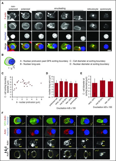
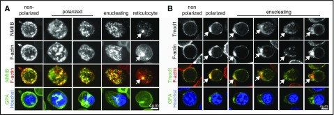
To determine whether the changes in F-actin organization, cell, and nuclear morphology observed during mouse erythroblast enucleation are conserved in human erythroblast enucleation, we visualized human bone marrow erythroblasts harvested directly from normal donors. Human bone marrow erythroblast enucleation is also characterized by a bright F-actin spot or spots in polarized cells, which follow the rear of the translocating nucleus throughout expulsion and are retained in the nascent reticulocyte (Figure 3A arrows; supplemental movies 4 and 5), similar to mouse FL and bone marrow erythroblast enucleation (Figure 1A; supplemental Figures 1-2). The cytoplasmic F-actin spots in enucleating human erythroblasts are associated with NMIIB and Tmod1, similar to mouse (Figure 4A-B). However, unlike mouse erythroblasts, which show robust F-actin foci at the Ter119 sorting boundary coincident with a cell surface constriction (Figure 1A arrowheads), human erythroblasts exhibit only a few small F-actin foci at the glycophorin A (GPA) sorting boundary, and a cell surface constriction is not observed (Figure 3A, arrowhead).
Figure 3.
Human bone marrow erythroblast enucleation is characterized by spherical nuclear morphology and prominent F-actin spots at the rear of the translocating nucleus. (A) Extended-focus projections of confocal Z-stacks of human bone marrow erythroblasts at various stages of enucleation were immunostained for GPA, phalloidin for F-actin, and Hoechst for nuclei. Arrows, prominent cytoplasmic F-actin spots. Arrowheads, small F-actin spot at GPA sorting boundary. Bar, 4 µm. (B) Schematic representation of quantitative image analysis of the geometry of the cell and nucleus at the GPA-sorting boundary. (C) Scatterplot of cell diameter at GPA-sorting boundary plotted as a function of nuclear protrusion for erythroblasts at all stages of nuclear expulsion. No significant correlation was observed. (D) Bar graph of nuclear long axis plotted as a function of extent of nuclear protrusion past the GPA-sorting boundary for erythroblasts binned according to extent of enucleation. The long axis of the nucleus remains unchanged during enucleation. (E) Bar graph of cell diameter at GPA-sorting boundary plotted as a function of nuclear protrusion for erythroblasts binned according to extent of enucleation. Diameter of the GPA-sorting membrane boundary remains constant and is the size of the nucleus. The nucleus does not need to deform to pass through this opening during expulsion from human erythroblasts. Images for quantification of human erythroblasts were obtained from 73 confocal Z-stacks of cells obtained from 6 separate human bone marrow samples. A total of 71 polarized or enucleating cells were analyzed. Twenty-four enucleating cells with protruding nuclei were analyzed in panel C and separated into mid, early, and late enucleation stages for panels D-E. Numbers (n) of polarized or enucleating (mid, early, late) cells or pyrenocytes indicated on bars in panels D and E. (F) 3D reconstructions of confocal Z-stacks of (a-f) human bone marrow erythroblasts at various stages of enucleation immunostained for GPA, phalloidin for F-actin, and Hoechst for nuclei. Also shown is F-actin in regions, defined by yellow brackets, of (a′-f′) human bone marrow erythroblasts rotated 90° around the axis running perpendicular to the direction of nuclear expulsion, providing a view through the “neck” of the enucleating erythroblast. Dashed yellow lines demarcate location of the nuclear perimeter at the GPA-sorting boundary. Few, small puncta of F-actin are associated with the sorting boundaries of the enucleating erythroblasts. 3D grid dimensions are as indicated.
Figure 4.
F-actin is associated with NMIIB and Tmod1 in human enucleating erythroblasts. (A) Extended-focus projections of confocal Z-stacks of human bone marrow erythroblasts at various stages of enucleation immunostained for NMIIB and GPA, phalloidin for F-actin, and Hoechst for nuclei. (B) Confocal single optical sections of human bone marrow erythroblasts at various stages of enucleation immunostained for Tmod1 and GPA, phalloidin for F-actin, and Hoechst for nuclei. Arrows, bright F-actin spots at the rear of the translocating nucleus and in the reticulocyte. Bar, 4 µm.
Moreover, primary human erythroblast nuclei retain their spherical morphology throughout expulsion (Figure 3A), unlike mouse erythroblast nuclei (Figure 1A). This morphology is also observed in histological sections and smears of human bone marrows (supplemental Figure 7). Quantitative image analysis confirmed the lack of cell and nuclear deformation, with the diameter across the GPA sorting boundary and the length of the nucleus along its long axis remaining roughly constant throughout expulsion (Figure 3B-D). Nuclear diameter occupied ∼95% of the GPA sorting boundary diameter throughout expulsion, greater than the ∼75% occupancy observed in mouse erythroblasts (supplemental Table 1), indicating reduced space for cytoskeletal components in human, as compared with mouse erythroblasts, consistent with minimal F-actin staining at the GPA sorting boundary in human erythroblasts (Figure 3A; supplemental movies 4 and 5). Visualization of F-actin at the GPA sorting boundary in 3D reconstructions further revealed F-actin was present in disconnected small foci, inconsistent with formation of a continuous ring during nuclear expulsion (Figure 3F), similar to mouse (Figure 2F).
Mouse and human erythroblasts express different Lamin isoforms
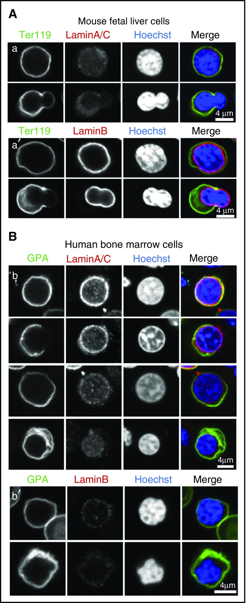
The differences observed in nuclear shapes adopted by mouse and human enucleating cells imply that mouse erythroblast nuclei may be more deformable than human erythroblast nuclei. Previous studies demonstrated that Lamins play an important role in nuclear stiffness, with increasing Lamin A/C to Lamin B ratio correlating with increased nuclear stiffness.37 We therefore stained mouse and human erythroblasts for Lamin A/C and Lamin B. Mouse enucleating erythroblasts exhibit undetectable Lamin A/C staining, but robust Lamin B staining (Figure 5A). By contrast, human erythroblasts have strong Lamin A/C staining but undetectable Lamin B (Figure 5B), consistent with stiffer nuclei in human erythroblasts and softer, more deformable nuclei in mouse.37
Figure 5.
Lamin B but not Lamin A/C is expressed in mouse FL erythroblasts, whereas human bone marrow erythroblasts have variable expression of Lamin A/C but no Lamin B. (A) Confocal single optical sections showing mouse FL erythroblasts immunostained for (a) Lamin A/C, or (a′) Lamin B along with Ter119 and Hoechst. (B) Confocal single optical sections of human bone marrow cells immunostained for (b) Lamin A/C or (b′) Lamin B along with GPA and Hoechst. Bars, 4 µm.
Tmod1 regulates cytoplasmic F-actin and enucleation
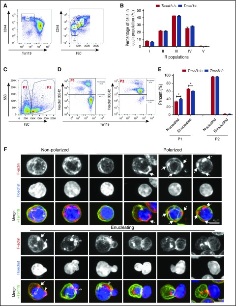
The presence of prominent cytoplasmic F-actin/NMIIB/Tmod1 spots that follow the nucleus during expulsion raised the possibility that this F-actin may play a conserved role in nuclear expulsion. First, we used our Tmod1−/− mouse model, which develops a mild anemia.24 We evaluated terminal erythroid differentiation and enucleation in the FL because the larger-sized erythroblasts (compared with adult) make it easier to evaluate cell morphologies and F-actin organization. Flow cytometry38,39 demonstrates that erythropoiesis is normal in wild-type and Tmod1−/− FL cells (Figure 6A-B). Measurement of enucleation in the small subpopulation of Ter119-positive cells (P1)15,40 identified a small but significant reduction in the percentage of enucleated cells in the Tmod1−/− FL, indicative of impaired enucleation in the absence of Tmod1 (Figure 6C-E). As control, we examined enucleation in the larger population (P2). Negligible enucleated cells were observed in these wild-type and Tmod1−/− erythroblasts, precluding premature enucleation (Figure 6C-E).
Figure 6.
Tmod1−/−FL erythroblasts differentiate normally but exhibit a slight enucleation defect and aberrant F-actin organization during polarization and enucleation. (A) Representative flow cytometry gating strategy of mouse FL cells from Tmod1+/+ and Tmod1−/− E14.5 embryos based on CD44 and Ter119 expression levels (left panel). High Ter119 cells are gated for CD44 and forward scatter (FSC) to reveal RII to RV populations (right panel). (B) Quantification of R populations from Tmod1+/+ and Tmod1−/− embryos. (C) Representative flow cytometry gating strategy of FL erythroblasts by forward scatter (FSC) and side scatter (SSC) identifies 2 populations of cells P1 and P2. (D) Enucleation in FL cells determined by Ter119 and Hoechst 33342 staining of P1 (left panel) or P2 population (right panel) to identify nucleated (HoechstHi) vs enucleated (HoechstLo) cells. (E) Quantification of the percent of enucleated cells in each population in FL cells from Tmod1+/+ and Tmod1−/− mouse embryos (N = 13 embryo FLs per group). The smaller P1 population of more mature cells shows impaired enucleation, whereas the larger P2 population representing larger more immature erythroid cells has negligible quantities of enucleated cells that do not change in the absence of Tmod1. (F) Extended focus projections of confocal Z-stacks of Tmod1−/− FL erythroblasts immunostained for Ter119, phalloidin for F-actin, and Hoechst for nuclei. Merges show Ter119 (green), F-actin (red), and Hoechst (blue). Arrows show mislocalized F-actin spots in polarized cells. Open arrowheads show abnormal F-actin accumulation at enucleating erythroblast neck. Protruding nuclear lobes show increased F-actin cables. Images of individual cells are representative of a total of 89 nucleated Ter119-stained erythroblasts, imaged in confocal Z stacks of FL cells obtained from 11 Tmod1−/− embryos (2 different litters). Of the 89 total Ter119-stained polarized and enucleating erythroblasts examined, 28 (∼31%) had abnormal F-actin (multiple foci in polarized cells, mislocalized F-actin spot near constriction, nonpolarized cells with large F-actin aggregates). Bars, 4 µm.
Confocal microscopy of polarized and enucleating Tmod1−/− FL erythroblasts indicates that F-actin organization is strikingly abnormal (Figure 6F). In polarized cells, instead of 1 prominent cytoplasmic F-actin spot, there are often several intense F-actin spots located around the nucleus in a nonpolarized fashion (Figure 6F arrows). In enucleating erythroblasts, the cytoplasmic F-actin spot is not always present and often appears in the wrong location near the neck, or over the emerging nuclear protrusion (Figure 6F arrowheads). The nucleus often assumes irregular shapes rather than the dumbbell-shaped morphology typical of wild-type cells. Because Tmod3 can nucleate F-actin assembly,36 and Tmod3 was previously shown to be important for mouse FL erythroblast enucleation,25 we tested whether increased Tmod3 might lead to abnormal F-actin and/or partly compensate for absence of Tmod1. However, Tmod3 levels appeared similar in wild-type and Tmod1−/− polarized or enucleating erythroblasts (supplemental Figure 6A). Thus, loss of Tmod1 in the mouse leads to abnormal F-actin organization and impaired enucleation.
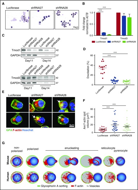
To assess the function of Tmod1 during human erythroid differentiation, we used lentivirally encoded short hairpin RNAs (shRNAs) to knockdown TMOD1 in CD34+ cells differentiating to erythroid cells in culture (Figure 7). Two different shRNAs corresponding to different regions of TMOD1 were used, both efficiently decreasing Tmod1 expression by >80% (Figure 7B-C). Using flow cytometry,26 we observed that TMOD1 knockdown leads to only a slight delay in the terminal stages of differentiation, suggesting a role for TMOD1 in the very late stages (supplemental Figure 8A). We sorted the differentiated cells (supplemental Figure 8B), and using SYTO60 for nuclei, we observed that the absence of TMOD1 severely impairs enucleation (Figure 7D). In TMOD1 knockdown conditions, TMOD3 levels are unchanged (Figure 7C), precluding compensatory mechanisms. Strikingly, TMOD1 knockdown erythroblasts exhibit a considerably greater accumulation of F-actin in the cytoplasmic spot at the rear of the nucleus (Figure 7E-F). This F-actin spot often colocalizes with GPA, suggesting that membrane vesicles may be associated with the abnormal F-actin spot. These observations suggest a new function for Tmod1 in the formation and the regulation of this F-actin spot during enucleation.
Figure 7.
TMOD1 knockdown during human erythroblast differentiation impairs enucleation and increases the amount of cytoplasmic F-actin in polarized erythroblasts. CD34+ cells were transduced with either luciferase (control) or TMOD1 (shRNA27 or shRNA28) lentivirus and induced toward erythroid differentiation. Puromycin selection was performed 48 hours posttransduction. (A) Wright Giemsa–stained cytospins of CD34+ cells at day 14. Bar, 10 µm. (B) Quantitative reverse transcription polymerase chain reaction of TMOD1 and TMOD3 levels normalized to ACTB in Luciferase control or TMOD1 knockdown. ***P < .001 (N = 3). (C) Representative western blot for Tmod1 and Tmod3 and glyceraldehyde-3-phosphate dehydrogenase (GAPDH) at day 11 and day 14 of differentiation. (D) Late orthochromatic erythroblast populations were flow sorted at day 16 based on the surface expression levels of GPA, α4-integrin, and Band3. SYTO60 was used to measure enucleation within these populations. ***P < .001. (E) Extended-focus projections of confocal Z-stacks of polarized, differentiated CD34+ cells (control or TMOD1 knockdown), immunostained for GPA, phalloidin for F-actin, and Hoechst for nuclei. Arrows show increased size of cytoplasmic F-actin spot in TMOD1 knockdown cells. Bar, 4 µm. (F) Average F-actin spot volume in polarized, or enucleating differentiated CD34+ cells in panel E. ***P < .001. N = 26 cells for Luciferase control, 29 for shRNA27, and 32 for shRNA28, obtained from 2 separate knockdown experiments. (G) Schematics of GPA sorting (green), F-actin reorganization (red), and nuclear (blue) expulsion during the progression of mouse and human erythroblast enucleation. In both mouse and human erythroblasts, the F-actin network undergoes dramatic reorganization during enucleation. As the erythroblasts polarize, an F-actin spot (the enucleosome) appears at the rear of the nucleus. The enucleosome follows the translocating nucleus and may drive expulsion. Mouse erythroblast nuclei adopt a dumbbell-shaped morphology during expulsion, with prominent F-actin foci associated with the cell surface and nuclear constriction at the Ter119-sorting boundary, whereas human erythroblast nuclei retain a spherical morphology, and only very small F-actin foci are present at the GPA-sorting boundary over the translocating nucleus. Small membrane vesicles are depicted near the rear of the translocating nucleus, based on our (eg, supplemental Figure 3B) and others’ observations.11,18 For simplicity, microtubules are not depicted.
Discussion
Enucleation is a key process of mammalian erythropoiesis and was first observed in 1907 by Jolly.41 Erythroblast nuclear expulsion has been studied in different species, by a variety of methods,30,31,42-44 and shares features of asymmetric cell division with the formation of 2 divergent daughter “cells”: an enucleated reticulocyte and a pyrenocyte (expelled nucleus). Observations by Koury et al20 and others2,11,12,14 that F-actin and NMIIB accumulate at the site of nuclear expulsion also suggested that a contractile ring provides force to expel the nucleus, similar to cytokinesis. Here we tested this model using high-resolution confocal fluorescence microscopy of enucleating primary mouse and human erythroblasts, revealing novel and unexpected details of F-actin and NMIIB organization during nuclear polarization and expulsion (Figure 7G). First, we discovered a novel F-actin structure in the cytoplasm we have named the “enucleosome,” associated with NMIIB and Tmod1, appearing initially in polarized erythroblasts and persisting throughout nuclear expulsion, following along with the rear of the translocating nucleus in both mouse and human erythroblasts. Second, we observed large F-actin/NMIIB foci at the Ter119-sorting boundary during nuclear expulsion in mouse erythroblasts, coinciding with a cell surface constriction and nuclear deformation. In human erythroblasts, a cell surface constriction does not form, the nucleus remains round, and only very small F-actin foci are present at the GPA-sorting boundary. Third, the F-actin foci at the Ter119/GPA-sorting boundary (or neck) do not coalesce into a continuous ring with a decreasing diameter during progression of nuclear expulsion in either mouse or human erythroblasts. Only at the completion of enucleation does F-actin appear to form a small contractile ring-like structure to finally separate the pyrenocyte from the reticulocyte in both mouse and human erythroblasts (Figures 1F and 3F).11,20
These data suggest that a contractile ring is unlikely to function in expelling the nucleus. It is also difficult to imagine how constriction of an F-actin contractile ring orthogonal to the direction of nuclear expulsion would provide forces to expel the nucleus, rather than cutting it in half. By contrast, the enucleosome located at the rear of the translocating nucleus is well positioned to exert forces in the direction of nuclear expulsion. This structure could provide force via actin polymerization-based mechanisms directly pushing on the nucleus, or by actomyosin contractility in the cytoplasm indirectly pushing on the nucleus,45 as initially proposed by Wang et al2 for mouse erythroblasts. In addition, F-actin polymerization and actomyosin contractility processes are likely to coordinate with microtubule- and vesicular trafficking-based mechanisms to expel the nucleus.2,4,11,16,18,20,46 Future studies of nuclear expulsion using live cell microscopy and fluorescent-tagged proteins will be required to explore these ideas further.
During enucleation, the nucleus adopts different shapes in human as compared with mouse primary erythroblasts (ie, spherical or dumbbell shapes, respectively), and this characteristic is species specific. Curiously, both dumbbell-shaped and round nuclei can be observed in enucleating erythroblasts derived from CD34+ progenitors using in vitro culture systems,29 but not in vivo, as shown here. In mouse erythroblasts, robust accumulation of F-actin and NMIIB at the Ter119-sorting boundary may provide contractile forces that constrict the cell surface and deform the nucleus during expulsion, whereas in human erythroblasts, the low abundance of F-actin and NMIIB at the GPA-sorting boundary may explain both the absence of cell surface constriction and the lack of nuclear deformation. Nuclear stiffness could also contribute to differences in nuclear deformation. Although biomechanical measurements have not been performed for mouse erythroblast nuclei, a useful predictor of erythroid nuclear stiffness is thought to be its Lamin A:B ratio, which increases with nuclear stiffness as observed by Discher and colleagues.37 We observed that mouse erythroblast nuclei exhibit little to no Lamin A signal in immunofluorescence, but show robust Lamin B staining in accordance with previous studies,37,47 implying a low Lamin A:B ratio, low predicted stiffness, and superior deformability. Although nuclear morphology during enucleation varies in different species, and in vitro cultures,29-31,42,43 elucidation of a direct role for Lamin A:B ratio awaits functional experiments.
Our observation that the F-actin–capping protein Tmod1 is associated with the enucleosome in mouse and human erythroblasts raised the possibility that Tmod1 could regulate its assembly, providing an approach to evaluate its importance for enucleation. Therefore, we studied effects of Tmod1 knockout or knockdown in mouse and human erythropoiesis using in vivo and in vitro models. In Tmod1−/− mouse FL erythroblasts, terminal differentiation is not affected, but enucleation is mildly impaired and F-actin organization is strikingly abnormal with extra F-actin spots in incorrect locations. Because Tmod3 is not detected in the enucleosome from wild-type or Tmod1−/− mice, and Tmod3 levels do not appear increased in Tmod1−/− erythroblasts, we speculate that the mild inhibition of enucleation in absence of Tmod1 may be due to actin-regulatory factors other than Tmod3, although we cannot rule out partial compensation by Tmod3, as observed in Tmod1−/− mature erythrocytes.24 Tmod3−/− FL erythroblasts also present abnormal F-actin organization and impaired enucleation.25 However, Tmod3 may operate via a different molecular mechanism than Tmod1, based on their different subcellular locations and different F-actin abnormalities in their absence.25 On the other hand, because erythropoiesis is abnormal in Tmod3−/− but not Tmod1−/− FL erythroblasts, Tmod3 might regulate F-actin before the orthochromatic stage, thereby leading subsequently to enucleation defects.
In human erythroblasts in which TMOD1 levels had been reduced by lentiviral shRNA knockdown, terminal differentiation is also unaffected, but enucleation is dramatically reduced. In addition, in TMOD1-depleted polarized or enucleating erythroblasts, the enucleosome appears larger and brighter than in control cells. Thus, absence of Tmod1 (in mouse) or reduced levels of TMOD1 (in human) leads to dysregulation of F-actin, likely due to impaired capping and increased actin polymerization at pointed ends, which in turn adversely impacts enucleation. The altered organization of the F-actin cytoplasmic spots in the Tmod1 knockout or TMOD1 knockdown conditions strongly suggests a role for the enucleosome in providing forces to expel the nucleus. In human erythroblasts, we can infer that there is no compensation from another Tmod family member, because the levels of TMOD3 (the only other Tmod expressed in the erythroid lineage) are unchanged in the TMOD1 knockdown.
In summary, our study describes the F-actin rearrangements during mouse and human erythroblast enucleation and highlights the presence of the enucleosome, a novel F-actin structure at the rear of the nucleus, enriched in F-Actin, NMIIB, and Tmod1. To our knowledge, such a structure has never been described in primary cells before. We further demonstrate that Tmod1 is necessary for the regulation of F-actin polymerization in the human and mouse enucleosome, thereby controlling enucleation. Future studies will seek to identify the trafficking, attachment/anchorage points, molecular composition, and ultrastructure of the enucleosome.
Supplementary Material
The online version of this article contains a data supplement.
Acknowledgments
The authors thank Indira Sahdev and Suzan Durham for their help in obtaining bone marrow samples and Mohandas Narla for scientific discussions and his gift of the Band 3 antibody.
This research was supported by National Heart, Lung, and Blood Institute, National Institutes of Health grants R01-HL083464 (V.M.F.), R01-HL079571 (J.M.L.), and R01-HL134043 (subcontract to L.B.); National Institute of Arthritis and Musculoskeletal and Skin Diseases, National Institutes of Health K99-AR066534 (D.S.G.); National Institute of Diabetes and Digestive and Kidney Diseases, National Institutes of Health R01-DK095112 and R01-DK090554 (S.R.); and by the Pediatric Cancer Foundation (J.M.L. and L.B.). L.B. is the recipient of a St. Baldrick’s Scholar award.
Footnotes
The publication costs of this article were defrayed in part by page charge payment. Therefore, and solely to indicate this fact, this article is hereby marked “advertisement” in accordance with 18 USC section 1734.
Authorship
Contribution: R.B.N. and D.S.G. designed and performed research, analyzed data, and wrote the manuscript; J.P. and C.C. performed research, analyzed data, and edited the manuscript; S.R. and J.M.L. analyzed data and edited the manuscript; and L.B. and V.M.F. designed research, analyzed data, and edited the manuscript.
Conflict-of-interest disclosure: The authors declare no competing financial interests.
Correspondence: Velia M. Fowler, Department of Molecular Medicine, The Scripps Research Institute, 10550 N Torrey Pines Rd, MB114, La Jolla, CA 92037; e-mail: velia@scripps.edu; and Lionel Blanc, Laboratory of Developmental Erythropoiesis, The Feinstein Institute for Medical Research, 350 Community Dr, Manhasset, NY 11030; e-mail: lblanc@northwell.edu.
References
- 1.Hebiguchi M, Hirokawa M, Guo YM, et al. . Dynamics of human erythroblast enucleation. Int J Hematol. 2008;88(5):498-507. [DOI] [PubMed] [Google Scholar]
- 2.Wang J, Ramirez T, Ji P, Jayapal SR, Lodish HF, Murata-Hori M. Mammalian erythroblast enucleation requires PI3K-dependent cell polarization. J Cell Sci. 2012;125(Pt 2):340-349. [DOI] [PMC free article] [PubMed] [Google Scholar]
- 3.Ji P, Murata-Hori M, Lodish HF. Formation of mammalian erythrocytes: chromatin condensation and enucleation. Trends Cell Biol. 2011;21(7):409-415. [DOI] [PMC free article] [PubMed] [Google Scholar]
- 4.Keerthivasan G, Wickrema A, Crispino JD. Erythroblast enucleation. Stem Cells Int. 2011;2011:139851. [DOI] [PMC free article] [PubMed] [Google Scholar]
- 5.Palis J. Ontogeny of erythropoiesis. Curr Opin Hematol. 2008;15(3):155-161. [DOI] [PubMed] [Google Scholar]
- 6.Koury MJ. Abnormal erythropoiesis and the pathophysiology of chronic anemia. Blood Rev. 2014;28(2):49-66. [DOI] [PubMed] [Google Scholar]
- 7.McGrath K, Palis J. Ontogeny of erythropoiesis in the mammalian embryo. Curr Top Dev Biol. 2008;82:1-22. [DOI] [PubMed] [Google Scholar]
- 8.Ji P. New insights into the mechanisms of mammalian erythroid chromatin condensation and enucleation. Int Rev Cell Mol Biol. 2015;316:159-182. [DOI] [PubMed] [Google Scholar]
- 9.Gnanapragasam MN, Bieker JJ. Orchestration of late events in erythropoiesis by KLF1/EKLF. Curr Opin Hematol. 2017;24(3):183-190. [DOI] [PMC free article] [PubMed] [Google Scholar]
- 10.Gnanapragasam MN, McGrath KE, Catherman S, Xue L, Palis J, Bieker JJ. EKLF/KLF1-regulated cell cycle exit is essential for erythroblast enucleation. Blood. 2016;128(12):1631-1641. [DOI] [PMC free article] [PubMed] [Google Scholar]
- 11.Konstantinidis DG, Pushkaran S, Johnson JF, et al. . Signaling and cytoskeletal requirements in erythroblast enucleation. Blood. 2012;119(25):6118-6127. [DOI] [PMC free article] [PubMed] [Google Scholar]
- 12.Ubukawa K, Guo YM, Takahashi M, et al. . Enucleation of human erythroblasts involves non-muscle myosin IIB. Blood. 2012;119(4):1036-1044. [DOI] [PMC free article] [PubMed] [Google Scholar]
- 13.Kalfa TA, Pushkaran S, Zhang X, et al. . Rac1 and Rac2 GTPases are necessary for early erythropoietic expansion in the bone marrow but not in the spleen. Haematologica. 2010;95(1):27-35. [DOI] [PMC free article] [PubMed] [Google Scholar]
- 14.Ji P, Jayapal SR, Lodish HF. Enucleation of cultured mouse fetal erythroblasts requires Rac GTPases and mDia2. Nat Cell Biol. 2008;10(3):314-321. [DOI] [PubMed] [Google Scholar]
- 15.Lu Y, Hanada T, Fujiwara Y, et al. . Gene disruption of dematin causes precipitous loss of erythrocyte membrane stability and severe hemolytic anemia. Blood. 2016;128(1):93-103. [DOI] [PMC free article] [PubMed] [Google Scholar]
- 16.Thom CS, Traxler EA, Khandros E, et al. . Trim58 degrades Dynein and regulates terminal erythropoiesis. Dev Cell. 2014;30(6):688-700. [DOI] [PMC free article] [PubMed] [Google Scholar]
- 17.Kobayashi I, Ubukawa K, Sugawara K, et al. . Erythroblast enucleation is a dynein-dependent process. Exp Hematol. 2016;44(4):247-256.e212. [DOI] [PubMed] [Google Scholar]
- 18.Keerthivasan G, Small S, Liu H, Wickrema A, Crispino JD. Vesicle trafficking plays a novel role in erythroblast enucleation. Blood. 2010;116(17):3331-3340. [DOI] [PMC free article] [PubMed] [Google Scholar]
- 19.Keerthivasan G, Liu H, Gump JM, Dowdy SF, Wickrema A, Crispino JD. A novel role for survivin in erythroblast enucleation. Haematologica. 2012;97(10):1471-1479. [DOI] [PMC free article] [PubMed] [Google Scholar]
- 20.Koury ST, Koury MJ, Bondurant MC. Cytoskeletal distribution and function during the maturation and enucleation of mammalian erythroblasts. J Cell Biol. 1989;109(6 Pt 1):3005-3013. [DOI] [PMC free article] [PubMed] [Google Scholar]
- 21.Yamashiro S, Gokhin DS, Kimura S, Nowak RB, Fowler VM. Tropomodulins: pointed-end capping proteins that regulate actin filament architecture in diverse cell types. Cytoskeleton. 2012;69(6):337-370. [DOI] [PMC free article] [PubMed] [Google Scholar]
- 22.Fowler VM, Dominguez R. Tropomodulins and leiomodins: actin pointed end caps and nucleators in muscles. Biophys J. 2017;112(9):1742-1760. [DOI] [PMC free article] [PubMed] [Google Scholar]
- 23.Fowler VM. Tropomodulin: a cytoskeletal protein that binds to the end of erythrocyte tropomyosin and inhibits tropomyosin binding to actin. J Cell Biol. 1990;111(2):471-481. [DOI] [PMC free article] [PubMed] [Google Scholar]
- 24.Moyer JD, Nowak RB, Kim NE, et al. . Tropomodulin 1-null mice have a mild spherocytic elliptocytosis with appearance of tropomodulin 3 in red blood cells and disruption of the membrane skeleton. Blood. 2010;116(14):2590-2599. [DOI] [PMC free article] [PubMed] [Google Scholar]
- 25.Sui Z, Nowak RB, Bacconi A, et al. . Tropomodulin3-null mice are embryonic lethal with anemia due to impaired erythroid terminal differentiation in the fetal liver. Blood. 2014;123(5):758-767. [DOI] [PMC free article] [PubMed] [Google Scholar]
- 26.Hu J, Liu J, Xue F, et al. . Isolation and functional characterization of human erythroblasts at distinct stages: implications for understanding of normal and disordered erythropoiesis in vivo. Blood. 2013;121(16):3246-3253. [DOI] [PMC free article] [PubMed] [Google Scholar]
- 27.Repasky EA, Eckert BS. The effect of cytochalasin B on the enucleation of erythroid cells in vitro. Cell Tissue Res. 1981;221(1):85-91. [DOI] [PubMed] [Google Scholar]
- 28.Watanabe S, De Zan T, Ishizaki T, et al. . Loss of a Rho-regulated actin nucleator, mDia2, impairs cytokinesis during mouse fetal erythropoiesis. Cell Reports. 2013;5(4):926-932. [DOI] [PubMed] [Google Scholar]
- 29.Bell AJ, Satchwell TJ, Heesom KJ, et al. . Protein distribution during human erythroblast enucleation in vitro. PLoS One. 2013;8(4):e60300. [DOI] [PMC free article] [PubMed] [Google Scholar]
- 30.Skutelsky E, Danon D. An electron microscopic study of nuclear elimination from the late erythroblast. J Cell Biol. 1967;33(3):625-635. [DOI] [PMC free article] [PubMed] [Google Scholar]
- 31.Skutelsky E, Danon D. Reduction in surface charge as an explanation of the recognition by macrophages of nuclei expelled from normoblasts. J Cell Biol. 1969;43(1):8-15. [DOI] [PMC free article] [PubMed] [Google Scholar]
- 32.Green RA, Paluch E, Oegema K. Cytokinesis in animal cells. Annu Rev Cell Dev Biol. 2012;28:29-58. [DOI] [PubMed] [Google Scholar]
- 33.Kalfa TA, Zheng Y. Rho GTPases in erythroid maturation. Curr Opin Hematol. 2014;21(3):165-171. [DOI] [PMC free article] [PubMed] [Google Scholar]
- 34.Watanabe S, Okawa K, Miki T, et al. . Rho and anillin-dependent control of mDia2 localization and function in cytokinesis. Mol Biol Cell. 2010;21(18):3193-3204. [DOI] [PMC free article] [PubMed] [Google Scholar]
- 35.Goley ED, Welch MD. The ARP2/3 complex: an actin nucleator comes of age. Nat Rev Mol Cell Biol. 2006;7(10):713-726. [DOI] [PubMed] [Google Scholar]
- 36.Yamashiro S, Speicher KD, Speicher DW, Fowler VM. Mammalian tropomodulins nucleate actin polymerization via their actin monomer binding and filament pointed end-capping activities. J Biol Chem. 2010;285(43):33265-33280. [DOI] [PMC free article] [PubMed] [Google Scholar]
- 37.Shin JW, Spinler KR, Swift J, Chasis JA, Mohandas N, Discher DE. Lamins regulate cell trafficking and lineage maturation of adult human hematopoietic cells. Proc Natl Acad Sci USA. 2013;110(47):18892-18897. [DOI] [PMC free article] [PubMed] [Google Scholar]
- 38.Liu J, Zhang J, Ginzburg Y, et al. . Quantitative analysis of murine terminal erythroid differentiation in vivo: novel method to study normal and disordered erythropoiesis. Blood. 2013;121(8):e43-e49. [DOI] [PMC free article] [PubMed] [Google Scholar]
- 39.Kusakabe M, Hasegawa K, Hamada M, et al. . c-Maf plays a crucial role for the definitive erythropoiesis that accompanies erythroblastic island formation in the fetal liver. Blood. 2011;118(5):1374-1385. [DOI] [PubMed] [Google Scholar]
- 40.Yoshida H, Kawane K, Koike M, Mori Y, Uchiyama Y, Nagata S. Phosphatidylserine-dependent engulfment by macrophages of nuclei from erythroid precursor cells. Nature. 2005;437(7059):754-758. [DOI] [PubMed] [Google Scholar]
- 41.Jolly J. Recherches sur la formation des globules rouges des mammiferes. Archives d'Anatomie Microscopique 1907;T9(Fasc 2).
- 42.Bessis M, Bricka M. [Dynamic aspect of blood cells; study by phase contrast microcinematography.] Rev Hematol. 1952;7(3):407-435. [PubMed] [Google Scholar]
- 43.Simpson CF, Kling JM. The mechanism of denucleation in circulating erythroblasts. J Cell Biol. 1967;35(1):237-245. [DOI] [PMC free article] [PubMed] [Google Scholar]
- 44.Skutelsky E, Farquhar MG. Variations in distribution of con A receptor sites and anionic groups during red blood cell differentiation in the rat. J Cell Biol. 1976;71(1):218-231. [DOI] [PMC free article] [PubMed] [Google Scholar]
- 45.Yi K, Li R. Actin cytoskeleton in cell polarity and asymmetric division during mouse oocyte maturation. Cytoskeleton. 2012;69(10):727-737. [DOI] [PubMed] [Google Scholar]
- 46.Chasis JA, Prenant M, Leung A, Mohandas N. Membrane assembly and remodeling during reticulocyte maturation. Blood. 1989;74(3):1112-1120. [PubMed] [Google Scholar]
- 47.Pajerowski JD, Dahl KN, Zhong FL, Sammak PJ, Discher DE. Physical plasticity of the nucleus in stem cell differentiation. Proc Natl Acad Sci USA. 2007;104(40):15619-15624. [DOI] [PMC free article] [PubMed] [Google Scholar]
Associated Data
This section collects any data citations, data availability statements, or supplementary materials included in this article.


