
Figure 2. Footprint fragment size-selection gel.
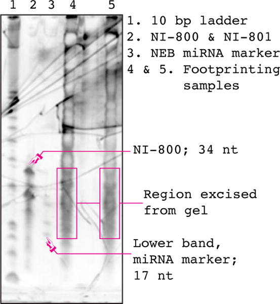
An image of a typical gel prepared according to Section 5.3. Footprinting samples were prepared from cultures of wild-type S. cerevisiae strain BY4742, as described in this protocol. NI-800 is used to determine the lower position on the gel to cut in order to isolate large (~28 nt) footprints. It is still routinely run, but has been made somewhat redundant by the discovery of small footprints and the use of the NEB miRNA marker.