Diaphragmatic paralysis can be either unilateral or bilateral. It is a rare condition and can have several causes [1]. As the diaphragm is the most important muscle of inspiration, diaphragmatic paralysis commonly complicates ventilation.
Short abstract
Mouthpiece ventilation can improve exercise tolerance in patients with unilateral or bilateral diaphragm paralysis http://ow.ly/X2Pd30dCT7n
Diaphragmatic paralysis can be either unilateral or bilateral. It is a rare condition and can have several causes [1]. As the diaphragm is the most important muscle of inspiration, diaphragmatic paralysis commonly complicates ventilation.
Unilateral diaphragmatic paralysis (UDP) does not usually cause any symptoms at rest but may cause dyspnoea during mild exertion [2]. Bilateral diaphragmatic paralysis (BDP) is associated with dyspnoea that worsens when the patient is recumbent, increased work of breathing and exercise intolerance. The aggravation of dyspnoea when recumbent results in marginal sleep quality with frequent awakenings. With disease progression, there is increasing ventilatory failure with hypoxaemia and hypercapnia, which may further worsen due to atelectasis and ventilation–perfusion mismatch [1].
Nocturnal respiratory failure in BDP is treated with noninvasive ventilation (NIV), with improvement of sleep quality and ventilation [3]. However, feasible options to treat daytime exercise-induced complaints are lacking. Here, we describe two cases of BDP and UDP treated with ventilatory support during exercise using mouthpiece ventilation (MPV).
Recently, MPV was described in the context of increased availability of MPV modes on portable ventilators [4]. When daytime NIV is necessary, MPV is the more comfortable and logical mode of ventilation to administer. MPV is an open system of ventilatory support. When set to the assist-control mode with a physiological backup rate, air delivery is triggered by placing the lips on the mouthpiece, thereby creating negative pressure by inhaling through the mouthpiece. Ventilators with volume cycling in assist-control mode also permit air stacking, thereby increasing lung volumes to improve speech or coughing.
Case presentation
Case 1
A 74-year-old male with BDP due to neuralgic amyotrophy presented with progressive night-time symptoms. He was unable to lie recumbent and daytime hypercapnia was noted. Lung function worsened excessively when recumbent (table 1).
Table 1.
Lung function results in sitting and supine positions
| Predicted | Sitting position | Supine position | Sitting/supine | |||||
| Actual | % pred | LLN | ULN | Actual | % pred | |||
| Case 1 | ||||||||
| FEV1 L | 3.49 | 1.47 | 42% | 76.0 | 124 | 0.79 | 23% | 54% |
| FVC IN L | 4.77 | 2.92 | 61% | 80.7 | 119 | 1.65 | 35% | 57% |
| FEV1 % VCmax | 74.4% | 50.5% | 68% | 84.2% | 116% | 39.3% | 53% | 78% |
| FVC L | 4.59 | 2.90 | 63% | 78.2 | 122 | 2.01 | 44% | 69% |
| Case 2 | ||||||||
| FEV1 L | 3.57 | 2.29 | 64% | 76.5 | 124 | 1.10 | 31% | 48% |
| FVC IN L | 4.82 | 2.82 | 59% | 80.9 | 119 | 1.53 | 32% | 54% |
| FEV1 % VCmax | 75.2 | 81.0 | 108% | 84.4 | 116 | 72.3 | 96% | 89% |
| FVC L | 4.63 | 2.60 | 56% | 78.4 | 122 | 1.13 | 24% | 44% |
LLN: lower limit of normal; ULN: upper limit of normal; FEV1: forced expiratory volume in 1 s; FVC: forced vital capacity; IN: inspiratory; VCmax: maximal vital capacity.
Nocturnal NIV was initiated (BiPAP A40; Phillips, Amsterdam, the Netherlands) using a facial mask as the interface. This was easily accepted with nearly immediate subjective benefits and reduction of hypercapnia. However, he still had progressive complaints of severe dyspnoea after minimal exercise, being unable to use the bathroom independently. Given this level of disability, daytime ventilatory support through MPV was considered. The Elisee 150 ventilator (Resmed, San Diego, CA, USA) was chosen as the assisted breathing device, mainly because of its portability during exercise, and its convenient size and weight (26×24×13 cm and 4 kg, respectively) (see table 2 for ventilator settings).
Table 2.
Ventilator settings
| Case 1 | Case 2 | |
| Mode | PACV | ACV |
| Backup rate | 2 | 0 |
| IPAP cmH2O | 13 | 0 |
| EPAP cmH2O | 0 | 0 |
| Volume guarantee mL | Trigger auto | 1200 |
PACV: pressure-assisted controlled volume; ACV: assisted controlled volume; IPAP: inspiratory positive airway pressure; EPAP: expiratory positive airway pressure.
A 6-min walking distance (6MWD) test was performed with and without MPV to measure the impact of MPV on exercise tolerance and exercise-induced dyspnoea. Without ventilatory support, the patient walked 106 m with 20 stops. After 1 h of rest, he performed a 6MWD test using MPV. A specialist homecare ventilation nurse carried the ventilator. His 6MWD improved to 291 m with only three stops, an improvement in distance of 175% together with a spectacular decrease in the number of stops (table 3).
Table 3.
Results of the 6MWD test without and with MPV
| Case 1 | Case 2 | |||
| Without MPV | With MPV | Without MPV | With MPV | |
| 6MWD m | 106 | 291 | 135 | 200 |
| Stops | 20 | 3 | 1 | 0 |
| Borg dyspnoea score | ||||
| Start of test | 7 | 5 | 5 | 7 |
| End of test | 9 | 9 | 7 | 5 |
| Saturation | ||||
| Start of test | 96% | 95% | 97% | 94% |
| End of test | 96% | 95% | 97% | 93% |
| Control after 2 min pause | 96% | 99% | 99% | 93% |
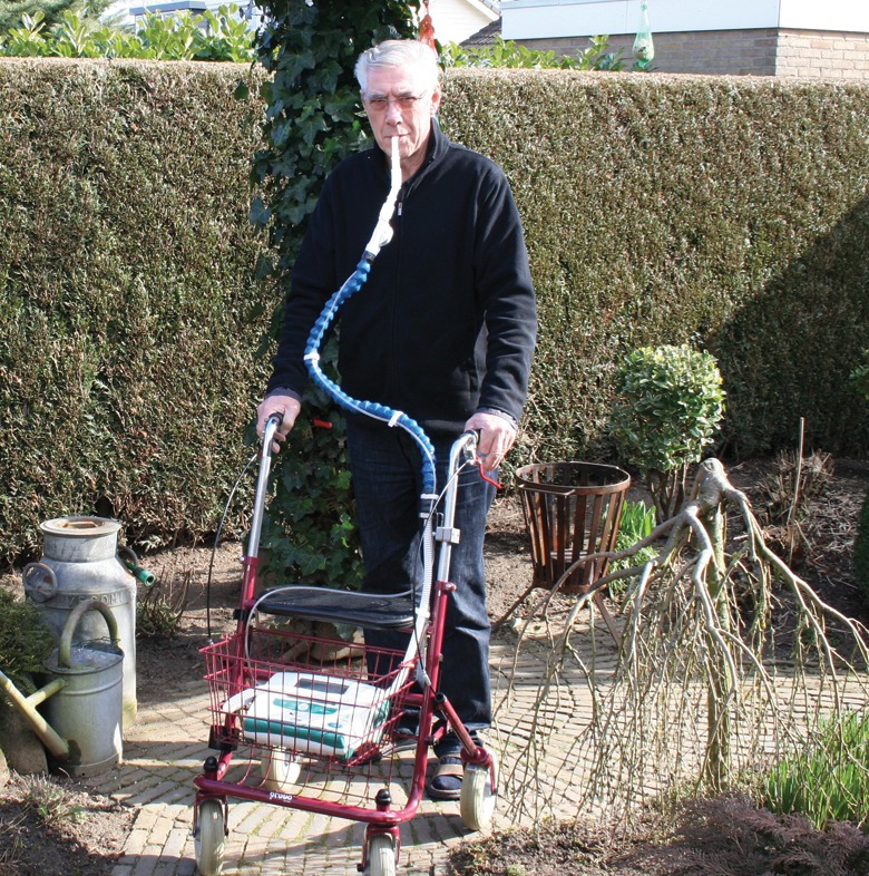
At the time of writing, he is able to function at a satisfactory level when using the device (figure 1).
Figure 1.

Patient with BDP using MPV during exercise.
Case 2
A 69-year-old male with neuralgic amyotrophy-related UDP, consulted with worsening sleep quality and intolerance of continuous positive airway pressure (CPAP) therapy, which he had been using in the past for obstructive sleep apnoea. His lung function was significantly worse when recumbent (table 1) and radioscopy of the diaphragm showed severe reduced movement of the right hemidiaphragm. His CPAP therapy was changed to NIV, which improved sleep quality and resulted in less fatigue.
In 2015, he returned with decrease of exercise tolerance due to heavy dyspnoea; he was almost unable to leave the house. His 6MWD was 135 m with one stop (table 3).
Consequently, it was decided to change his ventilator to NIV with the Astral 150 (Resmed), which is suitable for daytime MPV, and has favourable size and weight properties (28×21×9 cm and 3.2 kg, respectively) (see table 2 for ventilator settings). His 6MWD improved to 200 m with MPV, with no need to stop; an improvement 148% in distance (table 3).
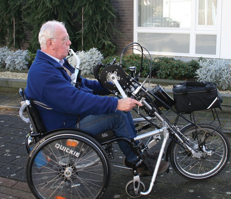
At the time of writing, he uses the device frequently in daytime, experiences less fatigue and reports a subjective increase in quality of life (QOL) (figure 2).
Figure 2.

Patient with UDP using MPV during exercise.
Discussion
This is the first report showing that MPV is a clinically beneficial treatment to improve exercise tolerance and exercise-induced dyspnoea in patients with UDP or BDP.
NIV reduces the work of breathing, improves gas exchange, relieves dyspnoea and rests the inspiratory muscles. The use of night-time NIV in patients with chronic respiratory failure has been proven to be successful [5, 6]. In addition, NIV during exercise is previously found out to be successful in chronic obstructive pulmonary disease patients and, with a lower level of evidence, in patients with restrictive lung disease [7]. An appropriate interface is crucial for successful NIV, both during the night and during exercise [8]. NIV for chronic respiratory failure is most commonly administered via (oro)nasal interfaces [8], which are useful for night-time NIV but less practical during the day or during (nonstationary) exercise. Most studies on NIV during exercise have investigated NIV via (oro)nasal or facial masks. One study investigated the effect of MPV on exercise endurance in eight patients with severe scoliosis, without significant improvement [9]. This is in contrast with our results but it is imaginable that, physiologically, patients with diaphragmatic paralysis but otherwise normal pulmonary volumes are more responsive to MPV than patients with restriction due to scoliosis.
In case 1, a nurse carried the ventilator, leaving us without objective results on exercise tolerance when the patient is carrying the device. The weight of the device can be heavy for patients with severe dyspnoea when carried in a backpack. Subjectively, however, he also has improved exercise tolerance while carrying the device on a rollator, which can be requested when the ventilator is too heavy. Both patients reported far fewer complaints during exercise and a subjective increase in QOL.
This report shows that daytime MPV should be considered for patients with BDP or severe UDP.
Task
1.When you suspect a patient of UDP, which diagnostic test(s) would you consider and why?
2.Chest radiography and/or a “sniff” test may not be as helpful in patients with BDP as in patients with UDP. Explain why.
Answers
1.Usually, an elevated diaphragm is noted on a conventional chest radiograph; paralysis of the diaphragm is then further confirmed using a fluoroscopic sniff test. Pulmonary function, including mouth pressures, can further confirm or identify functional consequences. More advanced and/or invasive techniques include transdiaphragmatic pressure (Pdi) measurements and electromyography (EMG).
Chest radiography can show an elevated hemidiaphragm. This is sensitive but not specific for the diagnosis of UDP. Diagnosis of UDP can be confirmed with a fluoroscopic sniff test. The patient sniffs forcefully and diaphragmatic movement is observed fluoroscopically. This will show paradoxical elevation of the paralysed hemidiaphragm with inspiration. This test is positive in >90% of patients with UDP.
The pulmonary function tests that should be performed if you suspect UDP are spirometry in the sitting and supine positions, lung volumes, maximal inspiratory pressure (MIP), and maximal expiratory pressure (MEP). The forced vital capacity (FVC) can decrease to the range of 70–80% predicted in UDP. This reduction is more pronounced in BDP. The FVC may decrease further by 15–25% in the supine position. The MIP reflects the strength of the diaphragm. It can decrease to around 60% predicted. The MEP is usually normal.
Measurement of Pdi is the gold standard for de diagnosis of paralysis of the diaphragm. Pdi is measured as the difference between the pleural pressure (Ppl) and gastric pressure (Pga). The test is performed by transnasal placement of two balloon-tipped catheters, one in the oesophagus above the diaphragm to assess changes in Ppl and one in the stomach to assess changes in Pga. Pdi can be measured during tidal breathing, at the end of inspiration or at the end of expiration with a sniff or maximal inspiratory force manoeuvre. At peak tidal volume inspiration, Pdi is negative in diaphragm paralysis and positive if the diaphragm is working normally. Reliability is an issue with this test because there is great variation even in the same individual.
EMG is usually not part of routine evaluation because it is more invasive and only performed in specialised centres. It will usually be performed when phrenic nerve pacing is considered.
Laboratory tests may be performed when a systemic neuromuscular disease is suspected. Anaemia can be established to assess whether it plays a role in dyspnoea. Thyroid tests can be performed, although thyroid disease is more commonly associated with BDP.
2.Chest radiography in patients with BDP usually shows bilateral, smooth elevation of the hemidiaphragms, with small lung volumes, and the costophrenic and costovertebral sulci are deep and narrow. Plate-like atelectasis may be present at the lung base. Fluoroscopy can show a false appearance of downward displacement of the diaphragm because of the upward movement of the ribs as the accessory muscles contract.
Acknowledgements
Both patients gave informed consent for publication of data and images regarding this case report. M. Koopman, R. Sprooten and L.E.G.W. Vanfleteren conceived and drafted this article. Additional important intellectual content was provided by E.F.M. Wouters. All co-authors critically revised the article and gave final approval of this version to be published.
Footnotes
Conflict of interest None declared.
References
- 1.Celli BR. Respiratory management of diaphragm paralysis. Semin Respir Crit Care Med 2002; 23: 275–281. [DOI] [PubMed] [Google Scholar]
- 2.Hart N, Nickol AH, Cramer D, et al. Effect of severe isolated unilateral and bilateral diaphragm weakness on exercise performance. Am J Respir Crit Care Med 2002; 165: 1265–1270. [DOI] [PubMed] [Google Scholar]
- 3.Khan A, Morgenthaler TI, Ramar K. Sleep disordered breathing in isolated unilateral and bilateral diaphragmatic dysfunction. J Clin Sleep Med 2014; 10: 509–515. [DOI] [PMC free article] [PubMed] [Google Scholar]
- 4.Garuti G, Nicolini A, Grecchi B, et al. Open circuit mouthpiece ventilation: concise clinical review. Rev Port Pneumol 2014; 20: 211–218. [DOI] [PubMed] [Google Scholar]
- 5.Simonds AK, Elliott MW. Outcome of domiciliary nasal intermittent positive pressure ventilation in restrictive and obstructive disorders. Thorax 1995; 50: 604–609. [DOI] [PMC free article] [PubMed] [Google Scholar]
- 6.Chen H, Liang BM, Xu ZB, et al. Long-term non-invasive positive pressure ventilation in severe stable chronic obstructive pulmonary disease: a meta-analysis. Chin Med J (Engl) 2011; 124: 4063–4070. [PubMed] [Google Scholar]
- 7.Ambrosino N, Cigni P. Non invasive ventilation as an additional tool for exercise training. Multidiscip Respir Med 2015; 10: 14. [DOI] [PMC free article] [PubMed] [Google Scholar]
- 8.Mehta S, Hill NS. Noninvasive ventilation. Am J Respir Crit Care Med 2001; 163: 540–577. [DOI] [PubMed] [Google Scholar]
- 9.Highcock MP, Smith IE, Shneerson JM. The effect of noninvasive intermittent positive-pressure ventilation during exercise in severe scoliosis. Chest 2002; 121: 1555–1560. [DOI] [PubMed] [Google Scholar]


