Figure 3.
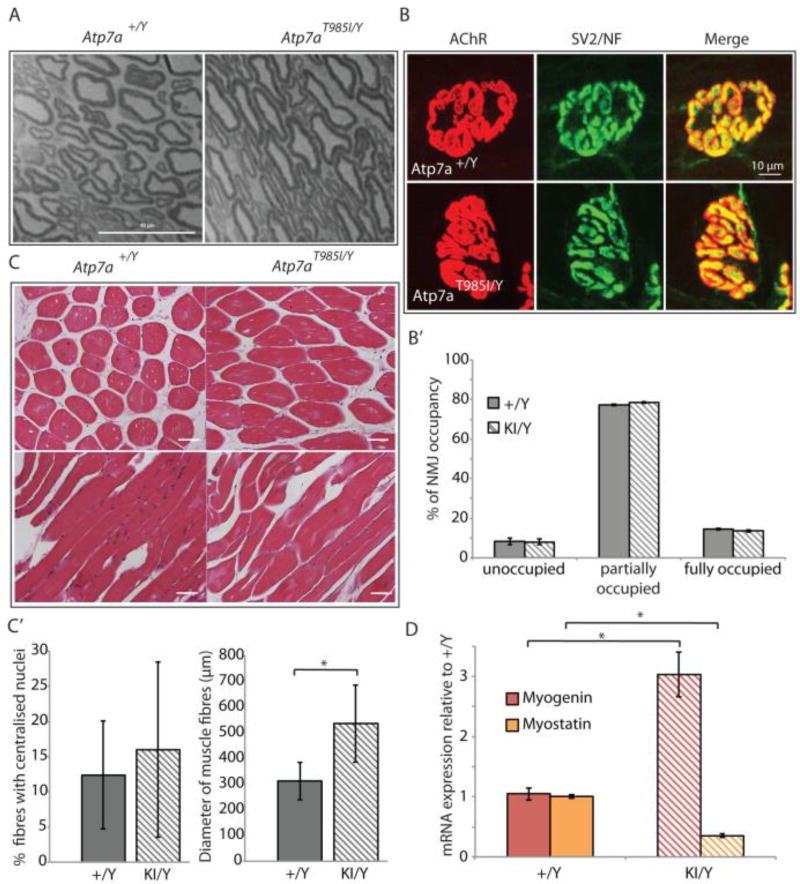
Nerve/muscle histology and NMJ analysis in Atp7aT985I aging mice. (A) Microtome sections (1 µm) from 18 month old Atp7aT985I/Y and Atp7a+/Y sciatic nerves stained with toluidine blue. Scale bar is 40 µm. (B) NMJ from hind limb lumbrical muscles from 24 month old knock in Atp7aT985I/Y (KI/Y) and wild type Atp7a+/Y (+/Y) mice, stained for post-synaptic (AChR) and pre-synaptic (SV2/NF) structures. The merge panel was used to quantify the NMJ occupancy (B’). Previously defined categories of NMJ percentage occupancy included 34: unoccupied (0–33%), partially occupied (34–66%) and fully occupied (67–100%). (C) 5 µm tibialis anterior sections from 24 month old Atp7aT985I/Y and Atp7a+/Y mice stained with H&E (top panels: cross sections; bottom panels: transverse sections). Stained sections were observed under transmission light at a 20X magnification and quantified (C’) for total and centralized nuclei, as well as fibre diameter. (D) Expression analysis of myogenin and myostatin from reversed transcribed template of soleus muscle by real time quantitative PCR. Mouse GAPDH was used to normalize expression of myogenin and myostatin.

