Figure 5.
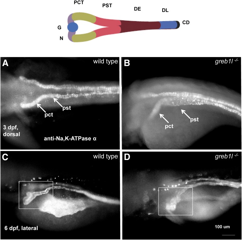
Renal morphology of zebrafish greb1l mutants. In all images, rostral is to the left and embryos are processed with anti-Na,K ATPase antibody (a6F) and a fluorescent secondary antibody. (A and B) Dorsal views of representative embryos at 3 dpf. Mutants present with swelling of the proximal convoluted tubule (PCT) and proximal straight tubule (PST). (C and D) Ventral-lateral views. Mutants have a deformed junction between the PCT and neck. Top, schematic modified from Drummond and Davidson (2016).

