Abstract
Objective
To report the effects of a primary tooth trauma on the underlying permanent tooth germ.
Clinical Presentation and Intervention
A 12-year-old girl was referred to our clinic with a complaint of poor aesthetic appearance. The crown of the permanent maxillary left central tooth exhibited an increased clinical crown height with an ‘enamel hyperplasia’ in the cervical third and had hypoplastic enamel with yellowish-brown discoloration extending from the middle third to the incisal edge. Radiographic examination revealed that the permanent maxillary left central tooth had abnormal root morphology with root dilaceration. The patient revealed a history of trauma at the age of 4 years. An aesthetic restoration with light-curing resin composite was performed on the vestibular surface of the maxillary left permanent central tooth.
Conclusion
Sequelae of a primary tooth trauma on the permanent tooth were restored. We recommend that parents should be aware of the consequences of untreated trauma to a primary tooth. Educational and preventive programmes on dental trauma are required to educate parents about emergency knowledge and sequelae of dental trauma.
Key Words: Tooth injury, Deciduous tooth, Sequelae
Introduction
Although dental injuries may occur at any age, one of the more likely times is from the age of 2–5 years, probably because at this age, children's coordination and judgement are not properly developed and falls are common. Consequently, many of these children suffer injuries to their primary teeth, generally fractured or displaced primary incisors [1,2]. Because of the close anatomic relationship between the apices of primary teeth and the germs of permanent successors, any trauma to the primary teeth may easily disturb the permanent dentition. Traumas may interfere with further odontogenesis, and, depending on the site and extent of the injury, different malformations may occur, ranging from a slight disturbance in the mineralization of enamel to a sequestration of the entire tooth germ [1,2]. In treating traumatic injuries to primary teeth, the objective is to deal with pain and to prevent sequelae in the underlying permanent tooth germ; however, there is no agreement on the ideal treatment [3]. We report the effects of a primary tooth trauma on the underlying permanent tooth germ.
Clinical Presentation and Intervention
A 12-year-old girl was referred to our clinic with a complaint of poor aesthetic appearance. The patient revealed a history of trauma at the age of 4 years. There was no record of the incident, but the girl's mother affirmed that the patient had fallen onto a sofa, and the primary maxillary left central tooth was dislocated from the socket with increased mobility. The girl was not seen by a dentist immediately after the trauma. Approximately 2 weeks later, when the tooth mobility increased, she was referred to a private dentist and the tooth was extracted. It was reported that the permanent maxillary left central tooth had erupted at 7 years of age with yellow-brown stains. The patient stated that initially there was no pain associated with this tooth, but she currently suffered from sensitivity to cold and sweets.
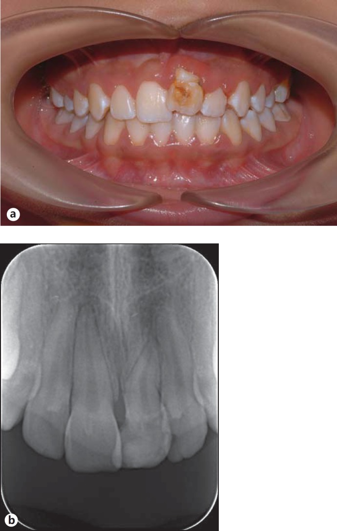
Her medical history did not reveal any important information. Intraoral examination showed that all permanent teeth were present with normal morphology in the mouth, except for the permanent maxillary left central tooth (fig. 1a). The crown of the permanent maxillary left central tooth exhibited an increased clinical crown height with an ‘enamel hyperplasia’ in the cervical third and hypoplastic enamel with yellowish-brown discoloration extending from the middle third to the incisal edge. Electrical pulp vitality tests proved that the tooth was vital. The labial gingival margins in relation to the permanent maxillary left central and lateral incisors appeared inflamed. Radiographic examination revealed that the permanent maxillary left central incisor had abnormal root morphology with root dilaceration (fig. 1b). There was no sign of any periapical pathology. An aesthetic restoration of light-curing resin composite was made on the vestibular surface of the maxillary left permanent central tooth. The patient has been scheduled for follow-up examinations.
Fig. 1.
a Intraoral view of the patient showing developmental disturbances in the permanent maxillary left central incisor. b Radiograph showing root dilaceration in the permanent maxillary left central tooth.
Discussion
Discoloration, alteration and hypoplasia of the enamel are the most frequent sequelae in successors after trauma to the primary tooth. Other sequelae that may occur less often are dilaceration of the crown and root, sequestering of the germ of the permanent tooth and even root duplication. The severity of sequelae is associated with different factors, such as age at the time of the accident, the degree of root resorption of the injured primary tooth, the type and extent of the traumatic lesion and the stage of development of the permanent tooth germ [1,2,4].
In the present case, there were both enamel hyperplasia and hypoplasia with yellowish-brown discoloration of the permanent maxillary left central incisor. Ameloblastic activity interrupted by the trauma contributed to the formation of areas of irregular and imperfect enamel on the buccal side of the permanent maxillary left central tooth, which probably caused the formation of hyperplastic enamel after the injury [4]. Yellow-brown discolorations are caused by the incorporation of breakdown products of hemoglobin from bleeding in the periapical area. The breakdown products of hemoglobin from bleeding can be incorporated into the tooth during tooth formation, even after the ameloblast activity is arrested [2].
Dilaceration of teeth is assumed to be a disturbance in the growth of the epithelial root sheath of Hertwig, and its true cause is unknown. Acute trauma, scar formation and primary tooth germ developmental anomalies may be contributing factors [5]. It has been reported that crown dilaceration occurs in cases with injury at an age between 1.5 and 3.5 years, and root malformation with injury between 4 and 5 years of age. The trauma in the present case, which occurred at 4 years of age, could have caused a direct impact on the root angulation [2].
Severe dental malformation resulting from injury to the primary dentition has been described previously [6]. However, most of the cases presented are the result of intrusive injuries to the primary teeth [6,7,8]. As our patient was not seen by a dentist at the time of injury, her mother's statements were taken into consideration, and it was thought that the primary tooth was extrusively luxated. No other case reports were found in the literature reporting the sequelae of extrusive luxation of primary teeth to their successors.
In the present case, multiple abnormalities in a permanent tooth following trauma to its predecessor are reported. It is a rare case demonstrating enamel hypoplasia, hyperplasia, discoloration and root dilaceration in the same tooth as sequelae after traumatic injury. Tewari and Pandey [9] reported similar multiple sequelae but not in the same tooth, rather in teeth in the same quadrant of the jaw.
Arıkan et al. [10] reported that in the absence of acute symptoms, parents tend not to visit a dental clinic for children's dental injuries, especially those affecting primary teeth. Consistent with their findings, in the present case the parents did not visit a dentist immediately. Traumatic injury to the primary teeth may directly affect the underlying permanent tooth germ; moreover, subsequent local infection may also hinder the regular formation of tooth tissues. When regular follow-up is carried out appropriately after a precise diagnosis, the chances of sequelae can be minimized.
Conclusion
In the present case, sequelae of a primary tooth trauma on the permanent tooth were restored. We recommend that parents should be aware of the consequences of untreated trauma to a primary tooth. Educational and preventive programmes on dental trauma are required to educate parents about emergency knowledge and sequelae of dental trauma.
References
- 1.Topouzelis N, Tsaousoglou P, Pisoka V, et al. Dilaceration of maxillary central incisor: a literature review. Dent Traumatol. 2010;26:427–433. doi: 10.1111/j.1600-9657.2010.00915.x. [DOI] [PubMed] [Google Scholar]
- 2.Andreasen JO, Sundstrom B, Ravn JJ. The effect of traumatic injuries to primary teeth on their permanent successors. I. A clinical and histologic study of 117 injured permanent teeth. Scand J Dent Res. 1971;79:219–283. doi: 10.1111/j.1600-0722.1971.tb02013.x. [DOI] [PubMed] [Google Scholar]
- 3.Gomes AC, Messias LPDA, Delbem ACB, et al. Developmental disturbance of an unerupted permanent incisor due to trauma to its predecessor. J Can Dent Assoc. 2010;76:a57. [PubMed] [Google Scholar]
- 4.Croll TP, Pascon EA, Langeland K. Traumatically injured primary incisors: a clinical and histological study. ASDC J Dent Child. 1987;54:401–422. [PubMed] [Google Scholar]
- 5.Von Arx TV. Developmental disturbances of permanent teeth following trauma to the primary dentition. Aust Dent J. 1993;38:1–10. doi: 10.1111/j.1834-7819.1993.tb05444.x. [DOI] [PubMed] [Google Scholar]
- 6.Ozdemir Y, Akın A, Eden E. Management of multiple sequelaes in permanent dentition: 3 years follow-up. Dent Traumatol. 2011;27:67–70. doi: 10.1111/j.1600-9657.2010.00954.x. [DOI] [PubMed] [Google Scholar]
- 7.Turgut MD, Tekçiçek M, Canoğlu H. An unusual developmental disturbance of an unerupted permanent incisor due to trauma to its predecessor – a case report. Dent Traumatol. 2006;22:28328–28326. doi: 10.1111/j.1600-9657.2006.00357.x. [DOI] [PubMed] [Google Scholar]
- 8.Güngör HC, Püşman E, Uysal S. Eruption delay and sequelae in permanent incisors following intrusive luxation in primary dentition: a case report. Dent Traumatol. 2011;27:156–158. doi: 10.1111/j.1600-9657.2011.00981.x. [DOI] [PubMed] [Google Scholar]
- 9.Tewari N, Pandey RK. Multiple abnormalities in permanent maxillary incisors following trauma to the primary dentition. J Indian Soc Pedod Prev Dent. 2011;2:161–164. doi: 10.4103/0970-4388.84691. [DOI] [PubMed] [Google Scholar]
- 10.Arıkan V, Sarı S, Sonmez H. The prevalence and treatment outcomes of primary tooth injuries. Eur J Dent. 2010;4:447–453. [PMC free article] [PubMed] [Google Scholar]


