Abstract
Objective
To investigate variations in the root canal morphology of mandibular first premolars in a population from the United Arab Emirates using micro-computed tomography (micro-CT) and conventional radiography.
Materials and Methods
Three-dimensional images of 50 extracted human mandibular first premolars were produced using a micro-CT scanner, and conventional radiography was also used to record the number of roots, the root canal system configuration, the presence of a C-shaped canal system and lateral canals, intercanal communications, and the number and location of apical foramina. The interpretations of micro-CT and conventional radiography were statistically analyzed using Fisher's exact test.
Results
Variable root canal configurations based on Vertucci's classification were observed in the teeth (i.e., types I, III, IV, V, and VII). The examined teeth exhibited the following 2 additional root canal configurations, which did not fit Vertucci's classification: type 1-2-3 and type 1–3. A C-shaped canal configuration was present in 14 (28%) cases, and lateral canals were present in 22 (44%) cases. Apical deltas were found in 25 (50%) cases, intercanal communications were seen in 6 (12%) cases, and apical loops were seen in 2 (4%) of the samples. Micro-CT and X-ray imaging identified 39 (78%) and 34 (68%) apical foramina, respectively. A single apical foramen was detected in 33 (66%) samples, and 2 or 3 apical foramina were detected in 14 (28 %) and 3 (6%) samples, respectively. In 18.5 (37%) samples the apical foramina were located centrally, and in 31 (62%) they were located laterally.
Conclusion
A complex morphology of the mandibular first premolars was observed with a high prevalence of multiple root canal systems.
Key Words: Micro-computed tomography, Root canal systems, Mandibular first premolars, Radiography, Dentistry
Introduction
The main objective of root canal therapy is to clean and shape the pulp space and comprehensively fill the root canal system with an inert material [1]. Endodontic treatment failure is frequently attributed to a direct or indirect microbial etiology [2,3] and to the inability to locate, debride, shape, and obturate all canals of the root system [1]. Teeth with 1 root commonly have single root canals; however, the presence of 2 root canal systems in single-rooted teeth is also not uncommon [4]. The endodontic treatment failure rate is highest in mandibular first premolars due to the frequent variations in their root canal morphology and the inability of the dentist to locate and access extra canal systems [5].
Over the years, various studies have been done to understand the root canal morphology of mandibular first premolars, utilizing different methods of investigation [6,7] and different populations [8,9,10]. The rate of occurrence of a single canal ranges from 54 to 88.5%; however, multiple canals have been reported in 11.5–46% of cases [11]. C-shaped root canals have also been observed in mandibular first premolars, and their prevalence is reportedly 10–24% [12,13,14,15].
Detailed knowledge of the internal and external anatomy of a tooth is essential to performing successful endodontic procedures [16], and micro-computed tomography (micro-CT) offers a noninvasive reproducible technique for 3-dimensional (3-D) assessment of the root canal system and can be applied quantitatively and qualitatively [17]. Racial and/or regional predispositions contribute to the wide internal anatomic variations in mandibular first premolars. Therefore, this study was performed to investigate different variations in the root canal system of mandibular first premolars in an Emirati population, utilizing micro-CT.
Materials and Methods
Fifty mandibular first premolars which had been extracted for orthodontic reasons were used in this study. It was confirmed through the patients' medical records that the teeth belonged to patients of only Emirati ethnic origin. This study was initiated after obtaining ethical approval from the Boston University Institute for Dental Research and Education and the Dubai (BUIDRE) Ethics Committee, and it was conducted in full accordance with the World Medical Association Declaration of Helsinki. The teeth were single rooted with completely formed apices, had not undergone endodontic treatment before, and were free from caries and large defective restorations. The reason for the extraction and the age and gender of the patients were not recorded.
The samples were placed in 5.25% sodium hypochlorite for 1 h to remove any organic material. Any remaining external soft-tissue or calculus was removed afterward by scaling. The outer surfaces of the samples were then examined cautiously for the presence of any developmental grooves or bifurcations. The samples were stored individually in small containers with normal saline (sodium chloride 0.9%) and labeled for identification before analysis.
The root canal configurations were determined by analyzing the reconstructed 3-D images and evaluating buccolingual and mesiodistal radiographs. Radiographs were taken in the buccolingual and mesiodistal aspects using a charge-coupled device sensor (Gendex, Des Plaines, IL, USA) and an X-ray generator (Gendex). The images were acquired using a paralleling technique with an exposure factor of 65 kVp at 7 mA for 0.063 s and a source-to-object distance of 5 cm.
Each sample was then scanned using a micro-CT scanner (SkyScan 1,172 X-ray micro-tomograph; SkyScan, Antwerp, Belgium) employing 70 kV at 142 μA and a scanning duration of 38 min. After completion of the scanning procedure, the specimens were replaced in the storage solution. Tagged image file format (TIFF) was selected to maximize and preserve the image quality.
Transmission X-ray images were recorded in 0.4° rotational steps for 180° of rotation. The resulting 2-D shadow/transmission images were used to reconstruct axial cross-sections. Each sample's raw data set was then reconstructed into images using cluster reconstruction software (NRecon/InstaRecon reconstruction engine; SkyScan).
The series of cross-sectional images with a voxel size of 11.94 × 11.94 × 11.94 μm were transferred to a 3-D visualization software package using SkyScan software (CTan version 1.11.10.0 for 2-D visualization and 2-D/3-D analysis, and CTvol version 2.2.1.0 for realistic 3-D visualization). The registered images were then processed to generate 3-D renderings of the external surface of the tooth and the internal root canal space. During reconstruction, the transparency was adjusted so that the internal morphology could be viewed. The reconstructed images obtained from the software were analyzed by a single observer. The observer (W.A.) was blinded in terms of the micro-CT scanned images and radiographs that were viewed. Root canal system configurations were determined in 2 ways: (a) the observer evaluated the 2-D digital radiographs and (b) 2 weeks later the same observer evaluated the 3-D videos and images obtained from the micro-CT. All of the findings of the observer were further confirmed by an endodontist who was not part of this study. The following characteristics were evaluated: the number of root canal system configurations and the types of configurations based on Vertucci's classification (types I-VIII) [7], extra classifications not explained by Vertucci, the number of apical foramina, the presence of apical deltas and lateral root canal systems, the presence of intercanal communications, and the presence of C-shaped root canal systems.
Statistical Analysis
SPSS software (version 19.0; SPSS Inc., Chicago, IL, USA) was used for analyses. Fisher's exact test was used to compare interpretations between micro-CT and X-ray images. p < 0.05 was considered statistically significant.
Results
Presence of Grooves
Of the 50 samples, deep mesiolingual radicular grooves and shallow depressions on the distal side were present in 10 (20%). In 12 (24%) samples, a groove was present on the mesial side or the distal side, and 12 (44%) had shallow depressions on either proximal side.
Root Canal System Types
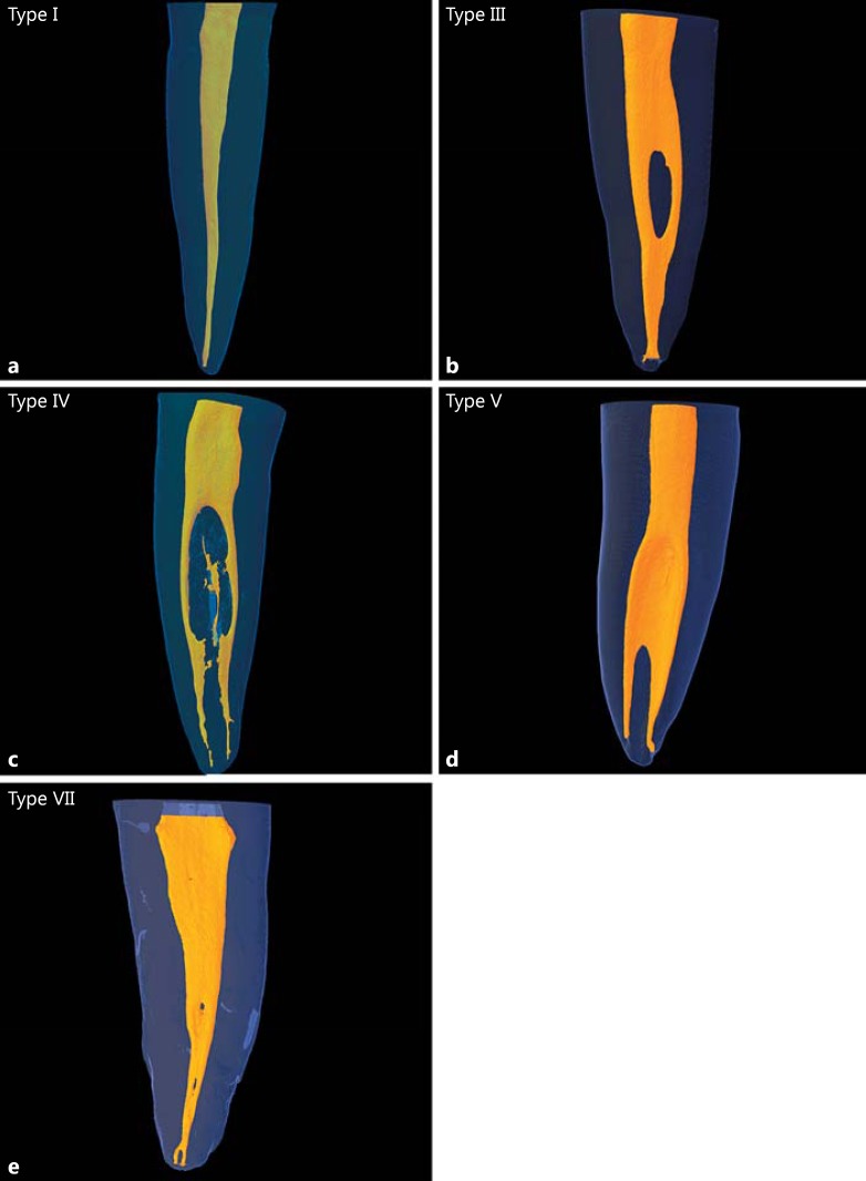
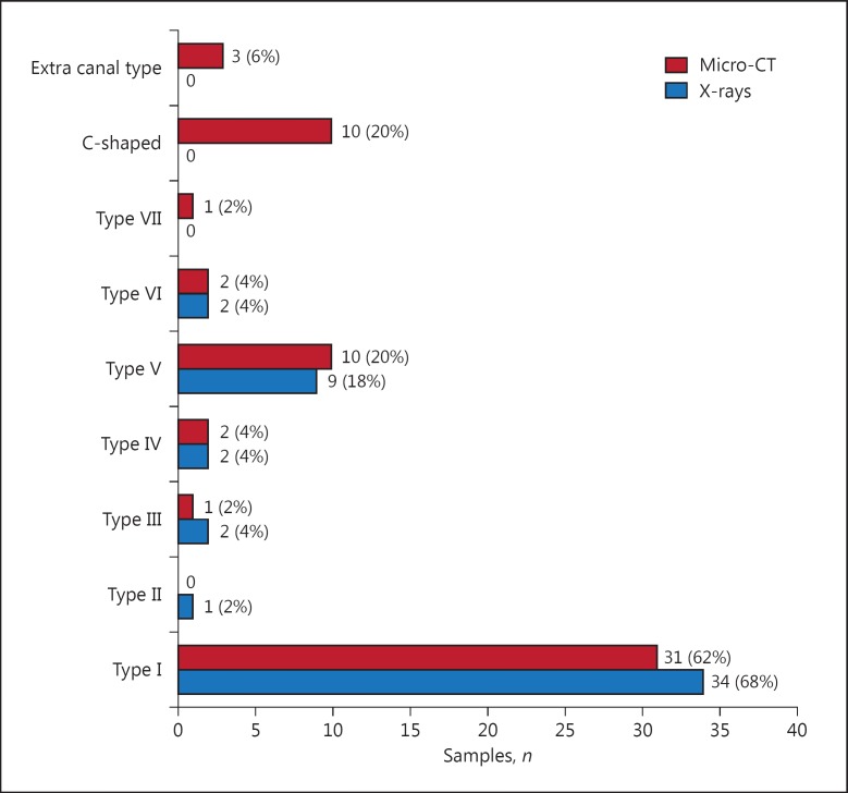
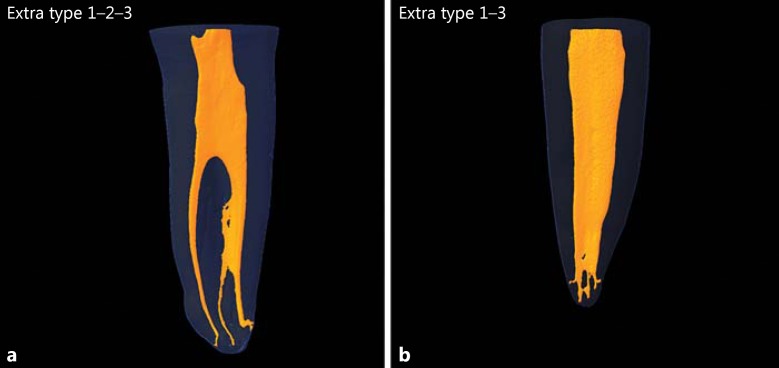
Based on Vertucci's classification, variable root canal configurations were observed in the mandibular first premolars as types I, III, IV, V, and VII (Fig. 1a-e). Although differences were present, no significant difference was found between the micro-CT interpretations of the images and X-rays (p > 0.05; Fig. 2). Some of the noteworthy differences included an extra canal type, C-shaped canals, and a type VII root canal configuration in micro-CT imaging, and these were not observed at all with X-rays. Three examined specimens exhibited 2 additional canal system configurations that did not fit Vertucci's classification. The other samples included type 1-2-3, where 1 system left the pulp chamber and divided in 2 within the root, and 1 of the canal systems further divided into 2 separate systems (giving rise to 3 separate apical foramina; Fig. 3a), and type 1–3, where 1 canal left the pulp chamber and divided into 2 separate systems (resulting in 3 separate apical foramina; Fig. 3b).
Fig. 1.
Root canal configuration types based on data obtained from micro-CT. a Type I. b Type III. c Type IV. d Type V. e Type VII.
Fig. 2.
Variable root canal configurations, based on Vertucci's classification, observed through X-rays and micro-CT.
Fig. 3.
Extra canal configuration. a Type 1-2-3. b Type 1–3.
In addition, C-shaped configurations existed at some point along the root length in 14 (28%) specimens (Fig. 4a-d). Ten teeth had C-shaped roots and 4 teeth did not have C-shaped roots and were categorized according to Fan's classification [18] (Table 1).
Fig. 4.
C-shaped canal configurations. a C1. b C2. c C4b. d C4c.
Table 1.
Fan's classification of root canal systems with C-shaped root configurations
| Category | Description |
|---|---|
| I (C1) | The shape is a continuous “C” with no separation or division |
| II (C2) | The canal shape resembles a semicolon resulting from a discontinuation in the “C” outline |
| III (C3) | Two separate round, oval, or flat canals are present |
| IV (C4) | Only one round, oval, or flat canal is present in the cross-section |
| IV (C4a) | Round canal; the long canal diameter is almost equal to the short diameter |
| IV (C4b) | Oval canal; the long canal diameter is at least 2 times shorter than short diameter |
| IV (C4c) | Flat canal; the long canal diameter is at least 2 times longer than the short diameter |
| V (C5) | Three or more separate canals are in the cross-section |
| VI (C6) | No canal lumen or no intact canal can be observed (which is usually seen near the apex only) |
Lateral Canal Systems
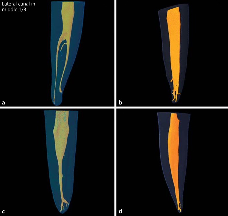
Lateral canals were observed on the X-ray image in 5 (10%) specimens and they were present on the micro-CT image in 22 (44%) specimens. The difference was statistically significant (p < 0.05). In addition, there was an increased prevalence of lateral canals towards the apical part of the root (Fig. 5a, b).
Fig. 5.
a Lateral canals observed under micro-CT. b Lateral canals observed under micro-CT. c Apical delta. d Apical loop.
Apical Foramina and Apical Deltas
Micro-CT scanning and radiography detected 63 apical foramina and 70 apical foramina, respectively (Table 2). With micro-CT imaging, a single apical foramen was detected in 33 (66%) cases, 2 apical foramina were present in 14 (28%) cases, and 3 apical foramina were found in 3 (6%) cases. An apical delta was detected by micro-CT in 25 (50%) samples in the apical third of the mandibular first premolars (Fig. 5c). With X-ray imaging, a single foramen was seen in 37 (74%) cases, and 2 foramina were found in 13 (26%) cases.
Table 2.
Number and percentage of apical foramina, their position, and incidence of apical delta observed under conventional radiography and micro-CT
| X-ray | Micro-CT | |
|---|---|---|
| Number of apical foramina | ||
| 1 | 37 (74) | 23 (76.6) |
| 2 | 13 (26) | 5 (16.6) |
| 3 | – | 3 (6) |
| Position of the apical foramina | ||
| Central | 45 (71.4) | 26 (37.2) |
| Lateral | 18 (28.5) | 44 (62.8) |
| Apical delta | – | 15 (50) |
| Total foramina, n | 63 | 70 |
Values are presented as numbers (%) unless otherwise stated. The total number of samples evaluated is 50.
Intercanal Communications and Loops
Intercanal communications were identified in 6 (12%) samples by micro-CT and in 4 (8%) samples by X-ray imaging. The difference was statistically significant (p < 0.05). Only 2 (4%) samples had an apical loop (Fig. 5d).
Discussion
In this study, using Vertucci's classification, the analyzed teeth were type I, type III, type IV, type V, and type VII. Detailed anatomical reconstruction through micro-CT revealed 2 canal system configurations that did not fit under Vertucci's classification that confirmed the detection of 2 “extra” canal system configurations (Fin) with use of a clearing technique on 900 mandibular premolars reported in a Jordanian population [9]. This finding could be related to the ethnic and/or regional background of the population studied. The 28% rate of C-shaped canals is a relatively high frequency because in a previous study in a Chinese population [19] C-shaped canals were reported in 18% of the sample. The probable reason for a high frequency in some populations has been explained previously by Manning [20], who reported that the presence of C-shaped canals is related to race, with Asians having a high incidence of C-shaped canals as compared to the other population groups.
In our study, developmental grooves, which could be of clinical importance, were observed in 20% of the samples and all of them exhibited a C-shaped canal. This 20% rate of C-shaped canals is similar to the rates of 14 and 18% reported by Baisden et al. [12] and Lu et al. [14], respectively. Lu et al. [14] reported that the grooves on mandibular first premolars were frequently present on the proximal lingual area of the middle root and did not always extend to the root apex.
The rates of lateral canals of 44 and 10%, observed using micro-CT imaging and X-ray imaging, respectively, are similar to those reported by Caliskan et al. [21]. Interestingly, 1 sample had 5 lateral canals in the apical region. When more than 1 lateral canal or intercanal communications were detected, most were present at the middle and apical levels of the root. Therefore, if evidence of lateral canals appears on the radiograph at these locations, the radiograph should be carefully inspected to exclude the presence of other branches at other locations along the root length.
The rate of isthmeses of 12% in our study was lower than the frequencies of 32.1 and 27.4% reported by others [9,24] and similar to the frequency reported by Caliskan et al. [21]. Previously, Cambruzzi and Marshall [22] called an intercanal communication or transverse anastomosis an ‘isthmus' and stressed the importance of preparing and packing it during endodontic treatment. Teixeira et al. [23] reported that an isthmus that is poorly accessible with root canal armamentarium could act as a bacterial reservoir and may reduce the success rate of surgical or nonsurgical endodontic procedures. Isthmuses reportedly exist in all types of roots in which 2 canal systems are normally found, which include the mesial roots of maxillary and mandibular molars and the distal roots of mandibular molars, the maxillary and mandibular first and second premolars, and mandibular incisors [1].
The rate of apical deltas found in the samples was 50%; this is higher than the values in previous studies, which range from 5.7 [23] to 15.5% in a Turkish population [8] and 29.2% in a Jordanian population [9]. The presence of apical deltas could account for some cases of persistent posttreatment disease because of possible inadequate cleaning and packing of these apical ramifications.
The finding of an “apical loop” in the apical third of the root in 4% of the samples is lower than that in a previous study which reported a 20% incidence of apical loops in the mesiobuccal root of maxillary first molars [25]. This wide variation between studies could be related to ethnic population differences. The same may be true for the presence of apical loops in mandibular premolars in the same population.
The findings of a centrally located apical foramen on micro-CT imaging (37.2%) and a centrally exiting apical foramen on X-ray imaging (71.4%) could be compared to the results of a previous laboratory study which demonstrated results comparable to our X-ray findings and reported a central location in 83% of cases in an Indian population [26].
The major limitation of the present study was the sample size; future studies with a greater sample size, targeting the same ethnicity, should be carried out to establish a clear picture of the current scenario.
Conclusion
In this study, mandibular premolars belonging to an Emirati population showed a high prevalence of multiple canals. Clinicians must be fully aware of the complexity of the root canal morphology of mandibular first premolars. They should consider a patient's ethnic origin when preoperatively evaluating a nonsurgical root canal treatment, and they should use all of the available armamentarium to achieve a successful root canal therapy.
References
- 1.Vertucci FJ. Root canal morphology and its relationship to endodontic procedures. Endod Topics. 2005;10:3–29. [Google Scholar]
- 2.Torabinejad M, Ung B, Kettering JD. In vitro bacterial penetration of coronally unsealed endodontically treated teeth. J Endod. 1991;16:566–569. doi: 10.1016/S0099-2399(07)80198-1. [DOI] [PubMed] [Google Scholar]
- 3.Siqueira JF, Jr, Lopes HP. Mechanisms of antimicrobial activity of calcium hydroxide: a critical review. Int Endod J. 1999;32:361–369. doi: 10.1046/j.1365-2591.1999.00275.x. [DOI] [PubMed] [Google Scholar]
- 4.Li X, Liu N, Ye L, et al. A micro-computed tomography study of the location and curvature of the lingual canal in the mandibular first premolar with two canals originating from a single canal. J Endod. 2012;38:309–312. doi: 10.1016/j.joen.2011.12.038. [DOI] [PubMed] [Google Scholar]
- 5.Ingle JI, Taintor JF. Endodontics: Modern Endodontic Therapy. ed 3. Philadelphia: Lea and Febiger; 1985. pp. 27–52. [Google Scholar]
- 6.Zillich R, Dowson J. Root canal morphology of mandibular first and second premolars. Oral Surg Oral Med Oral Pathol. 1973;36:738–744. doi: 10.1016/0030-4220(73)90147-3. [DOI] [PubMed] [Google Scholar]
- 7.Vertucci FJ. Root canal anatomy of the human permanent teeth. Oral Surg Oral Med Oral Pathol. 1984;58:589–599. doi: 10.1016/0030-4220(84)90085-9. [DOI] [PubMed] [Google Scholar]
- 8.Sert S, Bayirli GS. Evaluation of the root canal configurations of the mandibular and maxillary permanent teeth by gender in the Turkish population. J Endod. 2004;30:391–398. doi: 10.1097/00004770-200406000-00004. [DOI] [PubMed] [Google Scholar]
- 9.Awawdeh L, Al-Qudah AH. Root form and canal morphology of Jordanian maxillary first premolars. Int Endod J. 2008;34:956–961. doi: 10.1016/j.joen.2008.04.013. [DOI] [PubMed] [Google Scholar]
- 10.Khedmat S, Assadian H, Saravani AA. Root canal morphology of the mandibular first premolars in an Iranian population using cross-sections and radiography. J Endod. 2010;36:214–217. doi: 10.1016/j.joen.2009.10.002. [DOI] [PubMed] [Google Scholar]
- 11.Celikten B, Orhan K, Aksoy U, et al. Cone-beam CT evaluation of root canal morphology of maxillary and mandibular premolars in a Turkish Cypriot population. BDJ Open. 2016;15006:1–5. doi: 10.1038/bdjopen.2015.6. [DOI] [PMC free article] [PubMed] [Google Scholar]
- 12.Baisden MK, Kulild JC, Weller RN. Root canal configuration of mandibular first premolar. J Endod. 1992;18:505–508. doi: 10.1016/S0099-2399(06)81352-X. [DOI] [PubMed] [Google Scholar]
- 13.Sikri VK, Sikri P. Mandibular premolars: aberrations in pulp space morphology. Indian J Dent Res. 1994;5:9–14. [PubMed] [Google Scholar]
- 14.Lu TY, Yang SF, Pai SF. Complicated root canal morphology of mandibular first premolar in a Chinese population using the cross section method. J Endod. 2006;32:932–936. doi: 10.1016/j.joen.2006.04.008. [DOI] [PubMed] [Google Scholar]
- 15.Fan B, Yang J, Gutmann JL, et al. Root canal systems in mandibular first premolars with C-shaped root configurations. 1. Micro computed tomography mapping of the radicular groove and associated root canal cross sections. J Endod. 2008;34:1337–1341. doi: 10.1016/j.joen.2008.08.006. [DOI] [PubMed] [Google Scholar]
- 16.Alapati S, Zaatar EI, Shyama M, et al. Maxillary canine with two root canals. Med Princ Pract. 2006;15:74–76. doi: 10.1159/000089390. [DOI] [PubMed] [Google Scholar]
- 17.Kierklo A, Tabor Z, Pawińska M, et al. A microcomputed tomography-based comparison of root canal filling quality following different instrumentation and obturation techniques. Med Princ Pract. 2015;24:84–91. doi: 10.1159/000368307. [DOI] [PMC free article] [PubMed] [Google Scholar]
- 18.Verma P, Love RM. A micro CT study of the mesiobuccal root canal morphology of the maxillary first molar tooth. Int Endod J. 2011;44:210–217. doi: 10.1111/j.1365-2591.2010.01800.x. [DOI] [PubMed] [Google Scholar]
- 19.Baisden MK, Kulild JC, Weller RN. Root canal configuration of the mandibular first premolar. J Endod. 1992;18:505–508. doi: 10.1016/S0099-2399(06)81352-X. [DOI] [PubMed] [Google Scholar]
- 20.Manning SA. Root canal anatomy of mandibular second molars. 2. C-shaped canals. Int Endod J. 1990;23:40–45. doi: 10.1111/j.1365-2591.1990.tb00801.x. [DOI] [PubMed] [Google Scholar]
- 21.Caliskan MK, Pehlivan Y, Sepetcioglu F, et al. Root canal morphology of human permanent teeth in a Turkish population. J Endod. 1995;21:200–204. doi: 10.1016/S0099-2399(06)80566-2. [DOI] [PubMed] [Google Scholar]
- 22.Cambruzzi JV, Marshall FJ. Molar endodontic surgery. J Can Dent Assoc. 1983;1:61. [PubMed] [Google Scholar]
- 23.Teixeira FB, Sano CL, Gomes BP, et al. A preliminary in vitro study of the incidence and position of the root canal isthmus in maxillary and mandibular first molars. Int Endod J. 2003;36:276–280. doi: 10.1046/j.1365-2591.2003.00638.x. [DOI] [PubMed] [Google Scholar]
- 24.Vertucci F. Root canal morphology of mandibular premolars. J Am Dent Assoc. 1978;97:47–50. doi: 10.14219/jada.archive.1978.0443. [DOI] [PubMed] [Google Scholar]
- 25.Somma F, Leoni D, Plotino G, et al. Root canal morphology of the mesiobuccal canal root of maxillary first molar: a micro-computed tomographic analysis. Int Endod J. 2009;42:165–174. doi: 10.1111/j.1365-2591.2008.01472.x. [DOI] [PubMed] [Google Scholar]
- 26.Velmurugan N, Sandhya R. Root canal morphology of mandibular first premolars in an Indian population: a laboratory study. Int Endod J. 2009;42:54–58. doi: 10.1111/j.1365-2591.2008.01494.x. [DOI] [PubMed] [Google Scholar]


