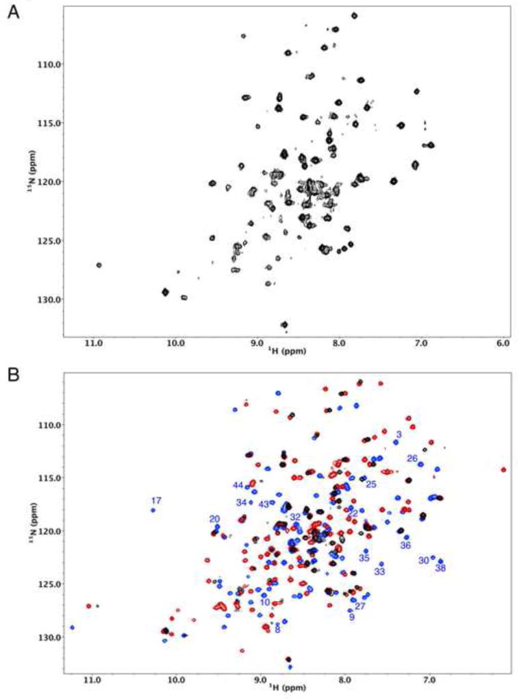
Figure 3.
Effects of bicarbonate on the 1H-15N HSQC spectrum of X1NTD. A) 1H-15N HSQC spectrum obtained for U-[15N]X1NTDox after removal of bicarbonate from the buffer. B) Overlay of the spectrum in A (black) with the spectrum of U-[15N]X1NTDox obtained in the presence of bicarbonate (blue) and with the spectrum of X1NTDred (red). Well-resolved resonances in the blue spectrum that correspond to residues in the N-terminal segment of the protein are annotated, and it is seen that nearly all of these are no longer observed after removal of bicarbonate. The buffer also contained 50 mM Tris-d11, pH 7.0, 140 mM NaCl. Sample temperature was 25 °C.

