

Abstract
Metacarpophalangeal pattern profile (MCPP) was determined on 34 Sotos syndrome individuals and compared with previous MCPP studies. The mean hand profile contained a major peak in the proximal phalangeal area and a smaller peak in the metacarpal area, while the distal hand bones were relatively short. There appear to be three recognized hand profiles in Sotos syndrome, which suggests heterogeneity of the syndrome, although correlation studies indicate clinical homogeneity of individuals in the younger age groups. Discriminant analysis of Sotos syndrome versus control subjects produced a function of two MCPP variables, plus age, which may be applied as another diagnostic tool.
Keywords: correlation studies, discriminant analysis, cerebral gigantism, hand pattern profile analysis
INTRODUCTION
Sotos syndrome, or cerebral gigantism, is characterized by large size at birth, large hands and feet, advanced osseous maturation, macrocephaly with prominent forehead and mild dilatation of lateral ventricles, down-slanting palpebral fissures, hypertelorism, prognathism, abnormal coordination, and variable degrees of mental retardation and language deficits. Early diagnosis is difficult; therefore, quantitative methods based on clinical or physical attributes such as the metacarpophalangeal pattern profile (MCPP) may be helpful.
Dijkstra [1985] reported that the hand profile of Sotos syndrome individuals changed with age, with more pronounced lengthening of the fingers around 2 years of age. Dijkstra [1985] and Wit et al. [1985] suggested that there were two distinguishable hand profiles in Sotos syndrome individuals. In their first profile type (I), identified in 12 of 17 individuals studied, the metacarpals were relatively longer before 3 years of age and the lengthening of the proximal phalanges was more pronounced after 3 years of age. Their second type (II), identified in 4 of 17 individuals, was described as a mirror image of the type I, with the fingers not as long and the profile less pronounced. The remaining patients had a normal hand profile (Z scores ranged from +2 to −2). After examination of additional Sotos syndrome individuals, a third profile type (III) emerged; it was considered “flat,” without up-and-down deviation.
Hence, we report a follow-up study of the MCPP analysis of 34 individuals whose clinical features were consistent with Sotos syndrome. Our initial report on Sotos syndrome suggested that MCPP analysis may be useful as a diagnostic tool, but additional testing was required [Butler et al., 1985; Butler and Meaney, 1986].
MATERIALS AND METHODS
Postero-anterior hand radiographs were obtained on 34 Sotos syndrome patients, 25 males and 9 females ranging in age from 0.8 to 24 years with a mean age of 5.9 years. The procedures for MCPP measurements, standardized Z score computations, correlation, and discriminant analyses were described previously [Garn et al., 1972; Poznanski, 1974; Butler et al., 1985; Butler et al., 1986].
RESULTS
The mean pattern profile, based on the 19 hand bones of 34 Sotos syndrome individuals, contains one major peak in the proximal phalangeal area and a smaller peak in the metacarpal area, with distal hand bones that are relatively short compared with the proximal bones (Fig. 1). The mean Z scores fall between 1.8 and 3.7; therefore, the hand bones were significantly longer than normal at the 5% level.
Fig. 1.

Mean MCPP for 34 individuals with Sotos syndrome. (★) indicates the bones that were selected in the discriminant analysis.
The hand profiles of the 34 patients were grouped according to the pattern type described by Dijkstra [1985] and by Wit et al. [1985]. Therefore, 17 of 34 patients had a type I profile, 8 of 34 had a type II or mirror-image profile, 5 of 34 had a type III profile, and 4 had a normal profile. The Pearsonian r test to assess similarity between the individual patterns and their group mean showed 23 of 34 individuals and 18 of 19 individuals before the age of 5 years with a significant positive correlation at the 5% level (Table I). There were 15 of 17 type I profiles; 4 of 8 type II profiles; 3 of 5 type III profiles, and 1 of 4 normal profiles, with significant positive correlation (P < .05).
TABLE I.
Correlations Between Sotos Syndrome Individual’s MCPP and Group Mean MCPP
| Age (years) | MCPP type† | Individual’s correlation with group mean |
|---|---|---|
| Males | ||
| 1.0 | I | 0.81*** |
| 1.0 | I | 0.78*** |
| 1.0 | I | 0.58*** |
| 1.2 | II | 0.39* |
| 1.7 | I | 0.83*** |
| 2.0 | I | 0.62*** |
| 2.1 | I | 0.78*** |
| 2.2 | I | 0.65*** |
| 3.0 | I | 0.84*** |
| 3.0 | I | 0.42* |
| 3.0 | I | 0.74*** |
| 3.0 | I | 0.88*** |
| 3.0 | I | 0.80*** |
| 3.2 | I | 0.69*** |
| 3.4 | III | 0.88*** |
| 4.6 | III | 0.54** |
| 5.0 | Normal | 0.11 |
| 5.0 | II | −0.22 |
| 7.3 | III | −0.14 |
| 8.0 | Normal | −0.41 |
| 12.1 | II | 0.65*** |
| 13.8 | II | 0.64*** |
| 14.0 | I | −0.04 |
| 19.0 | I | 0.70*** |
| 24.0 | Normal | −0.45 |
| Females | ||
| 0.8 | I | 0.48* |
| 3.0 | II | −0.00 |
| 4.0 | III | 0.47* |
| 5.0 | III | 0.26 |
| 6.0 | I | −0.22 |
| 8.0 | II | 0.12 |
| 8.3 | Normal | 0.43* |
| 9.0 | II | −0.45 |
| 10.0 | II | 0.74*** |
A forward stepwise method of discriminant analysis of 41 control subjects and the original 16 Sotos syndrome individuals resulted in a discriminant function based on two (third proximal phalanx and second middle phalanx) of the 19 hand bones plus age. Additionally, 18 Sotos syndrome individuals were used to test the method’s power to identify new Sotos syndrome patients. Fifteen of the 18 additional individuals (83%) were classified as Sotos syndrome based on the equation produced by the original discrimination of 16 Sotos syndrome patients.
A stepwise discriminant analysis of all 34 Sotos syndrome and 41 control subjects resulted in a correct classification rate of 93.3% (Fig. 2). Five Sotos syndrome and no control individuals were misclassified. This discriminant function was based on two MCPP variables, which were the Z scores representing 1) the third proximal phalanx (X8) and 2) the fourth proximal phalanx (X9).
Fig. 2.
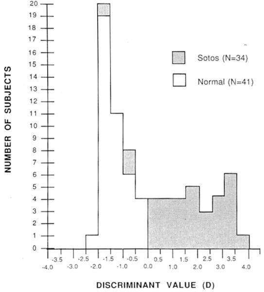
Histogram depicting normal and Sotos syndrome classification by discriminant analysis. D = 0.40 + 1.13(X8) − 0.64(X9) −0.12 (age in years, with 18 as maximum in an adult).
DISCUSSION
On physical examination, the individual with Sotos syndrome has an overall large hand size. The mean pattern profile based on our 34 patients confirms this characteristic in quantitative terms. Three distinct hand profiles apparently exist, supporting heterogeneity in this syndrome, although 68% of all Sotos individuals studied and 95% of individuals before age 5 years had a significant positive correlation at the 5% level. Therefore, a hand profile for Sotos syndrome apparently exists, particularly in the younger patient. The discriminant analysis and examination of the individual hand profiles suggest that effective classification of Sotos syndrome patients is possible and may be applied in a clinical setting as a diagnostic tool in patients of all ages in whom Sotos syndrome is suspected. MCPP analysis has been applied to at least 63 syndromes [Poznanski, 1984; Butler et al., 1986] and has been found useful as a diagnostic tool in several conditions. Therefore, the authors encourage the application of this methodology to patients suspected of having syndromes in which specific hand profiles exist.
Recently, Sotos syndrome patients have been reported with the fragile X chromosome [Beemer et al., 1986]. Therefore, the different hand profiles may derive from some individuals with Sotos syndrome and fragile X chromosome expression. Research is underway to compare the hand profiles of Sotos and fragile X syndrome individuals.
Acknowledgments
The authors acknowledge use of the facilities of Computing Services, Eastern Kentucky University, Richmond, and University of Kentucky, Lexington. The authors wish to thank Dr. William Wadlington, Sandy Cain, and Margaret Lane for their assistance in this study.
Contributor Information
Merlin G. Butler, Division of Genetics, Department of Pediatrics, Vanderbilt University School of Medicine, Nashville, Tennessee
Piet F. Dijkstra, Jan Van Breemen Instituut, Amsterdam, The Netherlands
F. John Meaney, Department of Medical Genetics, Indiana University School of Medicine and Genetic Diseases Section, Indiana State Board of Health, Indianapolis, Indiana.
David D. Gale, Eastern Kentucky University, Richmond, Kentucky
References
- Beemer FA, Veenema H, de Pater JM. Cerebral gigantism (Sotos syndrome) in two patients with fra(X) chromosomes. Am J Med Genet. 1986;23:221–226. doi: 10.1002/ajmg.1320230117. [DOI] [PubMed] [Google Scholar]
- Butler MG, Meaney FJ. Letter to the editor: Metacarpophalangeal pattern profile analysis in Sotos syndrome: An update. Am J Med Genet. 1986;24:761. doi: 10.1002/ajmg.1320240421. [DOI] [PMC free article] [PubMed] [Google Scholar]
- Butler MG, Meaney FJ, Kaler SG. Metacarpophalangeal pattern profile analysis in clinical genetics: An applied anthropometric method. Am J Phys Anthropol. 1986;70:195–201. doi: 10.1002/ajpa.1330700206. [DOI] [PMC free article] [PubMed] [Google Scholar]
- Butler MG, Meaney FJ, Kittur S, Hersh JH, Hornstein L. Metacarpophalangeal pattern profile analysis in Sotos syndrome. Am J Med Genet. 1985;20:625–629. doi: 10.1002/ajmg.1320200408. [DOI] [PMC free article] [PubMed] [Google Scholar]
- Dijkstra PF. Cerebral gigantism (Sotos syndrome). Metacarpophalangeal pattern profiles. Fortschr Geb Rontgenstr Nuklearmed Ergangzungsband. 1985;143:183–185. doi: 10.1055/s-2008-1052786. [DOI] [PubMed] [Google Scholar]
- Garn JC, Hertzog KP, Poznanski AK, Nagy JM. Metacarpophalangeal length in the evaluation of skeletal malformation. Radiology. 1972;105:375–381. doi: 10.1148/105.2.375. [DOI] [PubMed] [Google Scholar]
- Poznanski AK. The Hand in Radiological Diagnosis. 1st. Philadelphia: W.B. Saunders; 1974. [Google Scholar]
- Poznanski AK. The Hand in Radiological Diagnosis. 2nd. Philadelphia: W.B. Saunders; 1984. [Google Scholar]
- Wit JM, Beemer FA, Barth PG, Orthuys JWE, Dijkstra PF, van den Brande JL, Leschot NJ. Cerebral gigantism (Sotos syndrome). Compiled data of 22 cases. Analysis of clinical features, growth and plasma somatomedin. Eur J Pediatr. 1985;144:131–140. doi: 10.1007/BF00451898. [DOI] [PubMed] [Google Scholar]