
Figure 5.
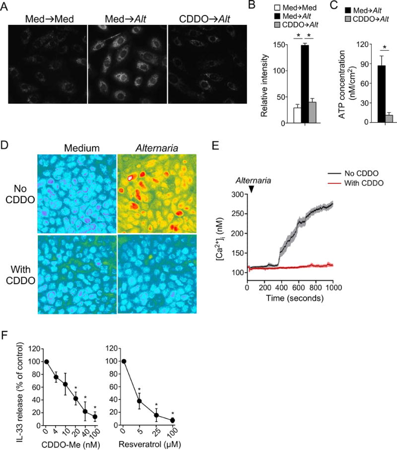
Treatment with Nrf2 activators inhibits oxidative stress and IL-33 release in human bronchial epithelial (hBE) cells. A, hBE cells were preincubated with medium alone or 50 nM CDDO-Me for 24 hours and exposed to Alternaria extract, followed by CellROX® Orange dye. Representative images are shown. B, Quantitative analysis of CellROX® Orange probe fluorescence intensities (mean±SEM, n=25). * p<0.05 between groups indicated by horizontal lines. C, hBE cells were treated similar to those in A. ATP release into extracellular medium was quantitated (mean±SEM, n=4). * p<0.05 between groups indicated by horizontal lines. D,E, hBE cells were preincubated with medium alone or CDDO-Me for 24 hours, and exposed to Alternaria extract. Representative images are shown in Panel D and kinetics changes in [Ca2+]i are shown in Panel E. Data are representative of four experiments. F, hBE33 cells were preincubated with the indicated concentrations of CDDO-Me or resveratrol for 24 hours and exposed to Alternaria extract. IL-33 concentrations in cell-free supernatants were normalized to the values without drugs as 100%. Data are presented as mean±SEM from three experiments. * p<0.05 compared to no drugs.