Abstract
The purpose of this study was to investigate the long-term effects of anterior cruciate ligament (ACL) resection on the morphological and contractile characteristics of rectus femoris (RF) and semimembranosus (SM) muscles in both injured and contralateral hindlimbs in rats. Wistar male rats (8-week old) were used. Rats were divided into two groups; ACL-resected and (sham-operated) control groups. Furthermore, right and left limbs of rats in the ACL-resected group were assigned as ACL-resected and contralateral groups, respectively, at 1 day, 1, 4, and 48 weeks after ACL resection. No ACL-resection-associated changes in the mass of both muscles were observed 1 week after ACL resection. On the other hand, ACL-resection-associated reduction on mean fiber cross-sectional area (fiber CSA) in RF muscle lasted 48 weeks after ACL resection. Furthermore, ACL-resection associated increase in fiber composition of type I fiber in RF muscle in contralateral limbs. In addition, long-term effects of ACL resection were observed in both ACL-resected and contralateral limbs. Evidences from this study suggested that ACL resection may cause to change in the morphological (fiber CSA) and contractile (distribution of fiber types) properties of skeletal muscles around the knee joint in not only injured but also contralateral limb. Rehabilitation for quantitative and qualitative muscle changes by ACL resection may be required a special care for a long-term period.
Key points.
ACL-resection-associated effects on mean fiber CSA as well as the relative distribution of fiber types lasted 48 weeks after ACL resection were investigated.
The responses of fiber CSA and fiber type composition following ACL resection in RF muscle were different from SM muscle.
Long-term effects of ACL resection were observed on the not only ACL-resected but also contralateral limbs.
Rehabilitation for quantitative and qualitative muscle changes by ACL resection may be required a special care for a long-term period.
Key words: Sports injury, ligament, quadriceps, hamstring, muscle fiber type, recovery
Introduction
Anterior cruciate ligament (ACL) injuries are often experienced in sports. After the ACL is injured, anterior laxity and rotatory instability of the tibia persist and ACL reconstruction is widely performed for professional, elite or competitive athletes (Johnson et al., 1992; Urbach et al., 2001). However, for general people in low-level sports activities, ACL reconstruction is avoided due to a long-term period of rehabilitation following the surgery and various social circumstances. In general, they receive a conservative treatment that consists of muscle strength training around injured knee joint (Bonamo et al., 1990; Konrads et al., 2016; Novak et al., 1996).
Patients having ACL resection are clinically allowed to resume sports activity using the muscle strength level in injured limb, compared with contralateral limb, several months after the injury. The returning to sport activity is generally permitted, when the recovery level of muscle strength in injured limb reaches or exceeds 80-85%, compared with the strength in contralateral limb, following ACL injury with or without its reconstruction (Colombet et al., 2006; Fu et al., 2008; Urbach et al., 2001). Although there are many reports investigating acute effects of ACL reconstruction on the contractile function of skeletal muscles around injured knee joint (Lee et al., 2013; Shim et al., 2015; Ucar et al., 2014), there is no clear evidence justifying this threshold for ACL-resected patients.
Furthermore, it has been suggested that the contractile properties of the contralateral limb might be changed by a compensational effect following ACL resection (Armstrong and Phelps, 1984; Gutmann et al., 1971; Yellin, 1974). It is generally accepted that the mechanoreceptor of ACL serves as a tension sensor and protects the knee joint by regulating muscles around the knee joint via the γ muscle spindle system (Barrack, 1989; Johansson et al., 1991; Lephart et al., 1997). It has been reported that the disruption of knee extension is observed in both ACL-injured and contralateral limbs of patients having ACL injury and is suggested to be attributed to the lack of afferent feedback from mechanoreceptors through the γ loop (Konishi et al., 2002a; 2002b; 2003; 2007). However, there are no reports investigating the muscular function of both ACL-injured and contralateral limbs following ACL rupture with or without its reconstruction. The details of the effects of ACL resection on the muscle properties of not only ACL-injured limb but also the contralateral limbs remain unclear.
Although the rehabilitation period to resume sport activity following ACL reconstruction is approximately 6 to 9 months (Feller et al., 2001), there is few reports regarding a long-term follow-up (several decades after ACL injury) of skeletal muscles around the knee joint (Ahmed et al., 2017; Konrads et al., 2016). Since patients with ACL resection often put an end of their clinical rehabilitation program without any permission, long-term effects of ACL resection on the morphological and contractile properties of skeletal muscles around knee joint remains unclear due to a small number of patients who complete their rehabilitation program (Konrads et al., 2016).
The morphological and contractile properties of skeletal muscles around the knee joint following ACL resection with or without its reconstruction are evaluated in an animal model. It is generally accepted that the morphological properties of skeletal muscle, such as the muscle wet weight, the relative muscle wet weight to body weight, and the fiber cross-sectional area (fiber CSA), are used as a key index for skeletal muscle mass, which reflects contractile force of the skeletal muscle (Fujiya et al., 2015; Ito et al., 2013; Yoshida et al., 2015). On the other hand, the contractile properties of a skeletal muscle could be estimated using the composition of fiber types of a skeletal muscle (Kishiro et al., 2013).
Detailed and long-term follow-up studies are useful for the improvements of the operation of ACL reconstruction and the rehabilitation program followed ACL resection with or without its reconstruction. Therefore, the purpose of this study was to investigate a long-term effects of ACL resection on the morphological and contractile properties of skeletal muscles around the knee joint, namely rectus femoris (RF) and semimembranosus (SM) muscles, among ACL-resected, contralateral and sham-operated sedentary control limbs of rats.
We hypothesized that long-term effects of ACL resection on muscles around the knee joint were observed on the not only ACL-resected but also contralateral limb.
Methods
Animal and grouping
The experimental procedures were carried out in accordance with St. Marianna University School of Medicine’s Animal Experiment Guidelines and were approved by the Animal Experiment Committee of the Experimental Animal Research Facilities of St. Marianna University School of Medicine (Approval No. 1502019).
Forty male Wistar rats (8-week old) were used. All rats were kept at a constant room temperature (24 ± 1ºC) and humidity (55 ± 10%) under a 12-hour light-dark cycle, and had free access to food and water. Rats were divided into two groups; ACL-resected (A, n = 20) and sham-operated control (SC, n = 20) groups. Furthermore, right and left limbs of rats in the A group were assigned as ACL-resected (AR) and contralateral control (AC) groups, respectively (Figure 1).
Figure 1.
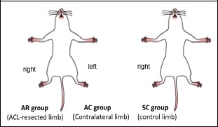
Grouping. Rats were divided into two groups; ACL-resected (A, n = 20) and sham-operated control (SC, n = 20) groups. Furthermore, right and left limbs of rats in the A group were assigned as ACL-resected (AR) and contralateral control (AC) groups, respectively.
ACL resection
The ACL resection was performed under the deep anesthesia with isoflurane (Forane®, Abbott Japan, Tokyo). The incision was made to the skin along the medial side of the patella from the anterior the knee of the right hindlimb, the joint capsule was deployed, and the patella was inverted lateral side. ACL was confirmed under direct view under the knee deeply flexed position. ACL was resected with a scalpel. The joint capsule and skin were sutured (the AR group). The contralateral left hindlimb of rats in the A group was similarly dissected to the skin and the joint capsule, and the joint capsule and the skin were sutured without resection the ACL (the AC group). In the SC group, rats received only incisions of the joint capsule of both hindlimbs without ACL resection. The right hindlimb was used for analyses. At 1 day, 1, 4, and 48 weeks after the treatment, RF and SM of five rats in each group were excised. Muscle wet weight was quickly measured, and frozen by liquid nitrogen, and stored at -80 ° C.
Histochemical analyses
The excised muscle was crossed at the center of the muscle width. Continuous frozen sections with a thickness of 10 μm were prepared by using a cryostat (CM 1900, Leica, Wetzlar, Germany). Hematoxylin & Eosin (HE) staining was performed according to the following procedure. After drying, the frozen section was fixed for 10 minutes using 4% paraformaldehyde. The nuclei were stained for 7 minutes with hematoxylin, the cytoplasm were stained with eosin for 5 minutes, dehydrated with ethanol, and covered with a cover glass. The samples were observed with an optical microscope (KEYENCE, Osaka, Japan). Mean fiber cross-sectional area (CSA) was calculated from ~200 muscle fibers of each section of each muscle using image analysis software Image J (Ver. 1.45i, Wayne Rasband, National Institutes of Health, USA.
Immunohistochemical analyses
Immunohistochemical staining was performed according to the following procedures. After drying, the frozen sections were incubated in Tris-buffered saline (TBS) for 5 minutes and soaked in blocking solution (Ultra V Block, Thermo, Cheshire, UK) for 1 hour. After completion of the blocking, the sections were allowed to react with the following diluted monoclonal antibodies at room temperature for 3 hours: anti-type I myosin heavy chain (MyHC) isoform (MyHCI, 1:200, BA-D5; Developmental Studies Hybridoma Bank [DSHB], Iowa, USA); anti-type IIa MyHC isoform (MyHCIIa, 1:100, SC-71, DSHB); and anti-type IIb MyHC isoform (MyHCIIb, 1:50, BF-F3, DSHB). After the primary antibody response, the sections were washed with TBS containing 0.1% Tween 20 (T-TBS) and allowed to react with the following fluorescent-labeled secondary antibodies at room temperature for 1 hour: Alexa Flour 488-labeled goat anti-mouse IgG2b (1:500, Invitrogen, California, USA), DyLight 405-labeled goat anti-mouse IgG1 (1:200, Invitrogen), and Alexa Flour 568-labeled goat anti-mouse IgM (1:500, Invitrogen). After the secondary antibody response, the sections were washed with T-TBS and covered with a cover slip. The frozen sections were observed under a fluorescence microscope (KEYENCE, Osaka, Japan). Using about image analysis software Image J (Ver. 1.45i, Wayne Rasband, National Institutes of Health, USA), 200 muscle fibers in three visual fields were counted for each section of each muscle. Muscle fiber type classification (%) (Figure 2) and CSA in each muscle fiber type (Kishiro et al., 2013) were calculated and measured.
Figure 2.
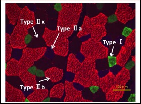
Classification of muscle fiber types. Muscle fiber type (Type I, IIa, IIb, and IIx) was classified by a standard immunohistochemical staining with anti-myosin heavy chain monoclonal antibodies.
Statistical analyses
Data are expressed as mean ± SEM. Significant difference was analyzed using two-way (treatment x time) analysis of variance (ANOVA) were performed followed by Tukey HSD post hoc test. Especially, Tukey HSD was employed to evaluate the effects of treatment within each level of “time” factor (by using SPSS Statistics 21.0J). The significance level was defined as p < 0.05.
Results
Muscle wet weight
In the present study, the relative muscle wet weight of RF and SM to body weight was evaluated. Relative RF and SM muscle weights are shown in Figures 3 and 4, respectively.
Figure 3.
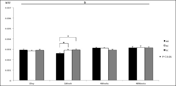
Muscle wet weight relative to body weight in rectus femoris muscle. 1 day: 1 day after ACL resection; 1 week: 1 week after ACL resection; 4 weeks: 4 weeks after ACL resection, 48 weeks: 48 weeks after ACL resection. Other abbreviations are the same as in Figure 1. Values are means ± SEM. n = 5 in each group. b: a significant main effect of time; *: p < 0.05.
Figure 4.
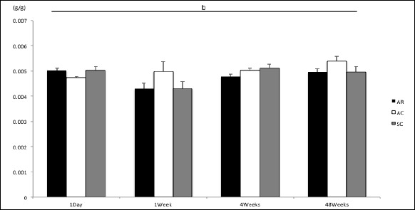
Muscle wet weight relative to body weight in semimembranosus muscle. Abbreviations are the same as in Figure 3. Values are means ± SEM. n=5 in each group. b: a significant main effect of time.
A significant main effect of time was observed in both RF and SM muscles (p < 0.05). One week after ACL resection, the relative RF wet weight in the AR group was significantly smaller than those in the AC and SC groups (p < 0.05) (Figure 3). On the other hand, there were no significant differences in the relative SM muscle weight among the groups (Figure 4).
Fiber CSA
Figures 5 and 6 show fiber CSA of RF and SM muscles, respectively. Significant main effects of both treatment and time in RF (Figure 5), and a significant main effect of time in SM (Figure 6) were observed (p < 0.05).
Figure 5.
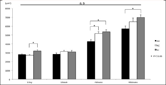
Fiber cross-sectional area (CSA) in rectus femoris muscle. Abbreviations are the same as in Figure 3. Unit: μm2. Values are means ± SEM. n=5 in each group. a: a significant main effect of treatment; b: a significant main effect of time; *: p < 0.05.
Figure 6.
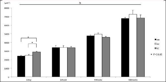
Fiber cross-sectional area (CSA) in semimembranosus muscle. Abbreviations are the same as in Figure 3. Unit: μm2. Values are means ± SEM. n=5 in each group. b: a significant main effect of time; *: p<0.05.
Mean fiber CSA of RF muscle in the AC group was significantly smaller than that in the SC group 1 day after ACL resection (p < 0.05), and RF muscle in the AR group was significantly smaller than that in the SC group 4 and 48 weeks after ACL resection (p < 0.05). Furthermore, fiber CSA of RF muscle in the AR group was significantly smaller than that in the AC group 4 week after the treatment (p < 0.05) (Figure 5). Mean fiber CSA of SM muscle in the AR and AC groups was significantly smaller than that in the SC group 1 day after ACL resection (p < 0.05) (Figure 6).
Relative distribution of muscle fiber types
Relative distribution of Type I, IIa, IIb, and IIx muscle fibers classified by MyHC phenotypes in RF and SM muscles 4 and 48 weeks after ACL resection are shown in Figures 7 and 8, respectively.
Figure 7.
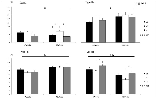
Relative distribution of muscle fiber type in rectus femoris muscle. Abbreviations are the same as in Figure 3. Unit: %. Values are means ± SEM. n=5 in each group. a: a significant main effect of treatment; b: a significant main effect of time; *: p<0.05.
Figure 8.
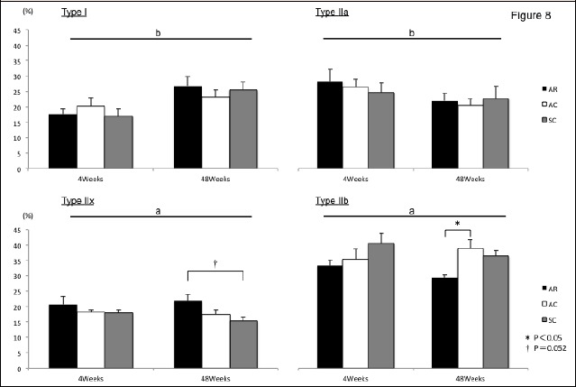
Relative distribution of muscle fiber type in semimembranosus muscle. Abbreviations are the same as in Figure 3. Unit: %. Values are means ± SEM. n=5 in each group. a: a significant main effect of treatment; b: a significant main effect of time; *: p<0.05.; †: p=0.052.
In RF muscle, significant main effects of treatment and time in Type IIb, and a significant main effect of treatment in Type I, and a significant main effect of time in Type IIa and IIx were observed, respectively (p 05).
The relative distribution of Type I of RF muscle in the AC group was significantly higher than that in the AR and SC groups 48 weeks after the treatment (p < 0.05). Relative distribution of Type IIb of RF muscle in the AC group was significantly lower than that in the SC group 4 and 48 weeks after the treatment (p < 0.05) (Figure 7).
A significant main effect of time in Type I and IIa, and a significant main effect of treatment in Type IIx and IIb of SM muscle were observed, respectively (p < 0.05). Four weeks after ACL resection, no significant differences in the relative distribution of fiber types of SM muscle were observed. Relative distribution of Type IIb fibers of SM muscle in the AR group was significantly lower than that in the AC group 48 weeks after the treatment (p < 0.05). Relative content of Type IIx fibers of SM muscle in the AR group showed a tendency to increase 48 weeks after ACL resection, compared with be the SC group (p = 0.052) (Figure 8).
Discussion
In this study, we investigated a long-term (48 weeks) effects of ACL resection on morphological and contractile characteristics of RF and SM muscles in both ACL-resected and contralateral limbs. No ACL-resection-associated changes in the mass of both muscles were observed 1 week after ACL resection. On the other hand, ACL-resection-associated effects on mean fiber CSA as well as the relative distribution of fiber types lasted 48 weeks after ACL resection were investigated. Furthermore, the responses of fiber CSA and fiber type composition following ACL resection in RF muscle were different from SM muscle. In addition, long-term effects of ACL resection were observed on the not only ACL-resected but also contralateral limbs. This study is the first report investigating long-term effects of ACL resection on the morphological and contractile properties of skeletal muscles around the knee joint in both injured and contralateral limbs.
Responses of the morphological and contractile properties of RF and SM muscles to ACL resection in injured limb
In the present study, a long-term effect of ACL resection on the morphological properties was observed in both RF and SM muscles. However, the responses of both muscles to ACL resection were different. In RF muscle, there were no significant changes in muscle wet weight. However, ACL resection associated decrease in mean fiber CSA was observed 4 and 48 weeks after ACL resection. In SM muscle, on the other hand, a significant reduction of mean fiber CSA was observed 1 day after ACL resection. These responses of both muscles to ACL resection are different. These results suggested that the functional and anatomical differences in both muscles might cause such different responses to ACL resection. In general, muscle weight is proportional to mean fiber CSA in the muscle. This discrepancy in the responses of muscle weight and fiber CSA in both muscles might be explained by the differences of contractile properties of muscle fiber types since peak force of slow Type I fibers is lower than that of fast Type II fibers. However, there have been no basic experiments, such as experiments using animal models, investigating a long-term study on the effects of ACL resection on morphological and functional characteristics of skeletal muscles around knee joint.
In this study, slight effects of ACL resection were observed in SM muscle, but not in RF muscle. In SM muscle, ACL-resection-associated increase of the relative distribution of Type IIx fibers was observed 48 weeks after ACL resection, compared with sham-operated control limb. In addition, there is a tendency to decrease in the relative distribution of Type IIb fibers in SM muscle. The mammalian skeletal muscle consist slow Type I and fast Type II (IIa, IIx, and IIb) muscle fibers. Type I is a slow-twitch and oxidative fiber with high fatigue resistance, which works to maintain posture (Nakatani et al., 2000). Fast Type IIb is classified as a fast-twitch and glycolytic fiber. Type IIa and IIx, which are intermediate fibers, have an intermediate function between Type I and IIb fibers (Botteinelli et al., 1991). Therefore, a fast-to-slow transition of MyHCs in SM muscle might be induced by ACL resection, but not RF muscle. SM muscle might get a fatigue resistance to maintain posture following ACL resection.
There is an anatomical difference between the knee extension muscle RF and knee flexion and internal rotation muscle SM (Kapandji, 1987). Generally, anterior and medial rotational laxity of the tibia occurs when the ACL is completely torn (Elias et al., 2003). In rats, a knee extensor RF is stretched during anterior laxity of the tibia after ACL resection. On the other hand, SM muscle is stretched by the tibia moving forward, and is shortened during the medial rotation of the tibia, respectively (Armstrong and Phelps, 1984; Kapandji, 1987). It is generally accepted that the shortening of skeletal muscle length induces a larger reduction of muscle mass, compared with a stretched skeletal muscle (Booth and Kelso, 1973). Therefore, the differences in the long-term lasting responses of RF and SM muscles to ACL resection might be attributed to muscle length, a stretched or shortened in resting position in rats.
It has been also suggested that ACL-resection-associated disruption of the afferent signals from the mechanoreceptor of ACL may modulate the muscle tone around the knee joint (Barrack et al., 1989; Barrett et al., 1991; Johansson et al., 1991; Kanemura et al., 2002; Ochi et al., 2002; Ochi et al., 1999). In fact, the lack of afferent signal from skeletal muscles reduces the firing rate of α-motor neurons (Macefield et al., 1993), resulting that the contractile characteristics of muscle fiber might be changed. Recently, however, it has been reported that the number of mechanoreceptors of ACL recovers 12 months after the rupture (Relph and Herrington, 2016). There is a possibility that the responses of the contractile properties of RF and SM muscles to ACL resection might be induced by the disruption of the afferent signal(s) from mechanoreceptors in ACL. Since the details for ACL-resection-associated changes in muscle length and the firing rate of α-motor neuron remains also unclear, we cannot explain the mechanism(s) of the ACL-mechanoreceptor-associated changes in the contractile properties of skeletal muscle of the knee joint in ACL resection, at present. Further studies are required.
Long-term effects of ACL resection on skeletal muscles
The life span of rats is generally approximately 48-72 weeks (Sengupta, 2013) and is considerably shorter than that of human. In the present study, the effects of ACL resection on the morphological and contractile properties of skeletal muscle around knee joint lasted until 48 weeks after the ACL resection were observed. This period (48 weeks) of rats may be equivalent over 50 years of human. Therefore, ACL injury may influence on the morphological and contractile properties of skeletal muscles around knee joint for ~50 years in human patient.
The results from this study revealed that the mean fiber CSA in skeletal muscles around the knee joint with ACL-resection was still smaller than that of control limb 48 weeks, which is a period of approximately half of rat’s life span, after ACL resection. Therefore, ACL resection may has long-lasting effects on the morphological and contractile properties of skeletal muscles around the knee joint in injured limbs, at least. Since this is the first report investigating long-term effects of ACL resection on skeletal muscles around the knee joint, we cannot explain the molecular mechanism for the long-term effects of ACL resection on skeletal muscle at present. Additional studies should be needed to elucidate this issue.
Effects of ACL resection on the skeletal muscles of contralateral limb
In the present study, long-term effects of ACL resection were observed on the not only ACL-resected but also contralateral limbs. Mean fiber CSA in contralateral limb was significantly increased in RF muscle 4 weeks after ACL resection. In contralateral RF muscle, furthermore, relative distribution of muscle fiber type I was significantly increased 48 weeks after the ACL resection, but was decreased in type IIb 4 and 48 weeks after the resection. In contralateral SM muscle, mean fiber CSA was significantly decreased 1 day after the ACL resection. Relative distribution of muscle fiber type IIb was increased 48 weeks after ACL resection. Knee extension function is disrupted in both the injured and contralateral limbs of ACL-injured patients (Konishi et al., 2002a; 2002b; 2003; 2007). However, there is no report investing long-term effects of ACL resection on skeletal muscles around the knee joint of the contralateral limb except the present study. On the other hand, patients are generally allowed to resume sports activity by using the relative muscle strength level of injured limb to contralateral limb clinically (Colombet et al., 2006; Fu et al., 2008; Urbach et al., 2001). Further studies to investigate the long-term effects of ACL resection with or without its reconstruction on skeletal muscle function in contralateral limb should be carried out.
Limitations of this study
This study has a few limitations. First of all, this study investigated only two muscles around the knee joint (RF and SM muscles) were evaluated. Both of knee extensor and flexor may be affected by ACL resection. In this study, furthermore, no functional evaluation of knee and muscle strength, and no investigation of ultrastructure as well as metabolic characteristics of muscle fibers were carried out. Further studies are necessary in the future to address these limitations.
Perspective
The present study demonstrated ACL-resection-associated atrophy of RF muscle persisted for a long period. Furthermore, the alteration of contraction characteristics of RF muscle was confirmed by the changes in fiber type distribution. These findings highlight that ACL-injured patients, even if they are asymptomatic and have no problems in ADL, are required to continue training for the long-term to address quantitative and qualitative muscle changes and to return to high-level sports activities. Taken together, the findings in the present study reaffirmed the importance of functional reconstruction of the knee joint through ACL reconstruction. In addition, we observed both morphological and functional changes of skeletal muscle in contralateral limb. Therefore, not only ACL-resected but also contralateral limbs should be trained with a special care for a long period.
Conclusions
Responses of fiber CSA and fiber type composition following ACL resection in RF muscle was different from SM muscle. Furthermore, the long-term effects of ACL resection were observed on the not only ACL-resected but also contralateral limbs. Evidences strongly suggest that ACL resection may cause to change in morphological and contractile properties of skeletal muscles around the knee joint in not only injured but also contralateral limbs. In addition, rehabilitation for quantitative and qualitative muscle changes by ACL resection may be required a special care for a long-term period.
Acknowledgements
There are no patents, products in development or marketed products to declare. The authors have no conflict of interest.
Biographies
Mahiro OHNO
Employment
Graduate student, St. Marianna University Graduate School of Medicine, Japan
Degree
MD
Research interests
Muscle fiber type, ACL injury
E-mail: m2ono@marianna-u.ac.jp
Hiroto FUJIYA
Employment
Professor, Department of Sports Medicine, St. Marianna University School of Medicine, Japan
Degree
MD, PhD
Research interests
Skeletal muscle injury, ACL
E-mail: fujiya-1487@marianna-u.ac.jp
Katsumasa GOTO
Employment
Professor, Department of Physiology, Graduate School of Health Sciences, Toyohashi SOZO University, Japan
Degree
PhD
Research interests
Plasticity of skeletal muscle, Muscle satellite cells
E-mail: gotok@sepia.ocn.ne.jp
Mitsutoshi KUROSAKA
Employment
Assistant, Department of Physiology, St. Marianna University School of Medicine, Japan
Degree
PhD
Research interests
Skeletal muscle injury
E-mail: fujiya-1487@marianna-u.ac.jp
Yuji OGURA
Employment
Assistant Professor, Department of Physiology, St. Marianna University School of Medicine, Japan
Degree
PhD
Research interests
Alpha-actin-3 and stress proteins
E-mail: yuji_ogura@marianna-u.ac.jp
Kanaka YATABE
Employment
Assistant, Department of Sports Medicine, St. Marianna University School of Medicine, Japan
Degree
MS
Research interests
Sports psychology, Statistics
E-mail: kanaka@mariannna-u.ac.jp
Takaaki KUDO
Employment
Graduate student, St. Marianna University Graduate School of Medicine, Japan
Degree
MD
Research interests
Muscle regeneration, Sports injury
E-mail: t2kudo@marianna-u.ac.jp
Hajime KOBAYASHI
Employment
Graduate student, St. Marianna University Graduate School of Medicine, Japan
Degree
MD
Research interests
Cartilage injury, Sports injury
E-mail: h17045@marianna-u.ac.jp
Hisateru NIKI
Employment
Professor, Department of Orthopaedic Surgery, St. Marianna University School of Medicine, Japan
Degree
MD, PhD
Research interests
Orthopaedic surgery, Foot and Ankle
E-mail: fujiya-1487@marianna-u.ac.jp
Haruki MUSHA
Employment
Professor Emeritus, St. Marianna University School of Medicine, Japan
Degree
MD, PhD
Research interests
Cardiac injury
E-mail: musha@marianna-u.ac.jp
References
- Ahmed I., Salmon L., Roe J., Pinczewski L. (2017) The long-term clinical and radiological outcomes in patients who suffer recurrent injuries to the anterior cruciate ligament after reconstruction. Bone and Joint Journal 99, 337-343. [DOI] [PubMed] [Google Scholar]
- Armstrong R. B., Phelps R.O. (1984) Muscle fiber type composition of the rat hindlimb. American Journal of Anatomy 171, 259-272. [DOI] [PubMed] [Google Scholar]
- Barrack R. L., Skinner H. B., Buckley S. L. (1989) Proprioception in the anterior cruciate deficient knee. American Journal of Sports Medicine 17, 1-6. [DOI] [PubMed] [Google Scholar]
- Barrett D. S. (1991) Proprioception and function after anterior cruciate reconstruction. Journal of Bone and Joint Surgery 73, 833-837. [DOI] [PubMed] [Google Scholar]
- Bonamo J.J., Fay C., Firestone T. (1990) The conservative treatment of the anterior cruciate deficient knee. American Journal of Sports Medicine 18, 618-623. [DOI] [PubMed] [Google Scholar]
- Booth F. W., Kelso J. R. (1973) Effect of hind-limb immobilization on contractile and histochemical properties of skeletal muscle. Pflügers Arch-European Journal of Physiology 342, 231-238. [DOI] [PubMed] [Google Scholar]
- Botteinelli R., Schiaffino S., Reggiani C. (1991) Force-velocity relations and myosin heavy chain isoform compositions of skinned fibres from rat skeletal muscle. Journal of Physiology 437, 655-672. [DOI] [PMC free article] [PubMed] [Google Scholar]
- Colombet P., Robinson J., Jambou S., Allard M., Bousquet V., de Lavigne C. (2006) Two-bundle, four-tunnel anterior cruciate ligament reconstruction. Knee Surgery, Sports Traumatology, Arthroscopy 14, 629-636. [DOI] [PubMed] [Google Scholar]
- Elias J. J., Faust A. F., Chu Y. H., Chao E. Y., Cosgarea A. J. (2003) The soleus muscle acts as an agonist for the anterior cruciate ligament. American Journal of Sports Medicine 31, 241-246. [DOI] [PubMed] [Google Scholar]
- Feller J. A., Cooper R., Webster K. E. (2001) Current Australian trends in rehabilitation following anterior cruciate ligament reconstruction. The Knee 9, 121-126. [DOI] [PubMed] [Google Scholar]
- Fu F. H., Shen W., Starman J. S., Okeke N., Irrgang J. J. (2008) Primary anatomic double-bundle anterior cruciate ligament reconstruction. American Journal of Sports Medicine 36, 1263-1274. [DOI] [PubMed] [Google Scholar]
- Fujiya H., Ogura Y., Ohno Y., Goto A., Nakamura A., Ohashi K., Uematsu D., Aoki H., Musha H., Goto K. (2015) Microcurrent electrical stimulation facilitates regeneration of injured skeletal muscle in mice. Journal of sports science and medicine 14, 297-303. [PMC free article] [PubMed] [Google Scholar]
- Gutmann E., Schiaffano S., Hanzlikova V. (1971) Mechanism of compensatory hypertrophy in skeletal muscle of the rat. Experimental Neurology 31, 451-464. [DOI] [PubMed] [Google Scholar]
- Ito T., Fujiya H., Goto K., Ogura Y., Kurosaka M., Yatabe K., Kishiro S., Yoshida A., Yoshioka H., Terauchi K., Beppu M., Funabashi T., Akema T., Musha H. (2013) Icing at early stage depresses skeletal muscle regeneration. Journal of St. Marianna University 4, 41-47. [Google Scholar]
- Johansson H., Sjolander P., Sojaka P. (1991) A sensory role for the cruciate ligament. Clinical Orthopaedics and Related Research 268, 161-178. [PubMed] [Google Scholar]
- Johnson R. J., Beynnon B. D., Nichols C. E., Renström F. H. (1992) The treatment of injuries of the anterior cruciate ligament. The Journal of Bone and Joint Surgery, 74, 140-151. [PubMed] [Google Scholar]
- Kanemura N., Kobayashi R., Kajihara H., Minematu A., Sasaki H., Tanaka S., Shirahama K., Miyamoto H., Watanabe M., Yata K., Moriyama H., Maejima H., Yoshimura O. (2002) Changes of mechanoreceptor in anterior cruciate ligament with hindlimb suspension rats. Journal of Physical Therapy Science 14, 27-32. [Google Scholar]
- Kapandji I. A. (1987) The knee. : The Physiology of the Joints. Volume Two Lower Limb. 5 th edition. Edinburgh London Melbourne and New York: Churchill Livingstone; 64-146. [Google Scholar]
- Kishiro S., Fujiya H., Goto K., Ogura Y., Kurosaka M., Yatabe K., Ito T., Yoshida A., Yoshioka H., Terauchi K., Beppu M., Funabashi T., Akema T., Musha H. (2013) Effects of STG tendon harvest and BTB harvest on knee joint muscles: An immunohistochemical evaluation. Journal of St. Marianna University 4, 49-59. [Google Scholar]
- Konishi Y., Aihara Y., Sakai M., Ogawa G., Fukubayashi T. (2007) Gamma loop dysfunction in the quadriceps femoris of patients who underwent anterior cruciate ligament reconstruction remains bilaterally. Scandinavian Journal of Medicine & Science in Sports 17, 393-399. [DOI] [PubMed] [Google Scholar]
- Konishi Y., Konishi H., Fukubayashi T. (2003) Gamma loop dysfunction in quadriceps on the contralateral side in patients with ruptured ACL. Medicine & Science in Sports & Exercise 35, 879-900. [DOI] [PubMed] [Google Scholar]
- Konishi Y., Fukubayashi T., Takeshita D. (2002a) Possible mechanism of quadriceps femoris weakness in patients with ruptured anterior cruciate ligament. Medicine & Science in Sports & Exercise 34, 1414-1418. [DOI] [PubMed] [Google Scholar]
- Konishi Y., Fukubayashi T., Takeshita D. (2002b) Mechianism of quadriceps femoris muscle weakness in patients with anterior cruciate ligament reconstruction. Scandinavian Journal of Medicine & Science in Sports 12, 371-375. [DOI] [PubMed] [Google Scholar]
- Konrads C., Reppenhagen S., Belder D., Goebel S., Rudert M., Barthel T. (2016) Long-term outcome of anterior cruciate ligament tear without reconstruction: a longitudinal prospective study. International Orthopaedics 40, 2325-2330. [DOI] [PubMed] [Google Scholar]
- Lee J. C., Kim Y., Park G. D. (2013) Effect of 12 weeks of accelerated rehabilitation exercise on muscle function of patients with ACL reconstruction of the knee joint. Journal of Physical Therapy Science 25, 1595-1599. [DOI] [PMC free article] [PubMed] [Google Scholar]
- Lephart S.M., Pincivero D.M., Giraldo J.L., Fu F.H. (1997) The role of proprioception in the management and rehabilitation of athletic injuries. American Journal of Sports Medicine 25, 130-137. [DOI] [PubMed] [Google Scholar]
- Macefield V. G., Gandevia S. C., Bigland-Ritchie B., Gorman R. B., Burke D. (1993) The firing rates of human motoneurones voluntarily activated in the absence of muscle afferent feedback. Journal of Physiology 471, 429-443. [DOI] [PMC free article] [PubMed] [Google Scholar]
- Mayer H.O., Brandt C.M., Weig T., Koehne M., Bernstein A., Suedkamp N.P., Hube R., Stoehr A. (2017) Long-term results of arthroscopic arthrolysis for arthrofibrosis after anterior cruciate ligament reconstruction. Arthroscopy 33, 408-414. [DOI] [PubMed] [Google Scholar]
- Nakatani T., Nakashima T., Kita T., Hirofuji C., Itoh K., Itoh M., Ishihara A. (2000) Cell size and oxidative enzyme activity of different types of fibers in different regions of the rat plantaris and tibialis anterior muscles. Japanese Journal of Physiology 50, 413-418. [DOI] [PubMed] [Google Scholar]
- Novak P.J., Bach B.R., Jr., Hager C.A. (1996) Clinical and functional outcome of anterior cruciate ligament reconstruction in the recreational athlete over the age of 35. American Journal of Knee Surgery 9, 111-116. [PubMed] [Google Scholar]
- Ochi M., Iwasa J., Uchio Y., Adachi N., Kawasaki K. (2002) Induction of somatosensory evoked potentials by mechanical stimulation in reconstructed anterior cruciate ligaments. Journal of Bone and Joint Surgery, 84-B, 761-766. [DOI] [PubMed] [Google Scholar]
- Ochi M., Iwasa J., Uchio Y., Adachi N., Sumen Y. (1999) The regeneration of sensory nurones in the reconstruction of the anterior cruciate ligament. Journal of Bone and Joint Surgery 81, 902-906. [DOI] [PubMed] [Google Scholar]
- Relph N., Herrington L. (2016) The effect of conservatively treated ACL injury on knee joint position sense. International Journal of Sports Physical Therapy 11, 536-543. [PMC free article] [PubMed] [Google Scholar]
- Sengupta P. (2013) The laboratory rat: relating its age with human’s. International Journal of Preventive Medicine 4: 624-630. [PMC free article] [PubMed] [Google Scholar]
- Shim J. K., Choi H. S., Shin J. H. (2015) Effect of neuromuscular training on knee joint stability after anterior cruciate ligament reconstruction. Journal of Physical Therapy Science 27, 3613-3617. [DOI] [PMC free article] [PubMed] [Google Scholar]
- Ucar M., Koca I., Eroglu M., Eroglu S., Sarp U., Arik H. O., Yetisgin A. (2014) Evaluation of open and close kinetic chain exercise in rehabilitation following anterior cruciate ligament reconstruction. Journal of Physical Therapy Science 26, 1875-1878. [DOI] [PMC free article] [PubMed] [Google Scholar]
- Urbach D., Nebelung W., Becker R., Awiszus F. (2001) Effects of reconstruction of the anterior cruciate ligament on voluntary activation of quadriceps femoris. Journal of Bone and Joint Surgery 83, 1104-1110. [DOI] [PubMed] [Google Scholar]
- Yellin H. (1974) Changes in fiber types of the hypertrophy in denervated hemi-diaphragm. Experimental Neurology 42, 412-428. [DOI] [PubMed] [Google Scholar]
- Yoshida A., Fujiya H., Goto K., Kurosaka M., Ogura Y., Yatabe K., Yoshioka H., Terauchi K., Funabashi T., Akema T., Niki H., Musha H. (2015) Regeneration of injured tibialis anterior muscle in mice in response to microcurrent electrical neuromuscular stimulation with or without icing. Journal of St. Marianna University 6, 159-169. [Google Scholar]


