

Abstract
Background:
Injury prevention training has been shown to be effective in reducing the incidence of noncontact anterior cruciate ligament (ACL) injury; however, the underlying reason for the success of these training programs is unclear.
Purpose:
To investigate whether an ACL injury prevention program that has been shown to reduce the incidence of ACL injury alters sagittal plane hip and knee biomechanics during a drop-landing task.
Study Design:
Descriptive laboratory study.
Methods:
Thirty female club soccer players (age range, 11-17 years) with no history of knee injury participated in this study. Kinematics and ground-reaction forces were collected while each participant performed a drop-landing task prior to and immediately after participation in a 12-week ACL injury prevention training program.
Results:
After ACL injury prevention training, participants demonstrated decreased knee extensor moments (P = .03), increased energy absorption at the hip (P = .04), decreased knee-to-hip extensor moment ratios (P = .05), and decreased knee-to-hip energy absorption ratios (P = .03).
Conclusion:
Participation in an ACL injury prevention training program decreased reliance on the knee extensor muscles and improved use of the hip extensor muscles, which may explain the protective effect of this type of training program on ACL injury.
Clinical Relevance:
Based on these findings, clinicians can better understand how ACL injury prevention training, such as the Prevent Injury and Enhance Performance (PEP) Program, may change movement behavior at both the hip and knee. Furthermore, the study findings may support the implementation of the PEP Program, or a similar program, for clinicians aiming to improve use of the hip in an effort to reduce knee loading and consequent injuries.
Keywords: knee joint, anterior cruciate ligament, PEP Program, kinematics, kinetics
Anterior cruciate ligament (ACL) injuries are more prevalent in female athletes compared to male athletes participating in similar sports.1,21,24 Previous research has identified sex-based differences in lower extremity mechanics that may predispose females to an increased risk for ACL injury. In general, there is considerable evidence that females exhibit increased knee valgus angles and moments during athletic tasks such as cutting16,19,20,33 and landing9,12,15,26 when compared to males. Furthermore, females consistently have been shown to exhibit higher knee extensor moments and decreased knee flexion angles compared to males during similar tasks.14,32,36 Such findings are relevant to ACL injury risk, as it has been shown that the resulting anterior shear forces caused by high quadriceps forces at small knee flexion angles, combined with knee valgus loading, results in elevated ACL strain.18
Although there appears to be consensus in the literature that females perform sport-specific tasks in a way that increases ACL injury risk, little is known of the underlying reasons for this movement behavior. In a previous study, we found that during the deceleration phase of a drop-landing task, females who exhibited greater knee valgus angles and moments had lower hip extensor moments and energy absorption at the hip and higher knee extensor moments and energy absorption at the knee.28 This type of drop-landing task is typically performed by having subjects step off an elevated platform, land on 2 adjacent force plates, and then proceed to immediately jump as high as they can after landing. We have also found that during a drop-landing task, females exhibit higher knee adductor (valgus) moments and disproportionate use of the knee extensors relative to the hip extensors (greater knee/hip extensor moment ratios and energy absorption ratios) when compared to males.32 Based on these findings, we have hypothesized that if the hip extensors are unable to adequately contribute to the deceleration of the body center of mass, females may compensate by relying more on the quadriceps and passive knee restraints in the frontal plane (ie, ligaments) to absorb impact forces.32
Given that diminished use of the sagittal plane hip musculature may underlie the altered movement behavior observed in females, it is plausible that the protective effect afforded by ACL injury prevention training (ie, decreased knee loading) may be achieved through improved hip function. If shown to be true, this would support the notion that altered hip function may play a contributory role in the higher rates of ACL tears in females. Therefore, the primary objective of the current study was to investigate whether an ACL injury prevention program that has been shown to reduce the occurrence of ACL injury in females by 74%10,17 alters sagittal plane hip and knee biomechanics during a drop-landing task. More specifically, we hypothesized that after training, female soccer athletes would exhibit greater sagittal plane moments and energy absorption at the hip and a relative decrease in the sagittal plane moments and energy absorbed at the knee during a drop-landing task. Results of this study will provide insight into how ACL injury prevention training may change movement behavior, particularly at the hip, in a way that may reduce ACL injury risk.
Methods
To better understand how ACL injury prevention training influences lower extremity mechanics, we examined sagittal plane hip and knee joint kinematics and kinetics before and after an established 12-week ACL injury prevention training program. In particular, we were interested in elucidating the influence of such a training program on hip function during a task such as drop landing, which is commonly associated with ACL injury. Each subject participated in 2 biomechanical testing sessions (pre- and posttraining), which took place at a biomechanics research laboratory. The first biomechanical testing session occurred within the 2 weeks before the initiation of the training program. The second biomechanical testing session occurred within 2 weeks immediately after completion of the training program. To better understand the influence of ACL injury prevention training on lower extremity mechanics, key hip and knee biomechanical variables that have been associated with increased risk for ACL injury were examined.
Subjects
Using data from the literature, sample size was estimated for a minimal statistical power of 80% (P = .05).6 Given the variation of the dependent measures that were included in this study, 25 participants were deemed adequate.
Participants consisted of 30 female club soccer players (group means: age, 13.5 years; height, 162 cm; weight, 55 kg; years playing club soccer, 3; years playing American Youth Soccer Organization soccer, 7). Participants were recruited from area club soccer teams that agreed to participate in the ACL injury prevention training program. All participants were healthy with no current complaints of lower extremity injury. Participants were excluded from the study if they reported any of the following: (1) history of previous ACL injury or repair; (2) previous injury that resulted in ligamentous laxity at the ankle, hip, or knee; (3) presence of any medical or neurological condition that would impair their ability to perform a landing task; or (4) previous participation in an ACL injury prevention training program.
Before testing, approval by a university ethics committee was obtained. Part of this approval process required that the research team members complete formal human subjects research ethics training (ie, Collaborative Institutional Training Initiative training). Before participation, all procedures were explained to each participant, and informed consent was obtained as approved by a university institutional review board.
Procedures
Biomechanics
Kinematic data were collected using an 8-camera, 3-dimensional motion analysis system (Vicon, Oxford Metrics Ltd) at a sampling frequency of 250 Hz. Kinetic data were obtained at a sampling frequency of 1500 Hz using 2 force plates (Advanced Mechanical Technologies Inc) (Figure 1). Kinematic data were obtained with the use of reflective markers that were placed bilaterally over the following anatomic landmarks: the first and fifth metatarsal heads, medial and lateral malleoli, medial and lateral femoral epicondyles, greater trochanters, iliac crests, and a single marker on the joint space between the fifth lumbar and the first sacral spinous processes. In addition, triads of rigid reflective tracking markers were securely placed bilaterally on the lateral surfaces of the participant’s thigh, leg, and heel counter of the shoe (Figure 1). To control for the potential influence of varying footwear, participants were fitted with the same style of cross-training shoe (New Balance).
Figure 1.
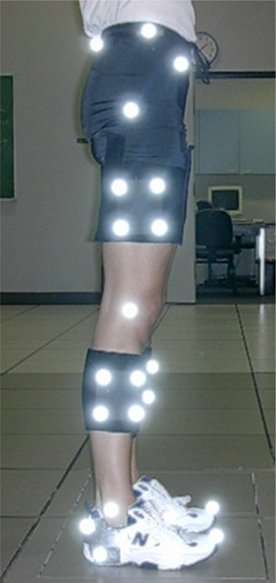
Lower extremity reflective marker placement.
After 3 practice trials, each participant performed 3 trials of a drop-landing task, which were recorded. Participants were instructed to step off a 36-cm platform (leading with their dominant limb) and land with their right foot on one force plate and their left foot on an adjacent force plate and then proceed to immediately jump as high as they could after landing (Figure 2). Participants were not given any verbal cues on landing or jumping technique.
Figure 2.

Laboratory setup.
ACL Injury Prevention Training
After the initial biomechanical testing session, participants participated in the Prevent Injury and Enhance Performance (PEP) Program developed by the Santa Monica Orthopaedic and Sports Medicine Research Foundation (Santa Monica, California, USA).10,17 Each training session took approximately 20 minutes and was overseen by an individual who was trained in the implementation of the program. All training took place at the site of the team’s soccer practice and consisted of a predetermined series of warm-up, stretching, strengthening, plyometrics, and sport-specific agility drills (Table 1). This training includes regular feedback regarding lower extremity movement patterns during the activities, with an emphasis on reduced knee valgus. The program was performed 2 times per week for 12 weeks during the course of the soccer season. Failure to complete 80% of the training sessions would have resulted in a subject being excluded from the study; however, all subjects completed at least 80% of the training sessions. Within 2 weeks of completion of the training program, all participants completed posttraining biomechanical testing using the identical procedures as described above.
TABLE 1.
Prevent Injury and Enhance Performance Program Exercises
| Phase | Drill |
|---|---|
| Warm-up (30-60 s each) | Jog line to line |
| Shuttle run | |
| Backward running | |
| Stretching (30 s × 2 repetitions) | Calf stretch |
| Quadriceps stretch | |
| Figure-four hamstring stretch | |
| Inner thigh stretch | |
| Hip flexor stretch | |
| Strengthening (30 s × 2 repetitions) | Walking lunges |
| Russian hamstring | |
| Single-toe raises | |
| Plyometrics (20 repetitions) | Lateral hops over cone |
| Forward/backward hops over cone | |
| Single-leg hops over cone | |
| Vertical jumps with headers | |
| Scissors jump | |
| Agilities | Shuttle run with forward/backward running |
| Diagonal runs | |
| Bounding run |
Data Processing
Coordinate data were digitized in Vicon Workstation software (Workstation, Oxford Metrics Ltd) and filtered using a fourth-order zero-lag Butterworth 12-Hz low-pass filter. Visual3D software (C-Motion Inc) was used to calculate 3-dimensional knee and hip kinematics. Joint kinematics were calculated using the gold standard, which is a joint coordinate system approach,11 and were reported relative to a static standing trial.
Kinematics, ground-reaction forces, and anthropometrics were used to calculate joint moments at the knee and hip using inverse dynamics equations in Visual 3D software.3 Kinetic data were normalized to body mass. The joint moments referred to in this investigation are the internal (muscle) moments. Sagittal plane joint power was computed for the hip and knee as the scalar product of angular velocity and net joint moment. Total energy absorbed at the hip and knee was calculated by integrating the respective power-time curves during the deceleration phase of landing.
The landing cycle was identified as the period from foot contact to toe-off, as determined by the force plate recordings. For the purposes of this study, only the deceleration phase of the drop-landing task was examined, as this timeframe corresponds to the period of greatest knee loading.2 The deceleration phase was defined as the time period from foot contact to peak knee flexion. Variables of interest during the deceleration phase of landing included mean knee extensor moment, mean hip extensor moment, and energy absorbed at the knee and hip of the subject’s dominant limb. The limb they would use to kick a soccer ball defined the dominant limb.
To further explore the relationship between the mean knee extensor and hip extensor moments, we examined the knee-to-hip extensor moment ratio (knee/hip extensor moment ratio = mean knee extensor moment/mean hip extensor moment). Using this ratio, a value greater than 1 was indicative of an increased knee extensor moment relative to the hip extensor moment, while a value less than 1 was indicative of an increased hip extensor moment relative to the knee extensor moment. A similar ratio was used to explore the relationship between the energy absorbed at the knee and hip (knee/hip energy absorption ratio = knee energy absorption/hip energy absorption).
Statistical Analyses
Pre- and posttraining differences for each dependent variable of interest were evaluated using paired t tests. Statistical analyses were performed using SPSS statistical software and a significance level of P ≤ .05.
Results
The pre- and posttraining values for all variables of interest are presented in Table 2. After ACL injury prevention training, participants exhibited decreased knee extensor moments (P = .03) but did not exhibit a significant change in hip extensor moments (P = .07). Furthermore, participants exhibited increased hip extensor energy absorption (P = .04) but did not exhibit a significant change in knee extensor energy absorption posttraining (P = .11). When examining ratios, participants exhibited a decreased knee/hip extensor moment ratio (P = .05) and a decreased knee/hip extensor energy absorption ratio (P = .03) after training.
TABLE 2.
Group Means (SD) for All Variables of Interest Pre- and Posttraining
| Variable | Pretraining | Posttraining | P |
|---|---|---|---|
| Mean knee extensor moment, Nm/kg | 1.30 (0.29) | 1.18 (0.25) | .03 |
| Mean hip extensor moment, Nm/kg | 0.68 (0.22) | 0.77 (0.24) | .07 |
| Knee extensor energy absorption, W/kg | 373.77 (87.09) | 346.43 (85.98) | .11 |
| Hip extensor energy absorption, W/kg | 129.79 (45.83) | 150.75 (47.55) | .04 |
| Knee-to-hip extensor moment ratio | 2.34 (1.82) | 1.76 (0.89) | .05 |
| Knee-to-hip extensor energy absorption ratio | 3.18 (1.30) | 2.58 (1.13) | .03 |
Discussion
Consistent with our proposed hypotheses, we found that participation in a 12-week ACL injury prevention training program resulted in a distinct change in the strategy used to attenuate impact forces (Figure 3). This posttraining adjustment in behavior was best illustrated by the change in the knee/hip extensor moment ratio and the knee/hip energy absorption ratio. Taken together, our results point to the modification of biomechanical/neuromuscular risk factors as being a potential mechanism by which ACL injury prevention programs are successful in decreasing the incidence of ACL tears.
Figure 3.
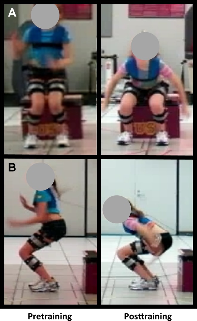
Lower extremity views in the (A) sagittal plane and the (B) frontal plane of a participant performing the drop-landing task pre– and post–injury prevention training.
We have previously reported that female athletes exhibit greater knee/hip extensor moment ratios and greater knee/hip energy absorption ratios compared to males during a drop-landing task, indicating that females rely more on the knee extensors to attenuate impact forces.28 Similar sex differences in sagittal plane mechanics have been observed during landing4,30 and cutting tasks.13,20,22,27 The ACL injury prevention training program evaluated in the current study appeared to result in a modification of this sex-specific tendency, as illustrated by less reliance of the knee extensors to decelerate the body center of mass during landing. Specifically, participants in the current study exhibited posttraining decreases in the knee extensor moment and a concurrent increase in hip extensor energy absorption. These changes resulted in a 25% reduction in the knee/hip extensor moment ratio and a 19% reduction in the knee/hip energy absorption ratio. Taken together, the observed changes after ACL injury prevention training are suggestive of increased use of the hip extensors as a means to control the deceleration phase of the landing task. The concurrent decrease in the knee extensor moment posttraining may have implications for ACL injury risk, as elevated quadriceps forces are thought to contribute to anterior tibial shear forces and increased loading of the ACL.7 Specifically, increased anterior tibial shear forces have been noted in individuals who perform a drop-landing task with less hip flexion and greater knee flexion range of motion, and greater knee extensor moments and quadriceps activation.31
The current study adds to the growing body of literature demonstrating that ACL injury prevention training alters lower extremity biomechanics.5,8,23,25 However, the majority of these studies have focused only on knee mechanics. Nagano et al25 reported an increase in knee flexion angles following participation in a jump and balance training program. They concluded that increased knee flexion may reduce ACL strain, which would in turn decrease the risk for ACL injury. Similarly, Myer et al23 examined 18 female high school athletes and found an increase in maximum knee flexion during a drop vertical jump task after 7 weeks of plyometric training. Cochrane et al5 examined knee joint kinetics after participation in a 12-week intensive balance training program and found a 10% reduction in the knee extensor moments. They concluded that balance training resulted in changes in knee joint loading and may reduce the risk of ACL injury. Our study is unique in that we evaluated sagittal plane hip and knee mechanics after ACL injury prevention training. As such, we were better able to examine changes in biomechanical strategies by analyzing the knee/hip moment and energy ratios as opposed to examining sagittal plane knee and hip mechanics in isolation.
Although we did find a difference in movement behavior after training, we can only postulate the underlying mechanism for this change. For example, it is possible that participants gained strength at the hip, as hip muscle weakness has been associated with knee mechanics associated with an increased risk for ACL injury.29,35 The work of Stearns et al34 supports this theory; these authors found that women who exhibited higher knee extensor moments relative to hip extensor moments during landing (compared to men) also exhibited relatively lower hip strength relative to knee strength. While evidence in the literature points to increased hip strength as a probable cause for the underlying mechanism of change, it could be the case that no change in strength occurred but that the subjects learned a new movement strategy that better used the hip musculature. As the PEP Program does emphasize instruction in movement mechanics (ie, “soft landing” and “no knee caving in”), this is a distinct possibility. Given the nature of the PEP Program, a likely explanation for the observed posttraining movement behavior is the combination of increased hip strength and a newly established movement pattern. Regardless of the cause of the observed posttraining change in landing strategy, our results point to the modification of biomechanical/neuromuscular risk factors as being a potential mechanism by which injury prevention programs are successful in decreasing ACL tears. Our findings are particularly compelling because the ACL injury prevention training program implemented in this study has been shown to reduce the incidence of ACL injuries in female soccer athletes.10,17
We found that participation in a 12-week ACL injury prevention program resulted in biomechanical changes that may be considered to be “ACL protective.” After training, participants demonstrated decreased knee extensor moments, increased energy absorption at the hip, an increase in the hip-to-knee moment ratio, and an increase in the hip-to-knee energy absorption ratio. Our findings suggest that the protective effect afforded by ACL injury prevention training may be achieved through decreased reliance on the knee extensors and improved use of the hip extensors.
When interpreting the findings of this study, it is important to keep in mind that although differences in lower extremity biomechanics were identified after participation in an ACL injury prevention training program, muscle strength was not measured. Ideally, future studies would include strength measurements pre- and posttraining to better understand the underlying reasons for the biomechanical changes. An additional limitation of this study is that it did not include a control group. While these athletes exhibited biomechanical changes after participation in an ACL injury prevention training program, it is unknown what changes would have occurred over that same time period if they had undergone their typical soccer practice and game schedule over 12 weeks without the ACL injury prevention training. Future studies should consider implementing a control group.
Conclusion
Participation in a 12-week ACL injury prevention training program changes the way athletes perform a landing task. Considerable attention has been placed on ACL injury prevention training, but little remains known as to how these programs modify lower extremity movement patterns that have been identified as placing athletes at an increased risk of ACL injury. In the current study, athletes performed a landing task in a way that better used their hip musculature, which likely, in turn, “protected” their knees after training by decreasing the knee extensor moments. Our findings support the implementation of the PEP Program, or a similar program, for clinicians aiming to improve lower extremity mechanics and use of the hip musculature. Furthermore, this study may provide clinicians with an overall impression of hip and knee movement strategies during the drop-landing task, which will inform their movement pattern assessments before and after ACL injury prevention training.
Footnotes
One or more of the authors has declared the following potential conflict of interest or source of funding: This study was funded by the National Institutes of Health (R01 AR053073-02).
Ethical approval for this study was obtained from the University of Southern California Health Sciences Campus Institutional Review Board (proposal HS-04B017).
References
- 1. Agel J, Arendt EA, Bershadsky B. Anterior cruciate ligament injury in National Collegiate Athletic Association basketball and soccer: a 13-year review. Am J Sports Med. 2005;33:524–531. [DOI] [PubMed] [Google Scholar]
- 2. Boden BP, Dean GS, Feagin JA, Jr, Garrett WE., Jr Mechanisms of anterior cruciate ligament injury. Orthopedics. 2000;23:573–578. [DOI] [PubMed] [Google Scholar]
- 3. Bresler B, Frankel JP. The forces and moments in the leg during level walking. Trans Am Soc Mech Eng. 1950;72:27–36. [Google Scholar]
- 4. Butler RJ, Willson JD, Fowler D, Queen R. Gender differences in landing mechanics vary depending on the type of landing. Clin J Sport Med. 2013;23:52–57. [DOI] [PubMed] [Google Scholar]
- 5. Cochrane JL, Lloyd DG, Besier TF, Elliott BC, Doyle TL, Ackland TR. Training affects knee kinematics and kinetics in cutting maneuvers in sport. Med Sci Sports Exerc. 2010;42:1535–1544. [DOI] [PubMed] [Google Scholar]
- 6. Cohen J. Statistical Power Analysis for the Behavioral Sciences. 2nd ed Mahwah, NJ: Lawrence Erlbaum; 1990. [Google Scholar]
- 7. DeMorat G, Weinhold P, Blackburn T, Chudik S, Garrett W. Aggressive quadriceps loading can induce noncontact anterior cruciate ligament injury. Am J Sports Med. 2004;32:477–483. [DOI] [PubMed] [Google Scholar]
- 8. Favre J, Clancy C, Dowling AV, Andriacchi TP. Modification of knee flexion angle has patient-specific effect on anterior cruciate ligament injury risk factors during jump landing. Am J Sports Med. 2016;44:1540–1546. [DOI] [PubMed] [Google Scholar]
- 9. Ford KR, Myer GD, Hewett TE. Valgus knee motion during landing in high school female and male basketball players. Med Sci Sports Exerc. 2003;35:1745–1750. [DOI] [PubMed] [Google Scholar]
- 10. Gilchrist J, Mandelbaum BR, Melancon H, et al. A randomized controlled trial to prevent noncontact anterior cruciate ligament injury in female collegiate soccer players. Am J Sports Med. 2008;36:1476–1483. [DOI] [PubMed] [Google Scholar]
- 11. Grood ES, Suntay WJ. A joint coordinate system for the clinical description of three-dimensional motions: application to the knee. J Biomech Eng. 1983;105:136–144. [DOI] [PubMed] [Google Scholar]
- 12. Haddas R, James CR, Hooper TL. Lower extremity fatigue, sex, and landing performance in a population with recurrent low back pain. J Athl Train. 2015;50:378–384. [DOI] [PMC free article] [PubMed] [Google Scholar]
- 13. Landry SC, McKean KA, Hubley-Kozey CL, Stanish WD, Deluzio KJ. Neuromuscular and lower limb biomechanical differences exist between male and female elite adolescent soccer players during an unanticipated side-cut maneuver. Am J Sports Med. 2014;35:1089–1095. [DOI] [PubMed] [Google Scholar]
- 14. Lephart SM, Ferris CM, Riemann BL, Myers JB, Fu FH. Gender differences in strength and lower extremity kinematics during landing. Clin Orthop Relat Res. 2002;401:162–169. [DOI] [PubMed] [Google Scholar]
- 15. Liederbach M, Kremenic IJ, Orishimo KF, Pappas E, Hagins M. Comparison of landing biomechanics between male and female dancers and athletes, part 2: influence of fatigue and implications for anterior cruciate ligament injury. Am J Sports Med. 2007;42:1888–1900. [DOI] [PubMed] [Google Scholar]
- 16. Malinzak RA, Colby SM, Kirkendall DT, Yu B, Garrett WE. A comparison of knee joint motion patterns between men and women in selected athletic tasks. Clin Biomech (Bristol, Avon). 2001;16:438–445. [DOI] [PubMed] [Google Scholar]
- 17. Mandelbaum BR, Silvers HJ, Watanabe DS, et al. Effectiveness of a neuromuscular and proprioceptive training program in preventing anterior cruciate ligament injuries in female athletes. Am J Sports Med. 2005;33:1003–1010. [DOI] [PubMed] [Google Scholar]
- 18. Markolf KL, Burchfield DM, Shapiro MM, Shepard MF, Finerman GA, Slauterbeck JL. Combined knee loading states that generate high anterior cruciate ligament forces. J Orthop Res. 1995;13:930–935. [DOI] [PubMed] [Google Scholar]
- 19. McLean SG, Huang X, van den Bogert AJ. Association between lower extremity posture at contact and peak knee valgus moment during sidestepping: implications for ACL injury. Clin Biomech (Bristol, Avon). 2005;20:863–870. [DOI] [PubMed] [Google Scholar]
- 20. McLean SG, Lipfert SW, van den Bogert AJ. Effect of gender and defensive opponent on the biomechanics of sidestep cutting. Med Sci Sports Exerc. 2004;36:1008–1016. [DOI] [PubMed] [Google Scholar]
- 21. Messina DF, Farney WC, DeLee JC. The incidence of injury in Texas high school basketball. A prospective study among male and female athletes. Am J Sports Med. 1999;27:294–299. [DOI] [PubMed] [Google Scholar]
- 22. Miranda DL, Fadale PD, Hulstyn MJ, Shalvoy RM, Machan JT, Fleming BC. Knee biomechanics during a jump-cut maneuver: effects of sex and ACL surgery. Med Sci Sports Exerc. 2013;45:942–951. [DOI] [PMC free article] [PubMed] [Google Scholar]
- 23. Myer GD, Ford KR, McLean SG, Hewett TE. The effects of plyometric versus dynamic stabilization and balance training on lower extremity biomechanics. Am J Sports Med. 2006;34:445–455. [DOI] [PubMed] [Google Scholar]
- 24. Myklebust G, Maehlum S, Holm E, Bahr R. A prospective cohort study of anterior cruciate ligament injuries in elite Norwegian team handball. Scand J Med Sci Sports. 1998;8:149–153. [DOI] [PubMed] [Google Scholar]
- 25. Nagano Y, Ida H, Akai M, Fukubyashi T. Effects of jump and balance training on knee kinematics and electromyography of female basketball athletes during a single limb drop landing: pre-post intervention study. Sports Med Arthrosc Rehabil Ther Technol. 2011;3(1):14. [DOI] [PMC free article] [PubMed] [Google Scholar]
- 26. Pappas E, Hagins M, Sheikhzadeh A, Nordin M, Rose D. Biomechanical differences between unilateral and bilateral landings from a jump: gender differences. Clin J Sport Med. 2007;17:263–268. [DOI] [PubMed] [Google Scholar]
- 27. Pollard CD, Sigward SM, Powers CM. Gender differences in hip joint kinematics and kinetics during side-step cutting maneuver. Clin J Sport Med. 2007;17:38–42. [DOI] [PubMed] [Google Scholar]
- 28. Pollard CD, Sigward SM, Powers CM. Limited hip and knee flexion during landing is associated with increased frontal plane knee motion and moments. Clin Biomech (Bristol, Avon). 2010;25:142–146. [DOI] [PMC free article] [PubMed] [Google Scholar]
- 29. Powers CM. The influence of abnormal hip mechanics on knee injury: a biomechanical perspective. J Orthop Sports Phys Ther. 2010;40:42–51. [DOI] [PubMed] [Google Scholar]
- 30. Schmitz RJ, Kulas AS, Perrin DH, Riemann BL, Shultz SJ. Sex differences in lower extremity biomechanics during single leg landings. Clin Biomech (Bristol, Avon). 2007;22:681–688. [DOI] [PubMed] [Google Scholar]
- 31. Shultz SJ, Nguyen AD, Leonard MD, Schmitz RJ. Thigh strength and activation as predictors of knee biomechanics during a drop jump task. Med Sci Sports Exerc. 2009;41:857–866. [DOI] [PMC free article] [PubMed] [Google Scholar]
- 32. Sigward SM, Pollard CD, Powers CM. The influence of sex and maturation on landing biomechanics: implications for anterior cruciate ligament injury. Scand J Med Sci Sports. 2012;22:502–509. [DOI] [PMC free article] [PubMed] [Google Scholar]
- 33. Sigward SM, Powers CM. The influence of gender on knee kinematics, kinetics and muscle activation patterns during side-step cutting. Clin Biomech (Bristol, Avon). 2006;21:41–48. [DOI] [PubMed] [Google Scholar]
- 34. Stearns KM, Keim RG, Powers CM. Influence of relative hip and knee extensor muscle strength on landing biomechanics. Med Sci Sports Exerc. 2013;45:935–941. [DOI] [PubMed] [Google Scholar]
- 35. Willson JD, Ireland ML, Davis I. Core strength and lower extremity alignment during single leg squats. Med Sci Sports Exerc. 2006;38:945–952. [DOI] [PubMed] [Google Scholar]
- 36. Yu B, Lin CF, Garrett WE. Lower extremity biomechanics during the landing of a stop-jump task. Clin Biomech (Bristol, Avon). 2006;21:297–305. [DOI] [PubMed] [Google Scholar]