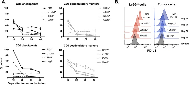
Figure 2. Expression of immune checkpoints and costimulatory markers in the MOC1 tumor microenvironment trended down with tumor progression.
A., Immune checkpoints (PD1, CTLA-4, Tim3 and Lag3) and costimulatory markers (CD27, 41BB, ICOS and OX40) were measured on CD4+ and CD8+ TIL from MOC1 tumors at day 10, 20, 30 and 40 after tumor implantation (n = 5/time point) via flow cytometry. * denotes a statistically significant change (p < 0.05) from day 10 to 20. B., representative histograms of PD-L1 expression on MOC1 tumor infiltrating Ly6Ghi myeloid and tumor cells with tumor progression. * denotes a statistically significant change (p < 0.05) from previous time point.

