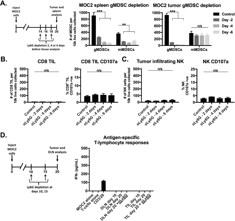
Figure 5. Depletion of gMDSCs from MOC2 tumor-bearing mice did not enhance effector immune cell activation.
A., schematic demonstrating a single injection of Ly6G depleting antibody (clone 1A8, 200 μg/injection) at either day 14, 16 or 18 (6, 4 or 2 days before tissue analysis, respectively) before tissue analysis on day 20. Right bar graphs demonstrate absolute numbers of splenic and tumor MDSC after Ly6G mAb administration. B., CD8+ TIL infiltration and degranulation (CD107a positivity) were assessed by flow cytometry following gMDSC depletion. C., NK cell tumor infiltration and degranulation (CD107a positivity) were assessed by flow cytometry. D., schematic demonstrating Ly6G depletion at days 10 and 15 with tissue analysis at day 20. Draining lymph node T-lymphocytes and TIL were isolated from mice treated with Ly6G depleting antibody or isotype control, pooled, and assessed for IFNγ production upon exposure to MOC2 tumor cells. **, p < 0.01; ***, p < 0.001. n/s, non-significant.

