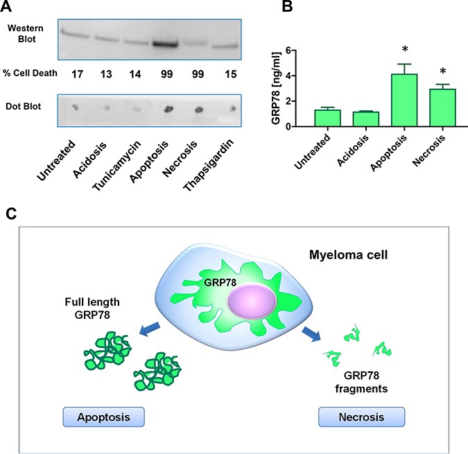
Figure 3. Plasma cells undergoing apoptosis or necrosis release full length or cleaved fragments of GRP78.

(A) Acidosis (pH 6.3), apoptosis, necrosis, and ER-stress were induced in NCI-H929 multiple myeloma cell line by treatment with acetic acid, 100 μM calcimycin, 100 mM H2O2, 30ng/ml tunicamycin, and 1nM thapsigargin for 72 h, respectively. Supernatants were analyzed by the sheep-anti human GRP78 polyclonal antiserum used in the sandwich ELISA. Apoptosis resulted in an increase of full length GRP78 in the Western Blot analysis. Necrosis gave no increased signal in Western Blot analysis, but on Dot Blot analysis immune-reactive GRP78 fragments increased significantly. (B) Supernatants of NCI-H929 cells under acidosis (pH 6.3), apoptosis and necrosis were measured in the GRP78 sandwich ELISA. Apoptosis as well as necrosis gave significant higher signals for immune-reactive GRP78 in the supernatants. * indicate p < 0.05 (C) Hypothetical model of GRP78 release and processing of MM cells under apoptotic and necrotic conditions. ELISA will measure apoptosis as well as necrosis of MM cells in bone marrow plasma samples.
