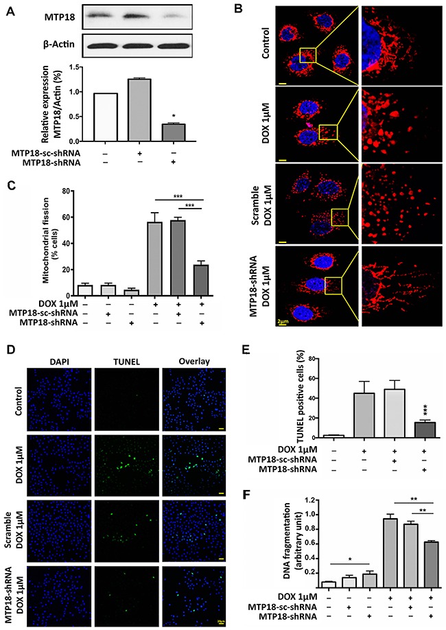
Figure 2. Knockdown of MTP18 interferes doxorubicin-induced mitochondrial fission and apoptosis.

(A) Analysis of mitochondrial protein 18 (MTP18) expression. Immunoblot shows MTP18 knockdown in AGS cells (upper panel). β-actin served as a loading control. The densitometry data were expressed as the mean±SEM of three independent experiments (lower panel). *P < 0.05 verses negative control or scramble (sc-shRNA). B and C. Knockdown of MTP18 interferes cells to undergo DOX-induced mitochondrial fission. AGS cells were exposed to high concentration (1μmol/L) doxorubicin (DOX). (B) Shows mitochondrial morphology. Bar=2μm. C shows percentage of cells with mitochondrial fission; ***P<0.001. (D) and (E) Knockdown of MTP18 interferes cells to undergo DOX-induced apoptosis. Apoptosis was analyzed by TUNEL assay. (D) Shows TUNEL positive cells. Bar=20μm. E shows percentage of TUNEL positive. (F) Knockdown of MTP18 interferes cells to undergo DOX-induced DNA fragmentation. DNA fragments were analyzed using the cell death detection ELISA. (D-F) AGS cells were exposed to high concentration (1μmol/L) DOX. *P<0.05, **P<0.01, and ***P<0.001 versus DOX alone or sc-shRNA treated with DOX. Data were expressed as the mean±SEM of three independent experiments. Figures presented are the representative of at least three independent experiments.
