

Abstract
Objective:
The objective of this study is to evaluate the effect of irrigating agents on push-out bond strength of resin postcemented with various adhesive systems at different radicular dentin sections.
Materials and Methods:
Sixty single-rooted premolar teeth were root canal treated, subsequently decorated at cementoenamel junction. The endodontic postspace was irrigated with 5.25% sodium hypochlorite (NaOCl) and 17% ethylenediaminetetraacetic acid (EDTA) for Group A (n = 30) and Group B (n = 30), respectively. The sample from each group was subdivided into three groups (10) according to luting protocol of etch-wash, self-etch, and self-adhesive. Individual teeth with cemented resin postsamples were sectioned into coronal, middle, and apical segments. Subsequently, subjected for pushout bond strength test by applying a load at 0.5 mm/min speed. The data were analyzed by three-way analysis of variance and Tukey comparison test at a significant difference level of 0.05.
Results:
The coronal section with 5.25% NaOCl irrigation and self-etch luting protocol provided the highest push out strength at 16.282 Mpa. The etch-wash luting protocol in both irrigations showed the lesser bond strength at 8.273 and 8.493 MPa, respectively, in coronal section.
Conclusions:
The self-etch adhesive system showed better push out bond strength and 17% EDTA had a negative influence on self-etch bond strength. The coronal sections had highest bond strength in comparison with apical radicular dentin sections.
Keywords: Composite resin cement, endodontic post, push-out bond strength
INTRODUCTION
The restoration of the endodontically treated teeth is an indispensable component of restorative dentistry. It is often necessary to use endodontic post to retain the core in a tooth with excessive coronal tooth structure loss.[1] The fiber post is preferred due to its esthetically favorable color[2] and reduced incidence of root fracture. Most commonly reported mode of clinical failure in fiber post is debonding from root canal.[3] The major factors influencing the bonding strength of resin post are the root canal shape, luting cements, radicular dentin pretreatment, negative C-factor,[4] difficulty in access and moisture control.[5]
There is a continuous effort to improve the adhesive bond strength between root dentin and fiber post. In the dental literature, the various irrigation, surface pretreatment, and adhesive system protocol are advocated to optimize the bonding strength.[6] Frequently suggested irrigation protocols are 5.25% sodium hypochlorite (NaOCl), 18% ethylenediaminetetraacetic acid (EDTA), and 2% chlorhexidine irrigant.
Previous studies have shown the improvement of bonding strength through the use of chelating agents like EDTA.[7] The influence of these irritation agents on the different adhesive protocols are yet to be ascertained. The constituent dentin morphology is varied in the different part of root dentin; consequently, the bonding quality is diverse across coronal, middle, and apical regions. Hence, the comparative bonding strength in the different root section also needs the further investigation. The objective of the study was to evaluate the effect of irrigation protocol on push-out bond strength of resin post at different root segments cemented with various adhesive systems.
MATERIALS AND METHODS
The ethical approval from the Institute Research Committee was obtained for the research proposal. Sixty single-rooted premolars extracted for orthodontic, periodontal, and prosthetic reasons were utilized for the study. The inclusion criterion for the teeth sample was the absence of caries, fractures, previous endodontic treatment and microcrack.
The teeth were decoronated at cementoenamel junction (CEJ) with thin diamond disc under copious water cooling. The root canals were assessed with 10-K file till the working length, subsequently instrumented up to apical master file size 40-K file with intermittent 2.5% of NaOCl irrigation. The root canals were obturated with gutta-percha through cold, lateral condensation, and sealer (Dentsply-Maillefer, Switzerland). The postspace was prepared with the sequential use of Gates-Glidden drills, Peeso reamer, and calibrated drills. Subsequent to postspace preparation, tooth samples were randomly divided into two groups of thirty each (n = 30). Irrigation protocol for Group I was with 5 ml of 5.25% NaOCl for 1 min. The Group II samples were irrigated with initial irrigation with 17% EDTA (File-Rite, Pulpdent Corporation, Watertown, USA) followed by 5.25% NaOCl for 1 min each. The last irrigation for both groups was 5 ml of distilled water.
The thirty samples from each group were further subdivided into three subgroups with ten samples each for total-etch wash (Rely X ARC, 3M ESPE, St. Paul, USA), self-etch (Panavia f 2.0, Kuraray Medical Inc. Okayama, Japan), and self-adhesive (Rely X Unicem, 3 M ESPE, St. Paul, USA) luting technique.
The top 1 mm thickness tooth sample slice at CEJ was discarded to avoid the influence of excess cement bonding to the outer dentin surface. Each sample was sectioned into three parts of 2 mm each with the help of thin diamond disc under water coolant. The three sections of root were identified as coronal (a), middle (b), and apical (c) portions [Figure 1]. The root slice was fixed to custom-made stainless steel testing platform and plunger size selected was 0.2 mm smaller than the postsize. The push out testing was conducted with universal testing machine (Instron, Norwood, MA, USA) by applying the load at 0.5 mm/min until postdislodged from the radicular dentin [Figure 2]. The force was applied from apical to coronal direction.
Figure 1.
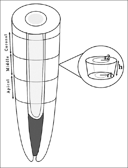
The sectioning of root and bonding area calculation
Figure 2.
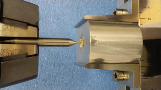
Testing of samples for push-out bond strength
The bonding area was calculated with the formula of 
The π is constant 3.14. R1 and R2 are postradius at larger and smaller radius. The “h” is the thickness of the post [Figure 1].[8] The digital caliper fino Pra Ceci caliper (FINO GmbH, Mangelsfeld Germany) was used to measure the dimensions of the samples.
The obtained data were analyzed with three-way analysis of variance (ANOVA) for conditioning versus location and irrigation protocol. The pair-wise multiple comparison test (Tukey) was used to determine the significance between the groups.
RESULTS
The highest push-out bond strength was observed [Table 1] in Group A with self-etch luting cement across all the radicular dentin sections. The coronal portion of this group showed the highest bond strength at 16.282 MPa. The least bonding strength was observed in etch-wash adhesive system in both irrigation systems. The substantial reduction in the bonding strength was recorded of a self-etch system with EDTA irrigation. The three-way ANOVA [Table 2] indicated no statistically significant three-way interaction between irrigation, cement, and root segment F = 0.51, P = 0.995.
Table 1.
Mean and standard deviations (MPa) of push-out bond strength

Table 2.
Three-way analysis of variance for mean bond strength at different sections of different bonding techniques

The pairwise analysis [Table 3] between etch-rinse bonding system versus irrigation shows the statistically significant difference between Group A1a and Group A1c (P = 0.036) and B1c (0.010). The significant difference was also observed between Group B1a and B1c with P = 0.017. Table 4 reveals all the self-etch groups had statistically significant difference except between Group A2a- A2b (P = 0.900), A2a-B2c (P = 0.083), and B2a-B2b (P = 0.662). Table 5 indicates the self-adhesive groups had statistically significant difference between A3a- A3c (P = 0.001), A3b-A3c (P = 0.009), A3c-B3a (P = 0.000), A3c-B3b (P = 0.001), and B3a-B3c (P = 0.004).
Table 3.
A Tukey honest significant difference comparisons between etch-wash adhesive system with irrigation protocol

Table 4.
A Tukey honest significant difference comparisons between self-etch adhesive system and irrigation protocol

Table 5.
A Tukey honest significant difference comparisons between self-adhesive and irrigation protocol

DISCUSSION
The debonding of fiber posts is the main mode of failures. Hence, the selection of the posts should be consistent with the bonding systems and resin cements. The bonding systems mainly incorporate two strategies, complete removal of smear layer with etch-wash technique[9] or incorporative smear layer as substrate.[10]
The results of the study showed the highest bonding strength with the self-etching luting technique at coronal segment. The corresponding values for Group A and Group B were 16.282 (2.073) and 11.383 (1.782) Mpa.
The results indicate the lesser bond strength in etch-wash technique in both irrigation protocols. The etch-wash adhesive system leads to complete removal of the smear layer along with highly mineralized peritubular dentin. It helps in opening of tubules and exposure of the Type-1 collagen dentin organic matrix.[11] The primer and bonding components of resin monomers interact with an organic matrix, leading to the formation of a hybrid layer. Since deeper layers of dentin comprised mainly of dentin tubules the hybridization of intertubular dentin is critical to the bonding strength.[12] The etch-wash system is known to depend on the micromechanical retention with the help of resin tags inside the funneled dentin tubules.[13] Reduced bond strength could also be due to the difficulty in maintaining the optimum dryness at inaccessible radicular dentin area.
The demineralization of smear layer in self-etch adhesive system depends on pH strength of acidic etchant component. The mild-moderate self-etch system alters the smear layer and demineralizes the dentin matrix. The few hydroxyapatite crystals remain over collagen fibers after the intermediate demineralization. The incorporated acidic component in self-etch technique is buffered by the mineral components released by substrate smear layer.[14] The residual inorganic minerals on the collagen matrix help in enhanced chemical bond lead to improved bond strength. The substantial reduction of the bond strength in self-etch group with the EDTA irrigation could be attributed to complete demineralization of collagen fibers and reduced chemical bonding process. The intraradicular irrigation of EDTA is also known to reduce the microhardness value of the root dentin. The modified soft dentin surface affects the adhesion and sealing ability of bonding agents.
The bonding procedure is simplified by incorporating phosphoric acid in the form of phosphoric esters in the self-adhesive luting system. The bonding to dentin is combined result of a formation of crosslinked polymer with composite fillers and micromechanical retention. The results from the study indicated the bond strength of the self-adhesive system was less compared to self-etch technique. This result is in agreement with the findings of previous researchers like Lührs et al.[15,16]
The coronal root section showed the highest bond strength in comparison with apical segments. The variation in bond strength is due to the structural difference in radicular dentin sections. The density of dentinal tubules is reduced toward the apical region. It affects the ratio between peritubular and intertubular dentin in comparison to coronal segment.[17] The reduced intertubular dentin is credited toward the lesser bond strength at an apical region. The previous studies report the progressive reduction in the hybrid layer thickens toward apical region.[18] The other clinical factors such as negative C-factors and limited access to the apical region also lead to reduced bond strength at apical region.
The limitations of the study are, though the study was performed in sound extracted natural teeth, change in moisture content, presence of invisible microcracks, functional age changes, morphological changes of pulp and dentin are difficult to standardize. The effect of thermal changes, and aging on the bonding strength needs further investigation.
CONCLUSIONS
The push-out bond strength values were significantly affected by irrigation protocol in self-etch luting group and not significantly affected by etch-wash, self-adhesive luting groups. The push-out bond strength was significantly higher in coronal segment in comparison to apical segment, and there was no significant difference between coronal and middle segments of all the groups.
Financial support and sponsorship
Nil.
Conflicts of interest
There are no conflicts of interest.
REFERENCES
- 1.Adanir N, Belli S. Evaluation of different post lengths’ effect on fracture resistance of a glass fiber post system. Eur J Dent. 2008;2:23–8. [PMC free article] [PubMed] [Google Scholar]
- 2.Vichi A, Ferrari M, Davidson CL. Influence of ceramic and cement thickness on the masking of various types of opaque posts. J Prosthet Dent. 2000;83:412–7. doi: 10.1016/s0022-3913(00)70035-7. [DOI] [PubMed] [Google Scholar]
- 3.Jongsma LA, Kleverlaan CJ, Feilzer AJ. Influence of surface pretreatment of fiber posts on cement delamination. Dent Mater. 2010;26:901–7. doi: 10.1016/j.dental.2010.05.005. [DOI] [PubMed] [Google Scholar]
- 4.Jongsma LA, Bolhuis PB, Pallav P, Feilzer AJ, Kleverlaan CJ. Benefits of a two-step cementation procedure for prefabricated fiber posts. J Adhes Dent. 2010;12:55–62. doi: 10.3290/j.jad.a17534. [DOI] [PubMed] [Google Scholar]
- 5.Malyk Y, Kaaden C, Hickel R, Ilie N. Analysis of resin tags formation in root canal dentine: A cross sectional study. Int Endod J. 2010;43:47–56. doi: 10.1111/j.1365-2591.2009.01631.x. [DOI] [PubMed] [Google Scholar]
- 6.Bitter K, Hambarayan A, Neumann K, Blunck U, Sterzenbach G. Various irrigation protocols for final rinse to improve bond strengths of fiber posts inside the root canal. Eur J Oral Sci. 2013;121:349–54. doi: 10.1111/eos.12057. [DOI] [PubMed] [Google Scholar]
- 7.Guerisoli DM, Marchesan MA, Walmsley AD, Lumley PJ, Pecora JD. Evaluation of smear layer removal by EDTAC and sodium hypochlorite with ultrasonic agitation. Int Endod J. 2002;35:418–21. doi: 10.1046/j.1365-2591.2002.00488.x. [DOI] [PubMed] [Google Scholar]
- 8.Bitter K, Meyer-Lueckel H, Priehn K, Kanjuparambil JP, Neumann K, Kielbassa AM, et al. Effects of luting agent and thermocycling on bond strengths to root canal dentine. Int Endod J. 2006;39:809–18. doi: 10.1111/j.1365-2591.2006.01155.x. [DOI] [PubMed] [Google Scholar]
- 9.Van Meerbeek B, De Munck J, Yoshida Y, Inoue S, Vargas M, Vijay P, et al. Buonocore memorial lecture. Adhesion to enamel and dentin: Current status and future challenges. Oper Dent. 2003;28:215–35. [PubMed] [Google Scholar]
- 10.Pashley DH, Horner JA, Brewer PD. Interactions of conditioners on the dentin surface. Oper Dent. 1992;Suppl 5:137–50. [PubMed] [Google Scholar]
- 11.Marshall GW, Jr, Marshall SJ, Kinney JH, Balooch M. The dentin substrate: Structure and properties related to bonding. J Dent. 1997;25:441–58. doi: 10.1016/s0300-5712(96)00065-6. [DOI] [PubMed] [Google Scholar]
- 12.Gwinnett AJ, Tay FR, Pang KM, Wei SH. Quantitative contribution of the collagen network in dentin hybridization. Am J Dent. 1996;9:140–4. [PubMed] [Google Scholar]
- 13.Nakabayashi N, Kojima K, Masuhara E. The promotion of adhesion by the infiltration of monomers into tooth substrates. J Biomed Mater Res. 1982;16:265–73. doi: 10.1002/jbm.820160307. [DOI] [PubMed] [Google Scholar]
- 14.Breschi L, Mazzoni A, Ruggeri A, Cadenaro M, Di Lenarda R, De Stefano Dorigo E, et al. Dental adhesion review: Aging and stability of the bonded interface. Dent Mater. 2008;24:90–101. doi: 10.1016/j.dental.2007.02.009. [DOI] [PubMed] [Google Scholar]
- 15.Lührs AK, Guhr S, Günay H, Geurtsen W. Shear bond strength of self-adhesive resins compared to resin cements with etch and rinse adhesives to enamel and dentin in vitro. Clin Oral Investig. 2010;14:193–9. doi: 10.1007/s00784-009-0279-z. [DOI] [PubMed] [Google Scholar]
- 16.Yazici AR, Yildirim Z, Ertan A, Ozgunaltay G, Dayangac B, Antonson SA, et al. Bond strength of one-step self-etch adhesives and their predecessors to ground versus unground enamel. Eur J Dent. 2012;6:280–6. [PMC free article] [PubMed] [Google Scholar]
- 17.Ferrari M, Mannocci F, Vichi A, Cagidiaco MC, Mjör IA. Bonding to root canal: Structural characteristics of the substrate. Am J Dent. 2000;13:255–60. [PubMed] [Google Scholar]
- 18.Mjör IA, Smith MR, Ferrari M, Mannocci F. The structure of dentine in the apical region of human teeth. Int Endod J. 2001;34:346–53. doi: 10.1046/j.1365-2591.2001.00393.x. [DOI] [PubMed] [Google Scholar]