Abstract
Objectives:
This article aimed to evaluate: (a) the agreement between a near-infrared light transillumination device and clinical and radiographic examinations in caries lesion detection and (b) the reliability of images captured by the transillumination device.
Methods:
Two calibrated examiners evaluated the caries status in premolars and molars on 52 randomly selected subjects by comparing the transillumination device with a clinical examination for the occlusal surfaces and by comparing the transillumination device with a radiographic examination (bitewing radiographs) for the approximal surfaces. Forty-eight trained dental hygienists evaluated and reevaluated 30 randomly selected images 1-month later.
Results:
A high concordance between transillumination method and clinical examination (kappa = 0.99) was detected for occlusal caries lesions, while for approximal surfaces, the transillumination device identified a higher number of lesions with respect to bitewing (kappa = 0.91). At the dentinal level, the two methods identified the same number of caries lesions (kappa = 1), whereas more approximal lesions were recorded using the transillumination device in the enamel (kappa = 0.24). The intraexaminer reliability was substantial/almost perfect in 59.4% of the participants.
Conclusions:
The transillumination method showed a high concordance compared with traditional methods (clinical examination and bitewing radiographs). Caries detection reliability using the transillumination device images showed a high intraexaminer agreement. Transillumination showed to be a reliable method and as effective as traditional methods in caries detection.
Keywords: dental caries, transillumination, bitewing radiography, reproducibility of results
Introduction
Caries detection, including the assessment of incipient and manifest lesions, is a central issue in everyday clinical practice.1,2 Traditional methods include a clinical evaluation supported by radiograph examination. Clinically, the International Caries Detection and Assessment System (ICDAS) is a visual scoring system to assess caries lesions at both initial and manifest thresholds.3–7 Radiographic examination is highly sensitive for detecting caries on surfaces that cannot be inspected visually, such as approximal surfaces. However, limitations in revealing the early stages of the disease have been reported,8 and the risk associated with radiographic exposure needs to be taken into consideration.9 As a complementary aid to visual examination, the near-infrared light transillumination method was designed and developed to support clinicians in the identification of caries lesions at different stages.10–12 Thanks to the specific optical properties of the caries tissue, the transillumination of the teeth amplifies the change in the scatter and absorption of light photons and thereby makes the lesion appear as a dark shadow.12 The diagnostic method was developed to facilitate the detection and localization of lesions in real time11 and since it is non-invasive, it can be used as frequently as needed. Clinical studies comparing the in situ extent of caries lesions and correlating the findings with those obtained by digital radiographs are fairly limited.13 A high concordance between transillumination and radiographic methods in enamel caries detection was described.12
Considering these precepts, the null hypothesis of this study was that the reliability of transillumination method in the detection of caries lesions corresponds to that obtained from clinical and radiographic examinations. To validate this hypothesis, an observational study was designed and carried out. Moreover, the reliability in the evaluation of transillumination images by trained dental hygienists was also assessed.
Materials and methods
The study was approved by the Ethical Committee at the University of Sassari (authorization number 389/2013), and it was conducted over 6 weeks from 9 June to 15 July 2014.
Comparison of detection methods
Examination tools and clinical procedure
The near-infrared transillumination device, KaVo® DIAGNOcam 2170 (KaVo Dental GmbH, Biberach/Riß, Germany), is a camera system (Class IIa medical device—European Community Directive 93/42/European Economic Community) which uses the light transmission through dental tissue to support the diagnosis of occlusal, approximal and secondary caries. A digital video camera records the image and displays it on a computer screen. The illumination corresponds to laser Class 1, according to EN 60825-1.
Planmeca intraoral radiographic equipment (Planmeca, Helsinki, Finland) and Kodak® UltraSpeed DF42 films KODAK GmbH/Dental, Stuttgart, Germany, with settings of 70 kV and 7 mA and an exposure time of 0.25 s, were used for bitewing radiographs. The radiographs were developed manually in conventional standard conditions.
The clinical examination was performed under standard circumstances in a dental chair using a plane mirror (Hahnenkratt, Königsbach-Stein, Germany) and a World Health Organization ballpoint probe (Asa Dental, Milan, Italy), under optimal light conditions.
Sample and detection methods
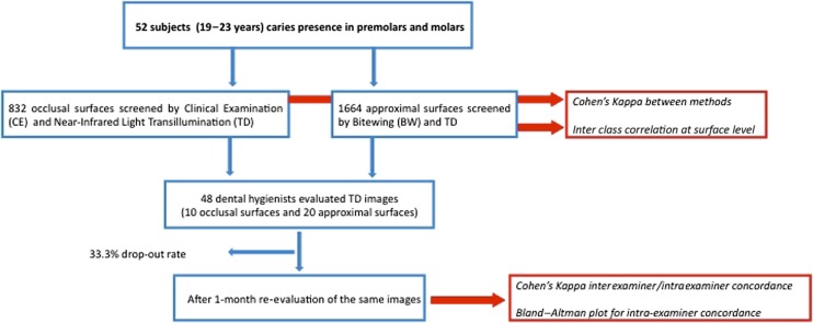
The study population consisted of students from the School of Medicine, University of Sassari, Italy. The survey aimed to include subjects with no missing teeth, no secondary caries and no fillings in premolars/molars. The exclusion criteria were subjects wearing fixed orthodontic appliances and subjects unable to be exposed to radiographs for medical/specific reasons. Considering that caries prevalence in young adults in Italy is generally high,14 a power analysis was performed to calculate an adequate sample size to compare the different caries detection methods. The number was fixed at 48 subjects. All students (age range 19–26 years; n = 1145) were invited to participate via email or a leaflet, in which the purpose of the study was described in detail. A total of 678 students accepted and were examined (59.2% acceptance rate), and 52 subjects fulfilled the inclusion/exclusion criteria and were enrolled. Each subject was coded with a number in order to protect his/her identity. The flow chart of the study is displayed in Figure 1.
Figure 1.
A flow chart of the study design.
For the occlusal surfaces, the transillumination method was compared with a clinical examination, while for the approximal surfaces it was compared with a radiographic examination. Two examiners, dentists from the Department of Surgery, Microsurgery and Medical Sciences, University of Sassari (Italy) with more than ten years of clinical experience, were calibrated in relation to ICDAS, transillumination method and radiographic examination. Calibration exercises for all three methods were carried out before the start of the study. One of the authors acted as the benchmark, training and calibrating the two examiners. 50 subjects not included in the sample and attending the dental clinic for recall were examined and re-examined after 72 h. Intraexaminer and interexaminer reliability was estimated through percentage agreement and Cohen's kappa statistics. Good interexaminer reliability was found, with no significant difference from benchmark values (p = 0.15) and a low mean square of error (0.47). Intraexaminer reliability was also high, Cohen's kappa = 0.88. 40 extracted human teeth (10 premolars and 30 molars), with a total of 80 approximal and 40 occlusal surfaces, were selected for the calibration of the transillumination and the radiographic examinations. Evaluations were carried out at 1-week intervals; kappa values for interexaminer and intraexaminer agreement were high for both methods (0.79 for transillumination and 0.83 for radiographic examination, respectively).
Before the clinical examination, each tooth was cleaned with a prophylactic paste (Clinpro™ Prophy Paste; 3M ESPE Dental Products 3MTM Italia Srl, Pioltello (MI), Italy) and then rinsed with a water spray for 10 s. The clinical examination was performed under standardized conditions as described above after drying the teeth for 5 s, recording both enamel and dentinal lesions using ICDAS.3–7 The subjects were examined on the same day, first by clinical and radiographic examination by one examiner, using an 8-inch round cone that was placed in contact with the ring of the film-holding system (RINN XCP; Dentsply, York, UK) placed in contact with the patient cheek during exposure. Then, the second examiner (blinded from the previous) performed the transillumination assessment placing the mouthpiece over the occlusal surfaces.
Scoring
The ICDAS scores were recorded on the following three surfaces: occlusal, distal and mesial. The presence of caries was classified as: (i) absent, (ii) in enamel (Scores 1, 2 and 3) or (iii) in dentine (Scores 4, 5 and 6).4
Radiographs were examined according to O'Mullane criteria, and mesial and distal surfaces were assessed according to the following criteria: 0—full thickness of the enamel and dentine visible and sound; 1—radiolucency confined to the outer half of the enamel; 2—radiolucency extending as far as the enamel–dentinal junction but not beyond; and 3—radiolucency in the enamel and dentine but not involving the pulp.15
The transillumination device was used for the detection of approximal and occlusal caries in the enamel or dentine according to the following criteria modified by the authors: 0 (sound)—no shadow was detected; 1—a defined approximal shadow in the enamel was present; and 2—a shadow reached the dentine.16 As it was impossible to measure the lesion vertically, all dark occlusal shadows were scored as 1.
Reliability in the evaluation of images captured using the transillumination device
48 dental hygienists with at least 7 years' professional experience and no familiarity with the transillumination method participated. They were engaged in a 60-min session describing the technology, run by one of the authors. Immediately after the training session, each participant was asked to diagnose caries from images of 10 teeth randomly obtained from the previous study, including a total of 10 occlusal and 20 approximal (10 mesial and 10 distal) surfaces. The participants were asked to fill in a form containing two possible answers (1—presence of caries and 2—absence of caries). 1 month later, the same images were mailed to the same participants, and a re-evaluation using the same criteria was proposed.
Statistical analysis
All data were analyzed using STATA 13 (Stata Corporation, College Station, TX). For all analyses, a p-value <0.05 was considered statistically significant. The general degree of agreement between the different detection methods was evaluated using Cohen's kappa, while the reproducibility of the two methods for each surface (occlusal or approximal) was assessed using intraclass correlation coefficients (ICCs). ICC values equal to 0 represent agreement equivalent to that expected by chance, while 1 represents full agreement.17
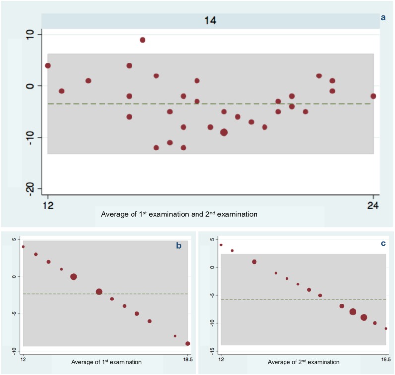
The interexaminer reliability for the transillumination method among dental hygienists was categorized using the kappa value of each participant following the criteria described by Landis and Koch.18 Values <0 indicate no agreement, 0–0.20 slight, 0.21–0.40 fair, 0.41–0.60 moderate, 0.61–0.80 substantial and 0.81–1 almost perfect agreement. For the intraexaminer reliability, the method created by Bland and Altman19 was used to show the variability of the two examinations. A plot of the two different evaluations and a comparison between them were performed in order to investigate the existence of any systematic difference between the measurements and to identify possible outliers.
Results
Comparison of detection methods
A total of 2496 surfaces (832 mesial, occlusal and distal surfaces, respectively) were analyzed.
The total number of occlusal caries lesions detected was similar: 149 lesions using transillumination device and 152 lesions using clinical examination with a Cohen's kappa of 0.99.
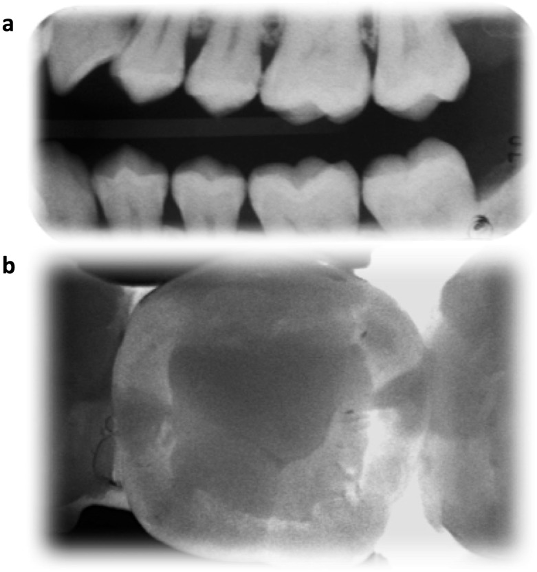
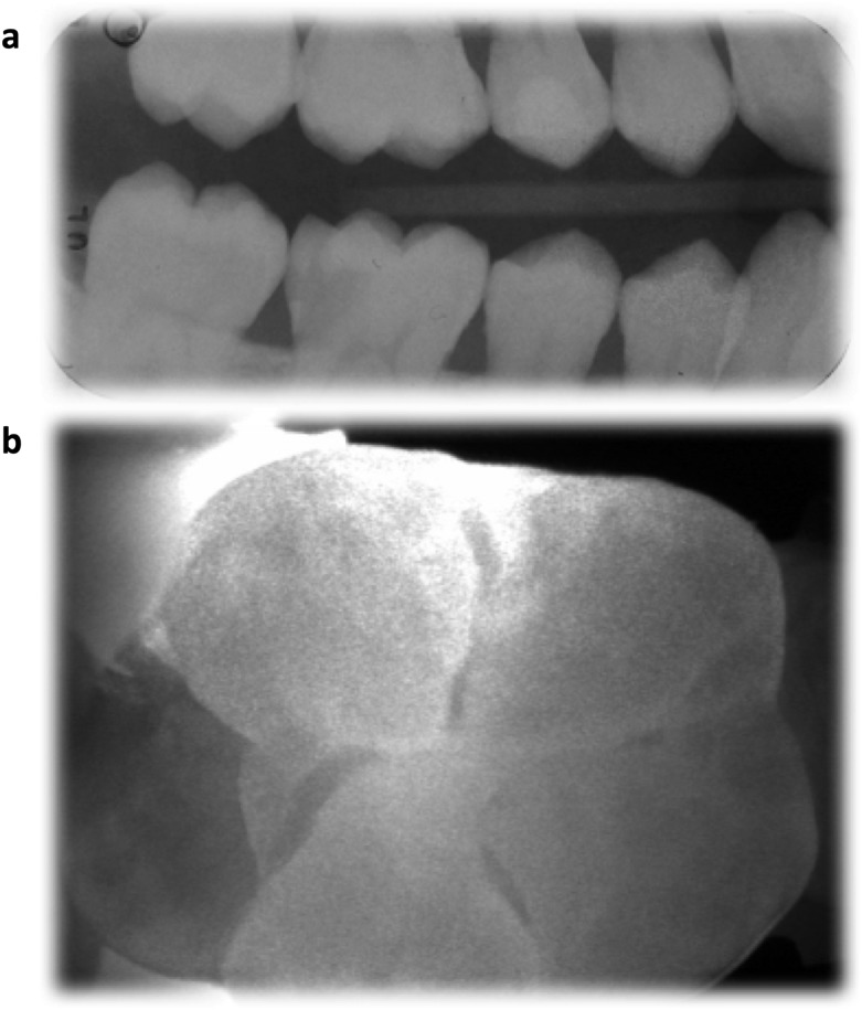
Approximal caries lesions identified using transillumination method were 83 and 70 using radiographic method (Cohen's kappa of 0.91). Transillumination and radiographic examination identified the same number of lesions (31) in the dentine. Cohen's kappa was 0.24 for enamel lesions, while complete agreement (kappa = 1) was observed for dentinal lesions (Table 1). In Figures 2 and 3, two illustrations of approximal surfaces are depicted.
Table 1.
Caries lesions on approximal surfaces in the enamel and dentine according to the near-infrared transillumination device and radiographic evaluations
| Caries severity | Transillumination n (%) | Radiography n (%) | Cohen's kappa value (SE) 95% CI |
|---|---|---|---|
| Enamel | 52 (3.1) | 39 (2.3) | 0.24 (0.06) 0.12–0.36 |
| Dentine | 31 (1.9) | 31 (1.9) | 1 |
CI, confidence interval; SE, standard error.
Percentages were calculated based on the total number of examined surfaces (n = 1664).
Figure 2.
The maxillary second premolar reveals proximal caries lesions in the dentin on the mesial and distal surfaces (a, b).
Figure 3.
The mandibular first molar reveals a proximal lesion in the dentin on the distal surface (a, b).
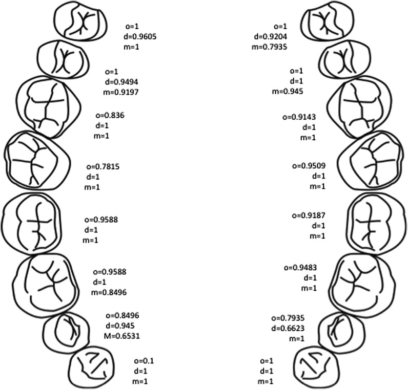
The mean ICC for the occlusal surfaces was 0.93, with the lowest value for the maxillary right second molar (ICC = 0.78), while perfect agreement (ICC = 1) was observed for several premolars. The mean ICC for approximal surfaces was 0.97 for the distal and 0.95 for the mesial surfaces (Figure 4). Regarding approximal enamel lesions, 17 lesions in molars were detected with the transillumination method, while 16 lesions were detected with the radiographic method (Cohen's kappa = 0.97), and 35 lesions were detected in premolars with transillumination compared with 23 lesions detected with radiographic methods (Cohen's kappa = 0.21). 29 affected mesial surfaces were registered with the transillumination device compared with 23 surfaces using radiographic examination (Cohen's kappa = 0.39). For the distal surfaces, 23 lesions were recorded with transillumination and 16 lesions with radiographic evaluation (Cohen's kappa = 0.34). Complete agreement was found for dentinal lesions using the two methods (Table 2).
Figure 4.
Comparison of the three detection methods: intraclass coefficient correlation between the transillumination method and the clinical evaluation for the occlusal surfaces (o) and between the transillumination device and bitewings for the approximal, mesial (m) and distal (d) surfaces is reported.
Table 2.
Caries lesions on approximal surfaces detected by the near-infrared transillumination device (TD) and the radiographic evaluation (BW): distribution of caries lesions in the enamel and dentine according to type of tooth and surfaces
| Lesions for teeth surfaces | Enamel |
Dentine |
||||
|---|---|---|---|---|---|---|
| DT n = 52, n (%) | BW n = 39, n (%) | Cohen's kappa value (SE) 95% CI | DT n = 31, n (%) | BW n = 31, n (%) | Cohen's kappa value (SE) 95% CI | |
| Molars | 17 (32.7) | 16 (41.03) | 0.97 (0.03) 0.91–1.00 | 13 (41.94) | 13 (41.94) | 1 |
| Premolars | 35 (67.3) | 23 (58.97) | 0.21 (0.08) 0.05–0.36 | 18 (58.06) | 18 (58.06) | 1 |
| Mesial | 29 (55.77) | 23 (58.97) | 0.39 (0.08) 0.23–0.56 | 11 (35.48) | 11 (35.48) | 1 |
| Distal | 23 (44.23) | 16 (41.03) | 0.34 (0.10) 0.15–0.54 | 20 (64.52) | 20 (64.52) | 1 |
CI, confidence interval; SE, standard error.
Reliability in the evaluation of images captured using the transillumination device
48 dental hygienists participated in the first evaluation and 32 dental hygienists (drop-out rate 33.3%) remained in the second. Cohen's kappa for each subject in terms of reliability between the two evaluations is shown in Table 3. Regarding interexaminer reliability in the first evaluation, the majority of the dental hygienists (87.5%) had either substantial (46.9%) or almost perfect agreement (40.6%), while in the second, a higher percentage had substantial agreement (75.0%) and a lower percentage almost perfect agreement (18.8%), with a shift towards a substantial level of agreement. 19 (59.4%) hygienists had substantial/almost perfect agreement, while 13 (40.6%) hygienists had fair/moderate agreement (Table 3). The Bland–Altman plot showed good intraexaminer reliability (Figure 5a) and a higher overrating of the number of the lesions in the second evaluation (Figure 5c) compared with the first (Figure 5b).
Table 3.
Reliability among dental hygienists (n = 32) using infrared transillumination images
| Evaluation | Fair agreement, n (%) | Moderate agreement, n (%) | Substantial agreement, n (%) | Almost perfect agreement, n (%) |
|---|---|---|---|---|
| EVA 1 | – | 4 (12.5) | 15 (46.9) | 13 (40.6) |
| EVA 2 | – | 2 (6.2) | 24 (75.0) | 6 (18.8) |
| Drop-out after EVA 1, n = 16 | – | 4 (25.0) | 6 (37.5) | 6 (37.5) |
| EVA 1/EVA 2 | 4 (12.5) | 9 (28.1) | 10 (31.2) | 9 (28.1) |
Interexaminer and intraexaminer reliability is categorized by following the scale of the degree of agreement in the two evaluations (immediately after training and 1 month later) reported in table as EVA 1 and EVA 2.
Figure 5.
Reliability in the evaluation of images captured using the transillumination device: intraexaminer reliability using a Bland–Altman plot of difference—each small dot is the average value of a single examiner observation, while larger dots are the sum of two or more examiners. The shaded region indicates 95% limits of agreement around the dashed line representing the mean.
Discussion
Accurate and early caries detection is crucial for an optimal treatment planning, helping the clinician to choose between surgical and non-surgical/medical approach.20 The purpose of this study was to assess the agreement in caries detection; a near-infrared light transillumination device, a relative new technology in the dental market, was compared against clinical evaluation and radiographic examination. Moreover, rating of the interreliability and intrareliability of the evaluation of transillumination images immediately after a training section and 1 month later was carried out.
Primary findings demonstrated the infrared transillumination technology to be as effective as clinical examination and bitewing radiographs in caries detection. Routinely, the evaluation procedure required both clinical and radiographic sessions, as they are regarded as the first choice for caries detection. However, radiographs are unable to identify the initial demineralization of the tooth, resulting in low sensitivity, with decalcification ranging from 40% to 60%, necessary to produce a radiographic image of caries, and this might result in false-negative tests.21–23 Conversely, the use of digital transillumination might lead to overdetection, as the device has a lower specificity compared with radiographs; besides, this method has been shown to be more sensitive than radiographs in detecting early changes in the enamel.12,13,20,24 These findings were confirmed in the present study; more approximal lesions in the enamel were detected using the transillumination device compared with those identified by the radiographic evaluation. The combination of these two methods might result in an improvement of the diagnostic accuracy.13 In everyday clinical practice, radiographic evaluation is usually limited to teeth in doubt for caries or routinely carried out in subjects at high risk of caries, increasing the probability of detecting a lesion, leading to a high specificity and low sensitivity.25 Instead, the near-infrared light transillumination might be used routinely over all surfaces particularly approximal surfaces, with no side effect, with an estimated high sensitivity.
An additional crucial point in caries detection is the interexaminer and intraexaminer reliability and in this study, it was calculated for the evaluation of data obtained by different operators unfamiliar with the transillumination technology. A high interexaminer and intraexaminer reliability among dental hygienists in the evaluation of the images obtained by the transillumination device was recorded. Kappa coefficients indicate a good repeatability in both evaluations even if a shift to overdetection from the first to the second examination was documented. Intraexaminer reliability was also high, showing that the images captured by the transillumination device are quite easy to “read” even for inexpert operators.
Dentists are responsible for caries diagnosis and management, while dental hygienists are responsible for referring the patient to the dentist if they detect caries.26 The digital transillumination device might represent a valid tool for not only dentists but also dental hygienists, which can, without radiation exposure to the patient, reach a high sensitivity in caries detection.
A primary limitation of the present study lies in the non-evaluation of the “true status” of the lesion, as the teeth were not extracted after examination. It is necessary to underline that the study was not aimed at determining the actual caries presence. So, even if it can be considered a factor, the true caries status was not an outcome.
A bias can probably be ascribed to the study design phase regarding the evaluation of the images obtained by the transillumination device, as the first evaluation was performed with a strict time limit, while the second evaluation was unrestricted, as dental hygienists received an email with the images and no time limit was given. A stochastic drift might also be postulated, as the misclassification made by professionals in the second evaluation occurred unconsciously, leading to a higher level of interexaminer agreement.
Some strengths of the present study also need to be considered. The transillumination technique was compared with the clinical examination for the occlusal surfaces and with the bitewing radiograph for the approximal surfaces, and this is the first study comparing the concordance of this technique in vivo with traditional methods. Moreover, the use of this device by dental hygienists opens up the likelihood of detection of initial caries lesion in order to send the patient to the dentist for further scrutiny.
Conclusion
The near-infrared transillumination device might be a useful tool for early caries detection. The images seem to be easily decodable even with low level of experience with the method and could be a useful diagnostic tool for preventive dental therapy and for the monitoring of lesions by different professionals in the field.
Acknowledgments
Acknowledgments
The authors thank the dental hygienists who participated in the study.
References
- 1.Pitts NB. Are we ready to move from operative to non-operative/preventive treatment of dental caries in clinical practice? Caries Res 2004; 38: 294–304. doi: https://doi.org/10.1159/000077769 [DOI] [PubMed] [Google Scholar]
- 2.Piovesan C, Moro BL, Lara JS, Ardenghi TM, Guedes RS, Haddad AE, et al. Laboratorial training of examiners for using a visual caries detection system in epidemiological surveys. BMC Oral Health 2013; 13: 49. doi: https://doi.org/10.1186/1472-6831-13-49 [DOI] [PMC free article] [PubMed] [Google Scholar]
- 3.International Caries Detection and Assessment System Coordinating Committee: Criteria Manual. International Caries Detection and Assessment System (ICDAS II) Workshop. Baltimore, MD, USA; March 2005.
- 4.Pitts NB, Ekstrand KR, ICDAS Foundation. International Caries Detection and Assessment System (ICDAS) and its International Caries Classification and Management System (ICCMS)—methods for staging of the caries process and enabling dentists to manage caries. Community Dent Oral Epidemiol 2013; 41: e41–52. doi: https://doi.org/10.1111/cdoe.12025 [DOI] [PubMed] [Google Scholar]
- 5.Assaf AV, de Castro Meneghim M, Zanin L, Tengan C, Pereira AC. Effect of different diagnostic thresholds on dental caries calibration—a 12 month evaluation. Community Dent Oral Epidemiol 2006; 34: 213–19. [DOI] [PubMed] [Google Scholar]
- 6.Agustsdottir H, Gudmundsdottir H, Eggertsson H, Jonsson SH, Gudlaugsson JO, Saemundsson SR, et al. Caries prevalence of permanent teeth: a national survey of children in Iceland using ICDAS. Community Dent Oral Epidemiol 2010; 38: 299–309. doi: https://doi.org/10.1111/j.1600-0528.2010.00538.x [DOI] [PubMed] [Google Scholar]
- 7.Ismail AI, Sohn W, Tellez M, Amaya A, Sen A, Hasson H, et al. The international caries detection and assessment system (ICDAS): an integrated system for measuring dental caries. Community Dent Oral Epidemiol 2007; 35: 170–8. [DOI] [PubMed] [Google Scholar]
- 8.Bader JD, Shugars DA, Bonito AJ. A systematic review of the performance of methods for identifying carious lesions. J Public Health Dent 2002; 62: 201–13. doi: https://doi.org/10.1111/j.1752-7325.2002.tb03446.x [DOI] [PubMed] [Google Scholar]
- 9.Ludlow JB, Davies-Ludlow LE, White SC. Patient risk related to common dental radiographic examinations: the impact of 2007 International Commission on Radiological Protection recommendations regarding dose calculation. J Am Dent Assoc 2008; 139: 1237–43. [DOI] [PubMed] [Google Scholar]
- 10.Keem S, Elbaum M. Wavelet representations for monitoring changes in teeth imaged with digital imaging fiber-optic transillumination. IEEE Trans Med Imaging 1997; 16: 653–63. doi: https://doi.org/10.1109/42.640756 [DOI] [PubMed] [Google Scholar]
- 11.Schneiderman A, Elbaum M, Shultz T, Keem S, Greenebaum M, Driller J. Assessment of dental caries with digital imaging fiber-optic transIllumination (DIFOTI): in vitro study. Caries Res 1997; 31: 103–10. [DOI] [PubMed] [Google Scholar]
- 12.Astvaldsdóttir A, Ahlund K, Holbrook WP, de Verdier B, Tranæus S. Approximal caries detection by DIFOTI: in vitro comparison of diagnostic accuracy/efficacy with film and digital radiography. Int J Dent 2012; 326401. doi: https://doi.org/10.1155/2012/326401 [DOI] [PMC free article] [PubMed] [Google Scholar]
- 13.Bin-Shuwaish M, Yaman P, Dennison J, Neiva G. The correlation of DIFOTI to clinical and radiographic images in Class II carious lesions. J Am Dent Assoc 2008; 139: 1374–81. [DOI] [PubMed] [Google Scholar]
- 14.O’Mullane DM, Kavanagh D, Ellwood RP, Chesters RK, Schafer F, Huntington E, et al. A three-year clinical trial of a combination of trimetaphosphate and sodium fluoride in silica toothpastes. J Dent Res 1997; 76: 1776–81. [DOI] [PubMed] [Google Scholar]
- 15.Senna A, Campus G, Gagliani M, Strohmenger L. Socio-economic influence on caries experience and CPITN values among a group of Italian call-up soldiers and cadets. Oral Health Prev Dent 2005; 3: 39–46. [PubMed] [Google Scholar]
- 16.Söchtig F, Hickel R, Kühnisch J. Caries detection and diagnostics with near-infrared light transillumination: clinical experiences. Quintessence Int 2014; 45: 531–8. doi: https://doi.org/10.3290/j.qi.a31533 [DOI] [PubMed] [Google Scholar]
- 17.Cohen J. A coefficient of agreement for nominal scales. Educ Psychol Meas 1960; 20: 37–46. [Google Scholar]
- 18.Landis JR, Koch GG. The measurement of observer agreement for categorical data. Biometrics 1997; 33: 159–74. doi: https://doi.org/10.2307/2529310 [PubMed] [Google Scholar]
- 19.Bland JM, Altman DG. Statistical methods for assessing agreement between two methods of clinical measurement. Lancet 1986; 1: 307–10. [PubMed] [Google Scholar]
- 20.Young DA, Featherstone JD. Digital imaging fiber-optic trans-illumination, F-speed radiographic film and depth of approximal lesions. J Am Dent Assoc 2005; 136: 1682–7. [DOI] [PubMed] [Google Scholar]
- 21.Machiulskiene V, Nyvad B, Baelum V. A comparison of clinical and radiographic caries diagnoses in posterior teeth of 12-year-old Lithuanian children. Caries Res 1999; 33: 340–8. doi: https://doi.org/16532 [DOI] [PubMed] [Google Scholar]
- 22.Chong MJ, Seow WK, Purdie DM, Cheng E, Wan V. Visual-tactile examination compared with conventional radiography, digital radiography, and diagnodent in the diagnosis of occlusal occult caries in extracted premolars. Pediatr Dent 2003; 25: 341–9. [PubMed] [Google Scholar]
- 23.Yang J, Dutra V. Utility of radiology, laser fluorescence, and transillumination. Dent Clin North Am 2005; 49: 739–52. doi: https://doi.org/10.1016/j.cden.2005.05.010 [DOI] [PubMed] [Google Scholar]
- 24.Young DA. New caries detection technologies and modern caries management: merging the strategies. Gen Dent 2002; 50: 320–31. [PubMed] [Google Scholar]
- 25.Chu CH, Chung BT, Lo EC. Caries assessment by clinical examination with or without radiographs of young Chinese adults. Int Dent J 2008; 58: 265–8. [DOI] [PubMed] [Google Scholar]
- 26.Barnes CM. Dental hygiene participation in managing incipient and hidden caries. Dent Clin North Am 2005; 49: 795–813. doi: https://doi.org/10.1016/j.cden.2005.05.013 [DOI] [PubMed] [Google Scholar]


