Abstract
Mounting evidence indicates that hotspot p53 mutant proteins often possess gain-of-function property in promoting cell mobility and tumor metastasis. However, the molecular mechanisms are not totally understood. In this study, we demonstrate that the hotspot mutation, p53-R273H, promotes cell migration, invasion in vitro and tumor metastasis in vivo. p53-R273H significantly represses expression of DLX2, a homeobox protein involved in cell proliferation and pattern formation. We show that p53-R273H-mediated DLX2 repression leads to upregulation of Neuropilin-2 (NRP2), a multifunctional co-receptor involved in tumor initiation, growth, survival and metastasis. p53-R273H-induced cell mobility is effectively suppressed by DLX2 expression. Furthermore, knockdown of NRP2 significantly inhibits p53-R273H-induced tumor metastasis in xenograft mouse model. Together, these results reveal an important role for DLX2-NRP2 in p53-R273H-induced cell mobility and tumor metastasis.
Tumor suppressor protein p53 is activated in response to various cellular stresses to transactivate a set of target genes involved in cell cycle arrest, apoptosis, senescence, DNA repair and cancer cell metabolism.1 It is well-documented that more than 50% of human tumors and cancers contain a mutation in p53 gene with the vast majority of these mutations occur in the DNA-binding domain.2, 3 The primary cancer-associated alterations in p53 gene are a single amino acid substitution.4 Several mutations clustered within the central DNA-binding domain are frequently found in cancers, refereed as ‘hotspot’ mutations, which include R175, G245, R248, R249, R273 and R282.5 Mutant p53 proteins usually have lost its DNA sequence-specific transcription function, they can also inhibit wild-type p53 function in a dominant negative fashion,6 since p53 protein usually exerts as a tetramer. It is now known that mutant p53 proteins not only lost their transcriptional function (loss of function), but also commonly obtain new function (gain of function).7 For instance, p53-R175H exhibits a gain of oncogenic function in driving cell migration and invasion.4, 8 Studies from animal models have confirmed that mice bearing a mutant p53 allele show more aggressive and metastatic tumor than p53 null.9, 10, 11 In human cancers, mutant p53 expression has been linked to poorer prognosis.12
Distal-less homeobox 2 (DLX2) plays an important role in control of cell migration,13 proliferation,14 differentiation,15 neurogenesis16, 17 and pattern formation.18 Downregulation of DLX expression has been reported in human solid and hematologic malignancies, and in lymphoblastic leukemias.19, 20, 21
Neuropilins (NRPs) are transmembrane surface glycoproteins consisting of two members, Neuropilin-1 (NRP1) and Neuropilin-2 (NRP2). NRP2 functions as a co-receptor of SEMA3, vascular endothelial growth factor and transforming growth factor β 1 and is important for cell proliferation, survival, epithelial-mesenchymal transition and metastasis.22 It has recently become widely acknowledged that upregulation of NRP2 expression is associated with tumorigenesis and, in particular, tumor metastasis.23
Here, we investigated the functions of mutant p53 protein (R273H, R175H, R248W) in regulation of cell migration and invasion. We demonstrated that p53-R273H upregulates NRP2 expression via suppression of DLX2 transcription, leading to increased cell mobility. In addition, knockdown of NRP2 significantly inhibits p53-R273H-induced tumor metastasis in vivo. Together, this study demonstrates the importance of DLX2-NRP2 pathway in p53-R273H-induced cell mobility, epithelial-mesenchymal transition and tumor metastasis.
Results
Expression of hotspot mutant p53 promotes scattering cell growth and accelerates cell migration
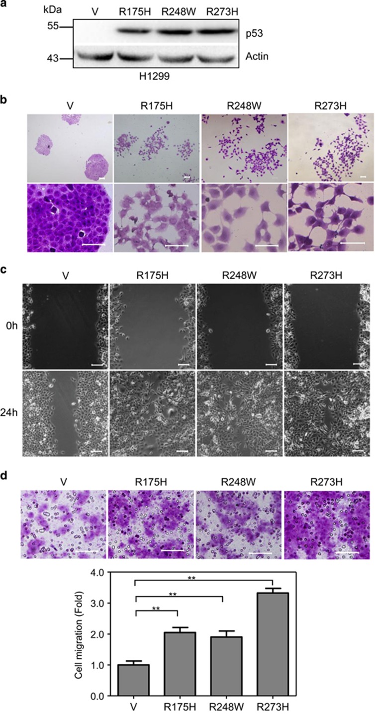
It is well documented that hotspot mutations of p53 gene, in particular, R175H, R248W and R273H, are loss of function alleles as they are unable to transactivate downstream genes involved cell growth control and apoptosis.24 However, it becomes clear that these mutant alleles, in addition, exhibit gain of new functions in promoting tumorigenesis, including promoting cancer metastasis,25 yet the molecular mechanisms are not totally understood. To investigate the molecular mechanisms by which the hotspot p53 mutations promote cancer metastasis, we first examined the effects of these mutant p53 alleles on the ability of cell migration and invasion. As shown in Figure 1, while human non-small cell lung cancer H1299 cells, which do not express endogenous p53, grew into typical epithelial morphology exhibiting tightly packed colonies and formed a continuous sheet with no intercellular spaces, expression of either p53-R175H, p53-R248W or p53-R273H led to scattering cell growth with mobile cell morphologies showing lamellipodia on the leading edge of the cells (Figures 1a and b). In addition, expression of mutant p53 significantly promoted cell migration, as evidenced by wound healing assay (Figure 1c) and transwell assays (Figure 1d). Notably, cells expressing p53-R175H and p53-R248W migrated approximately two times faster than that of control cells as assessed by transwell assays. By contrast, cells expressing p53-R273H migrated approximately three times faster than that of control cells, although the protein expression levels were comparable (Figure 1a). Expression of mutant p53 in human colon cancer HCT116 p53−/− cells also promoted scattering cell growth and accelerated cell migration (Supplementary Figures S1A–C). These observations indicate that hotspot p53 mutations promote scattering cell growth and accelerate cell migration.
Figure 1.
Expression of mutant p53 promotes cell morphological changes and cell migration. (a–d) H1299 cells stably expressing vector control (V) or a mutant p53 allele (p53-R175H, p53-R248W or p53-R273H) were subjected to western blot analyses using p53 (DO-1) antibody (a); subjected to photograph under a phase-contrast microscope (b); subjected to wound-healing assay (c); or subjected to transwell assays (d). Scale bar =100 μm. ** indicated P<0.01. Results are presented as means and S.E. from three independent experiments in triplicates
To examine whether expression of mutant p53 is indeed responsible for cell morphological changes, scattering cell growth and accelerated cell migration, we used an shRNA specific for p53 to knock-down p53-R273H in H1299 cells. As shown in Supplementary Figures S2A and B, p53-R273H was effectively knocked-down, which led to almost complete reverse of scattering cell growth and mobile cell morphology induced by p53-R273H (Supplementary Figure S2C). In addition, knockdown of p53-R273H resulted in total reverse of cell migration and invasion compared to control cells (Supplementary Figures S2D–F). Together, these results indicate that p53-R273H is responsible for scattering cell growth, morphological changes and accelerated cell migration/invasion in H1299 cells.
Downregulation of DLX2 promotes scattering cell growth, cell migration and invasion
Several studies have attempted to explore the molecular mechanisms by which mutant p53 exerts its function.26 It has been reported that p53-R175H can downregulate expression of DLX2,27 a homeobox protein important in control of cell migration.13 We therefore investigated whether DLX2 plays a role in p53-R273H- induced cell mobility.
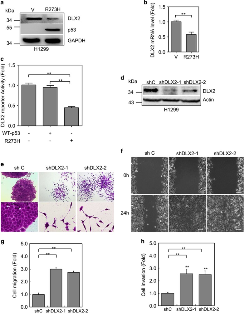
As shown in Figures 2a and b, p53-R273H significantly downregulated DLX2 in H1299 cells, as assayed by western blot analyses and Q-PCR analyses. Furthermore, p53-R273H but not wild-type p53 downregulated DLX2 reporter activity (Figure 2c). Next, we examined the effects of knockdown of DLX2 on cell morphology and cell migration/invasion. Two specific DLX2 shRNAs were used to knockdown endogenous DLX2 in H1299 cells, respectively (Figure 2d). Knockdown of DLX2 dramatically induced scattering cell growth (Figure 2e), cell migration (Figures 2f and g) and invasion (Figure 2h). Taken together, these results indicate that R273H specifically downregulated DLX2 expression, and reduced DLX2 expression significantly promotes scattering cell growth, cell migration and invasion.
Figure 2.
p53-R273H inhibits DLX2 expression in promoting cell scattering growth, migration and invasion. (a,b) Stable H1299 cells expressing p53-R273H (R273H) or a vector control (V) were subjected to western blot analyses for DLX2 expression (a) or Q-PCR analysis (b). Results are presented as means and S.E. from three independent experiments in triplicates. ** indicated P<0.01. (c) H1299 cells were co-transfected with DLX2-Lux, TK-Renilla, and a plasmid encoding either wild-type p53, p53-R273H or vector. Luciferase activities was normalized to Renilla activity and presented as fold of activation (mean±S.E.) from three independent experiments in triplicates. (d–h) Stable H1299 cells expressing shRNA specific for DLX2 (shDLX2-1, shDLX2-2) or GFP8 were subjected to western blot analyses (d). Cells were also subjected to photograph using microscopy (e); to wound-healing assay (f) and to transwell migration (g) or invasion (h) assays. Results are presented as means and S.E. from three independent experiments in triplicates. Scale bar =100 μm. ** indicated P<0.01
DLX2 plays a critical role in p53-R273H-induced scattering cell growth, cell migration and invasion
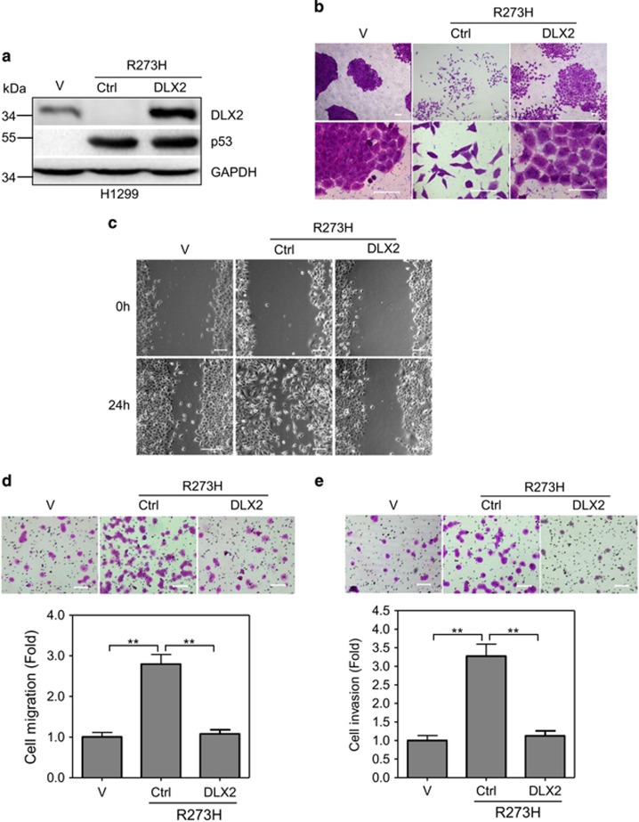
Since expression of p53-R273H in H1299 induces scattering cell growth and cell migration, concomitant with decreased expression of DLX2, we therefore investigated whether downregulation of DLX2 is responsible for p53-R273H-mediated changes of cell motility. In the rescuing experiments, ectopic expression of DLX2 significantly, but not completely, inhibited p53-R273H-induced scattering cell growth (Figures 3a and b). However, ectopic expression of DLX2 completely inhibited cell migration and invasion induced by p53-R273H (Figures 3c–e). Taken together, these results demonstrate that p53-R273H promotes scattering cell growth, cell migration and invasion via downregulation of DLX2.
Figure 3.
Ectopic expression of DLX2 in H1299 cells inhibits p53-R273H-induced scattering cell growth, migration and invasion. (a–e) H1299 cells stably expressing p53-R273H were infected with recombinant lentivirus encoding DLX2. Cells were subjected to western blot analyses (a) or photograph using microscopy (b); to wound-healing assay (c) and to transwell migration (d) or invasion (e) assays. Results are presented as means and S.E. from three independent experiments in triplicates. Scale bar =100 μm. ** indicated P<0.01
p53-R273H promotes scattering cell growth, cell migration and invasion via upregulation of NRP2 expression
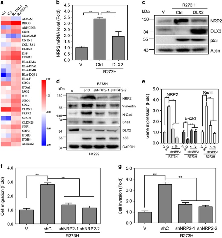
It has been shown that DLX2 functions as a transcription factor to regulate a set of genes involved in variety of different biological processes.18 Notably, microarrays analyses showed that NRP2, a co-receptor of several membrane receptors including transforming growth factor β1, PDGF, HGF and FGF receptors,28 is a direct downstream target that is inhibited by DLX2.13 Consistent with previous report, our RNA-Seq analyses also indicated that expression of p53-R273H in H1299 cells significantly upregulated NRP2 (Figure 4a). Indeed, knockdown of DLX2 significantly upregulated NRP2 expression in H1299, A549 or HepG2 cells (Supplementary Figures S3A–D).
Figure 4.
p53-R273H upregulates NRP2 to promote cell migration. (a) H1299 cells stably expressing mutant p53-R273H were subjected to RNA-seq based digital gene expression (DGE) analyses. Differentially expressed genes were identified by a P-value ⩽ 0.01 and an expression change of twofold or more between the two samples. (b,c) H1299 cells stably expressing p53-R273H were infected with recombinant lentivirus encoding DLX2. Cells were subjected to Q-PCR analysis (b) for NRP2 expression, and to western blot analyses (c) for NRP2, DLX2 and p53 expression. Results are presented as means and S.E. from three independent experiments in triplicates. ** indicated P<0.01. (d–g) Stable H1299 cells expressing p53-R273H were infected with recombinant lentivirus encoding a shRNA specific for NRP2 (shNRP2-1 or shNRP2-2) or a shGFP control were subjected to western blot analyses, as indicated (d); or to Q-PCR experiments to measure expression of NRP2, E-cadherin or Snail, as shown (e); or to transwell migration (f) or invasion (g) assays. Results are presented as means and S.E. from three independent experiments in triplicates. Scale bar =100 μm. ** indicated P<0.01
We therefore investigated whether NRP2 plays a role in p53-R273H- DLX2-mediated induction of scattering cell growth and cell mobility. First, we examined the effects of p53-R273H on NRP2 expression. Again, expression of p53-R273H in H1299 cells downregulated DLX2 protein expression, concomitant with upregulation of NRP2. Further analyses showed that p53-R273H significantly stimulated NRP2 transcription, which was repressed by expression of DLX2 (Figure 4b). Ectopic expression of DLX2 significantly attenuated p53-R273H induced upregulation of NRP2 (Figure 4c).
Second, since NRP2 has been reported promoting cell mobility,29 we therefore investigated the effects of p53-R273H-NRP2 axis on expression of several genes involved in epithelial-mesenchymal transition. As shown in Figure 4d, expression of p53-R273H in H1299 cells upregulated NRP2, concomitant with increased regulation of Snail, N-cadherin and Vimentin, which was significantly suppressed by knocked-down of NRP2 using two different NRP2 shRNAs.
Since E-cadherin expression is very low and barely detectable in H1299 cells, we employed Q-PCR analyses to examine the effects of p53-R273H on E-cadherin expression. As shown in Figure 4e, p53-R273H upregulated both NRP2 and Snail expression concomitant with significantly reduced E-cadherin expression, which were completely rescued by knockdown of NRP2. Furthermore, knockdown of NRP2 effectively inhibited p53-R273H-induced scattering cell growth (Supplementary Figure S4), cell migration and invasion (Figures 4f and g). Taken together, these results indicate that NRP2 plays a critical role in p53-R273H-induced cell mobility.
Ablation of endogenous p53-R273H reduces cell migration via modulation of DLX2 and NRP2 expression
To determine the role of endogenous mutant p53-R273H in cell mobility, we used human lung adenocarcinoma H1975 and human triple negative breast adenocarcinoma MDA-MB-468 cells, both of which express endogenous p53-R273H, and the protein levels of endogenous mutant p53-R273H are compared to the ectopic expression p53-R273H in H1299 (Supplementary Figure S1D).
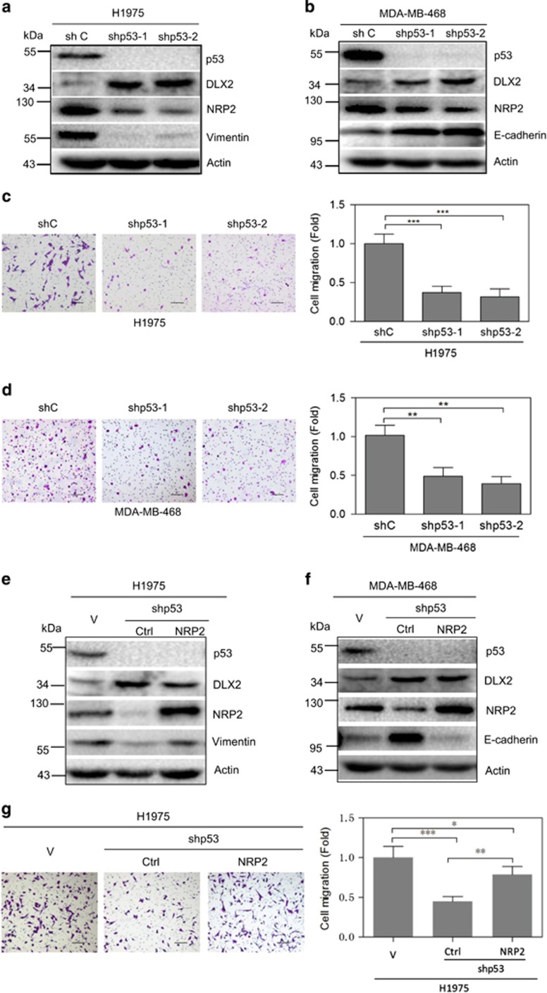
As shown in Figure 5, knockdown of endogenous p53-R273H by two different shRNAs resulted in significant upregulation of DLX2 and downregulation of NRP2 in both cells. Importantly, knockdown of endogenous p53-R273H leads to significant reduction of cell migration.
Figure 5.
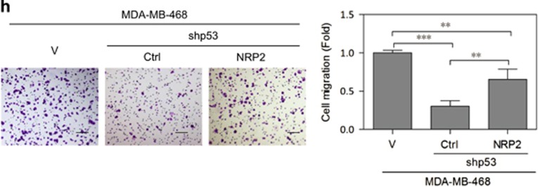
Ablation of endogenous p53-R273H reduces cell migration via modulation of DLX2 and NRP2 expression. (a–d) H1975 or MDA-MB-468 cells stably expressing a shRNA specific for p53 (shp53-1 or shp53-2) were subjected to western blot analyses (a,b) and to transwell migration assays (c,d). (e–h) H1975 or MDA-MB-468 cells stably expressing a shRNA specific for p53 (shp53-1) were infected with recombinant lentivirus encoding NRP2. Cells were subjected to western blot analyses (e,f) and to transwell migration assays (g,h). Results are presented as means and S.E. from three independent experiments in triplicates. Scale bar =100 μm. * indicated P<0.05, ** indicated P<0.01, *** indicated P<0.005
To demonstrate that reduced NRP2 expression is critical in depletion of p53-R273H-mediated reduction of cell mobility, we performed rescuing experiments. Ectopic expression of NRP2 significantly, but not completely, reversed migration in both H1975 and MDA-MB-468 cells expressing shRNA specific for p53 (Figures 5e–h).
We then examined whether p53-R273H-NRP2 axis can impact cell proliferation. Our data showed that knockdown of p53-R273H reduced cell proliferation as accessed by 3-(4,5-dimethylthiazol-2-yl)-5-(3-carboxymethoxyphenyl)-2-(4-sulfophenyl)-2H-tetrazolium) assays. However, restoration of NRP2 was unable to restore cell growth of both H1975 and MDA-MB-468 cells (Figures 5e and f; Supplementary Figures S5A and B). Thus, these data indicate that p53-R273H regulates NRP2 to primarily impact on cell mobility.
Reduced expression of NRP2 inhibits p53-R273H-induced tumor metastasis in vivo
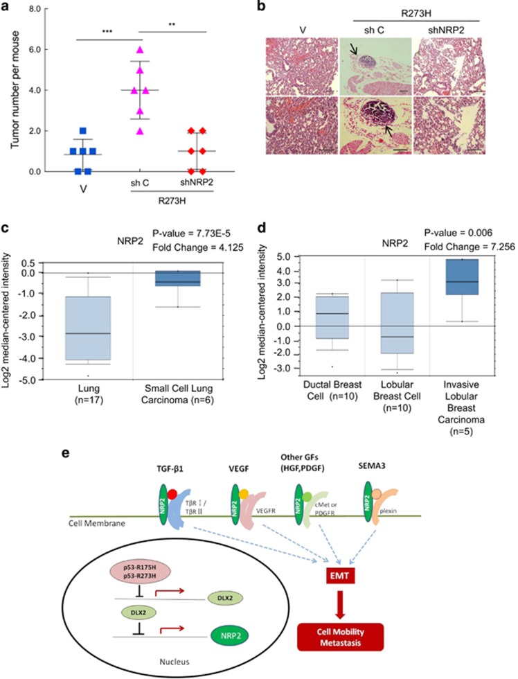
To determine the role of NRP2 in p53-R273H-induced tumor metastasis in vivo, we used stable H1299 cells expressing p53-R273H, or simultaneously expressing p53-R273H and shNRP2 in a mouse lung metastasis model. As shown in Figure 6a, p53-R273H promoted lung metastasis, as evidenced by increased tumor nodules on the lung surface, whereas simultaneous expression of shNRP2 effectively inhibited p53-R273H-induced metastasis. These results were further supported by pathological examinations using hematoxylin and eosin staining (Figure 6b). These results demonstrate that NRP2 is critical in p53-R273H-induced tumor metastasis in vivo.
Figure 6.
NRP2 is critical in p53-R273H-induced tumor metastasis in vivo and is upregulated in human clinical cancer samples. (a,b) Stable H1299 cells expressing p53-R273H were infected with recombinant lentivirus encoding an shRNA specific for NRP2 (shNRP2-1) or shGFP were tail-vain injected into female nude mice (6 mice per group). Mice were observed daily and killed after 60 days. Lungs were dissected and fixed, and were inspected for metastatic nodules on their surface. The graph represents the number of metastatic nodules in the lung from each mouse. A horizontal line indicates the mean of each group (a). Lungs were fixed, embedded in paraffin, sectioned, and stained by H&E for histological analysis (b). Metastatic nodules are indicated by arrows. Scale bars=100 μm. ** indicated P<0.01, *** indicated P<0.005. (c,d) NRP2 gene expression from Radvanyi Breast and Lung data set. Oncomine (Compendia Bioscience, Ann Arbor, MI, USA) was used for analysis and visualization. (c) Box-and-whisker plots representing NRP2 expression in human lung cancer samples. NRP2 fold change: 4.125; P=7.73E-5; (d) Box-and-whisker plots representing NRP2 expression in human breast cancer samples. NRP2 fold change: 7.256; P=0.006. (e) A working model in that mutant p53 R273H and NRP2 function in cell motility and tumor metastasis. H&E, hematoxylin and eosin
To further explore clinical relevance of NRP2 expression in human cancer specimen, we analyzed Oncomine Database. The data showed that upregulation of NRP2 significantly correlated in human lung cancer and breast cancer specimens (Figures 6c and d).
Taken together, our study reveals a new pathway with which mutant p53-R273H suppresses DLX2 transcription, resulting in downregulation of NRP2 expression, which in turn leads to attenuation of signaling transduction pathway involved in TGFβ, SEMA3 and vascular endothelial growth factor in epithelial-mesenchymal transition, cell mobility and cancer metastasis (Figure 6e).
Discussion
It is well documented that more than 50% of human tumors/cancers harbor point mutations on p53 gene. In particular, hotspot mutations, including R175H, R248W and R273H, are closely associated with human tumorigenesis. While these mutations have lost their transcription activity in regulation of genes involved in cell cycle progression and apoptosis, it is now clear that these mutations have exhibited gain of functions associated with cancer development, most strikingly, with cancer metastasis. In this study, we demonstrate that all three p53 hotspot mutations, R175H, R248W and R273H, can increase cell mobility with characteristics of scattering cell growth, spindle-like cell morphology and increase cell migration/invasion. We further show that p53-R273H downregulates expression of DLX2, resulting in upregulation of NRP2. In addition, we show that NRP2 plays a critical role in p53-R273H-induced cell mobility in vitro and tumor metastasis in vivo. Notably, although the levels of ectopically expressed p53-R273H in stable H1299-R273H cells are moderately higher than the endogenous p53-R273H protein levels in H1975 and MDA-MB-468 cells (Supplementary Figure 1D), the biological effects of the ectopically expressed p53-R273H in impacting expression of DLX2 and NRP2 is consistent with the function of endogenous p53-R273H.
Most p53 hotspot mutations are missense mutations located in exons 4–9 encoding the DNA binding domain. These mutations can be divided into DNA contact mutations directly involved in DNA binding, such as R273H and R248W,25 and conformational mutations, such as R175H, that alter global conformation of the protein. In our study, either DNA contact mutation (R273H/R248W) or conformational mutation (R175H) can enhance cell mobility, raising an interesting possibility that hotspot p53 mutations share a common gain of function in promoting cancer metastasis.
Notably, restorations of NRP2 can significantly, but not completely, restore cell mobility affected by ablation of endogenous p53-R273H, suggesting that there are alternative targets that contribute to p53-R273H-mediated cell mobility. Keep with this notion, we also found that restoration of NRP2 is unable to rescue cell grow defects coursed by ablation of endogenous p53-R273H. It has been reported that p53-R175H facilitates integrin recycling to promote cell invasion.8 p53-R175H can activate EGFR/PI3K/AKT pathway to promote migration and invasion.30 p53-R175H can also elevate Twist1 expression to induce epithelial-mesenchymal transition-like transition.31 In addition, it has been shown that p53-R273H promotes sustained EGF-induced ERK activation via the miR-27a/EGFR axis to facilitate cell proliferation and tumorigenesis.32 Furthermore, p53-R273H can upregulate CXC chemokines and enhances cell migration.33 Our study clearly demonstrates that p53-R273H is a gain of function mutant in upregulating NRP2, thereby promoting cell mobility and tumor metastasis.
The DLX2 gene encodes a homeobox transcription important for morphogenesis and development. Deregulation of DLX2 gene expression has been found in human solid tumors and hematologic malignancies.19, 20 DLX2−/− mice exhibit defective cell migration, resulting in increased neurite length.13, 34 It has reported that p53-R175H can downregulate DLX2 in H1299 cells, although the detail mechanism remains unknown.27 Additionally, DLX2 has been implicated in ATM-p53 signaling by modulating protein components of the TTI1/TTI2/TEL2 complex.35 In this study, we clearly demonstrate at the molecule level that p53-R273H-mediated downregulation of DLX2 significantly contributes to increased cell migration and invasion.
Multiple reports have indicated a role of NRP2 in cancer metastasis.36 For instance, NRP2 is shown to promote metastasis in melanoma37 and to enhance migration of hepatocellular carcinoma cells.29 In addition, NRP2 promotes metastasis in oesophageal squamous cell carcinoma through deregulation of ERK-MAPK-ETV4-MMP-E-cadherin.38 Notably, knockdown of wild-type p53 in colorectal cancer LS174T cells reduces NRP2 expression and tumor vessel information and cell growth.39 In this study, we found that p53-R273H suppresses DLX2, resulting in upregulation of NRP2, consistent with a previous study showing that DLX2 directly inhibits NRP2 transcription.40 Interestingly, DLX1 can also transcriptionally suppress NRP2.40 Whether mutant p53 also regulates NRP2 through DLX1 needs further investigation.
Mutant p53 proteins execute gain of function through multiple mechanisms, at both transcriptional and non-transcriptional levels. For instance, mutant p53 can directly bind to regulatory regions of target genes, such as MDR-1,41, 42 proliferating cell nuclear antigen43 or vascular endothelial growth factor.44 Mutant p53 can also physically interact with other p53 family members, such as p63 and p73, and inhibit transactivation of their respective target genes.45, 46, 47, 48 In addition, mutant p53 protein can modulate gene expression through interaction with cellular transcription factors, such as NF-Y,49, 50 SREBP2,51 Sp1,52, 53, 54, 55 ETS1,56, 57 and VDR.58 On the other hand, mutant p53 protein can interact with other cellular proteins, which are not transcription factors, such as TopB1,50 Pin1,12 MRE11,59 or PML60 to modulate various biological processes. The precise mechanism with which p53-R273H suppresses DLX2 transcription must wait for further investigation.
Together, our study provides additional evidence that mutant p53-R273H is a gain of function mutant that promotes cell migration, invasion and tumor metastasis. This study reveals that p53-R273H-DLX2-NRP2 axis may play an important patho-physiological role in tumor development.
Materials and methods
Cell culture, generation of stable cell lines and viral infections
Human NSCLC H1299, H1975 and A549, human hepatocellular carcinoma HepG2 and human triple negative breast cancer MDA-MB-468 cells were obtained from ATCC. Human colon cancer HCT116-p53−/− cell line was a generous gift from Dr. Bert Vogelstein. H1975 cells were maintained in RPMI-1640 media and other cells were maintained in Dulbecco’s modified Eagle’s medium (DMEM), supplemented with 10% fetal bovine serum (Hyclone Inc, Logan, UT, USA), 1% L-glutamine, 100U penicillin–streptomycin at 37 °C in a humidified incubator under 5% CO2.
For the generation of stable cell lines, H1299 and HCT116-p53−/− cells were transfected with either pCMV-neo-Bam-p53-R273H, pCMV-neo-Bam-p53-R175H, or pCMV-neo-Bam-p53-R248W by Lipofectamine 2000 (Invitrogen Inc, Hampshire, England). Twenty-four hours after transfection, cells were selected using fresh growth medium containing 1000 μg/ml G418 (sigma Inc, Louisiana, USA).
To generate recombinant lentiviruses expressing pLVX-Puro-DLX2, pLenti-M3-Blasticidin-NRP2, pLKO.1-Puro-shp53, pLKO.1-Puro-shDLX2, pLKO.1-Puro-shNRP2 or pLKO.1-Puro-shGFP, which was used to co-transfected HEK-293T cells with psPAX2 and pMD2G packaging plasmids using Lipofectamine 2000 (Invitrogen Inc). Supernatants containing recombinant lentiviruses were used in subsequent viral-infection experiments. Pool of drug-resistant cells stably expressing the desired gene or shRNA was selected against either puromycin or blasticidin, respectively.
Expression constructs
p53-R273H, p53-R175H, p53-R248W plasmids were gifts from Dr. Bert Vogelstein. Human DLX2 was cloned by PCR amplification and sub-cloned into pLVX-Puro. Human NRP2 gene was purchased from Vigene Biosciences Inc (Shangdong, China) and sub-cloned in pLenti-M3-Blasticidin. All constructs used in this study were confirmed by DNA sequencing. Specific shRNA sequences are listed below:
p53: shRNA-1: 5′-AAG-ACTCCAGTGGTAATCTACT-3′
shRNA-2: 5′-CACCATCCACTACAACTACAT-3′
DLX2: shRNA-1: 5′-GCACCATCT- ACTCCAGTTT-3′
shRNA-2: 5′-AGAGACCACTTATCCTCATTGCTTA-3′
NRP2: shRNA-1: 5′-AGATTGTCCTCAACTTCAA-3′
shRNA-2: 5′-ACACGACTGCAAGTATGAC-3′
Western blot analyses
Cells were lysed in EBC250 lysis buffer. (250 mM NaCl, 25 mM Tris, pH 7.4, 0.5% Nonidet P-40, 50 mM NaF, 0.5 mM Na3VO4, 0.2 mM phenylmethylsulfonyl fluoride, 20 μg/ml aprotinin and 10 μg/ml leupeptin). Equal amounts of total proteins were separated by SDS-PAGE, transferred to PVDF membrane and hybridized to an appropriate primary antibody and horseradish peroxidase-conjugated secondary antibody for subsequent detection by enhanced chemiluminescence.
Antibody used in this study were as follows: p53 (DO-1; 1:2000 dilution; Cat # sc-126; Santa Cruz Inc, CA, USA), NRP2 (1:200; Cat#Ab155680; Abcam, Cambridge, MA, USA), N-cadherin (1:1000; Cat#2447-1; Abcam, Cambridge, MA, USA), Vimentin (C-20, 1:1000, Cat#sc-7557; Santa Cruz Inc, CA, USA), Snail (1:1000; 3895, Cat#, Cell Signaling, MA, USA), Actin (C-11; 1:2000; Santa Cruz), GAPDH (1:2000; Cell Signaling, MA, USA) and DLX2 (1:200; generated from Zhengneng Biotechnology, Chengdu, China).
Quantitative RT-PCR
For quantitative RT-PCR (Q-PCR) analyses, total RNAs were isolated from cells using an RNA extraction kit (Qiagen, Hilden, Germany) according to the manufacturer’s instruction. Complementary DNAs (cDNA) were generated using the first-strand cDNA kit (Invitrogen Inc). Q-PCR analyses were performed in CFX96 Real-Time System (Bio-Rad, Hercules, CA, USA) using SYBR Green Master Mix (Bio-Rad) with the following PCR conditions: 95 °C annealing 2 min, 30–40 cycles of 30 s at 95 °C, 30 s at 55 °C and 1 min at 72 °C, followed by a 10 min 72 °C incubation. After finishing these cycles, a melting curve was generated to verify specificity. GAPDH was used as a reference. DNA sequences of the Q-PCR primers are list below:
GAPDH fw 5′-TGGACTCCACGACGTACTCA-3′
GAPDH rev 5′-AATCCCATCACCATCTTCCA-3′
p53 fw 5′-CCTCACCATCATCACACTGG-3′
p53 rev 5′-GCTCTCGGAACATCTCGAAG-3′
DLX2 fw 5′- GCACATGGGTTCCTACCAGT-3′
DLX2 rev 5′-ACTTTCTTTGGCTTCCCGTT-3′
NRP2 fw 5′-GGATGGCATTCCACATGTTG-3′
NRP2 rev 5′- TGTGAAAGGTCAGGGAGAGGAT-3′.
Colony formation assay
H1299 cells were seeded at single cell density in six-well plates and were allowed to grow in a humidified incubator under 5% CO2 for 5–10 days. Colonies were fixed for 20 min with methanol and were stained for 30 min with crystal violet (0.4% in methanol). Cell morphology was captured using a phase-contrast microscope.
Wound healing assay
To examine cell migration, live-cell imaging was performed on a Leica AF6000 inverted microscope at 37 °C under 5% CO2. H1299 cells were seeded in six-well plates and grown to 100% confluence. A ‘wound’ was created using pipette tips. Cells were washed with 1 × PBS and maintained in DMEM with 1% FBS. The cell images at exact same place were recorded every 30 min using a time-lapse microscopy (Leica AF6000).
Transwell assays for cell migration and invasion
Transwell assays were performed as described.61 Briefly, cell migration was measured using 6.5 mm, 8.0 μm-pore polycarbonate membrane transwell inserts (BD Biosciences, San Jose, CA, USA). Cell invasion was measured using matrigel-coated inserts (BD Biosciences, San Jose, CA, USA). Cells (5.0 × 10⁴ or 3 × 10⁵ for MDA-MB-468) were suspended in serum-free DMEM media and seeded into the inner chamber. The outer chamber was filled with normal growth media. Cells were incubated for 12–24 h. Non-migrating cells were carefully removed with a cotton swab. Migrating cells were stained with 0.4% crystal violet in methanol for 10–20 min at room temperature, and photographed under a Zeiss light microscope. At least 100 cells in total from five random fields were counted.
In vivo metastasis assays
2.0 × 10⁶ stable H1299 cells [p53-R273H (R273H), (R273H +shNRP2) or (R273H+shC)] in 100 μl PBS were injected into the lateral tail vein of 6-week-old female BALB/c nude mice (DaShuo Biotechnology, Chengdu, China). Mice were observed daily and killed after 60 days. The lungs were dissected and observed for metastatic nodules. Lungs were fixed overnight in 4% formaldehyde, embedded in paraffin and sectioned. Lung sections were stained using hematoxylin and eosin.
Luciferase assay
H1299 cells grown in 24-well plates were co-transfected with plasmids expressing wild-type p53 or p53-R273H in the presence of DLX2-luciferase and pRL-TK-Renilla luciferase reporter constructs. Twenty-four hours after transfection, luciferase activity was measured using a Dual Luciferase Reagent (Promega, Madison, WI, USA) according to the manufacturer’s instructions. Luciferase activity was normalized to Renilla luciferase activity.
Oncomine analyses and statistical analyses
Oncomine database (Compendia Bioscience, Ann Arbor, MI, USA; https://www.Oncom ine.org/resource/login.html) was used for analyses of gene expression. Quantitative data were analyzed statistically using Student’s t-test to assess significance. Data are presented as means±s.e., as noted on figure legends.
3-(4,5-dimethylthiazol-2-yl)-5-(3-carboxymethoxyphenyl)-2-(4-sulfophenyl)-2H-tetrazolium) Assay
To examine cell growth/viability, we used 3-(4,5-dimethylthiazol-2-yl)-5-(3-carboxymethoxyphenyl)-2-(4-sulfophenyl)-2H-tetrazolium) assay according to the manufacturer’s instruction. Briefly, 5000 H1975, or MDA-MB-468 cells per well were seeded in triplicates in a 96 well-plate, and incubated at 37 °C under 5% CO2. Ten μl of Dye solution (Promega) was added to each well at indicated time intervals (0, 24, 48, 72 and 96 h) after seeding.62 Absorbance at 490 nm was recorded using a 96-well plate reader (Thermo Scientific Varioskan flash).
Acknowledgments
We thank Dr. Hu Chen, Yong Yi, and Cheng Wang for critical reading of this manuscript. We are grateful for members of Xiao lab for stimulating discussion during the course of this study. We thank Dr. Bert Vogelstein for kindly providing p53-R273H, p53-R175H, p53-R248W expression plasmids. This work was supported by National Natural Science Foundation of China (NSFC) grant (81330054 and 81520108020) to ZX, and the National Key Basic Research Program (973 program) of China (2012CB910700) to ZX.
Publisher’s Note
Springer Nature remains neutral with regard to jurisdictional claims in published maps and institutional affiliations.
Footnotes
Supplementary Information accompanies this paper on Cell Death and Disease website (http://www.nature.com/cddis).
Edited by Y Shi
The authors declare no conflict of interest.
Supplementary Material
References
- Bode AM, Dong Z. Post-translational modification of p53 in tumorigenesis. Nat rev Cancer 2004; 4: 793–805. [DOI] [PubMed] [Google Scholar]
- Hollstein M, Sidransky D, Vogelstein B, Harris CC. p53 mutations in human cancers. Science 1991; 253: 49–53. [DOI] [PubMed] [Google Scholar]
- Kandoth C, McLellan MD, Vandin F, Ye K, Niu B, Lu C et al. Mutational landscape and significance across 12 major cancer types. Nature 2013; 502: 333–339. [DOI] [PMC free article] [PubMed] [Google Scholar]
- Muller PA, Trinidad AG, Timpson P, Morton JP, Zanivan S, van den Berghe PV et al. Mutant p53 enhances MET trafficking and signalling to drive cell scattering and invasion. Oncogene 2013; 32: 1252–1265. [DOI] [PMC free article] [PubMed] [Google Scholar]
- Hanel W, Marchenko N, Xu S, Yu SX, Weng W, Moll U. Two hot spot mutant p53 mouse models display differential gain of function in tumorigenesis. Cell death differ 2013; 20: 898–909. [DOI] [PMC free article] [PubMed] [Google Scholar]
- Muller PA, Vousden KH. Mutant p53 in cancer: new functions and therapeutic opportunities. Cancer cell 2014; 25: 304–317. [DOI] [PMC free article] [PubMed] [Google Scholar]
- Gofman L, Fernandes NC, Potula R. Relative role of Akt, ERK and CREB in alcohol-induced microglia P2X4R receptor expression. Alcohol alcohol 2016; 51: 647–654. [DOI] [PMC free article] [PubMed] [Google Scholar]
- Muller PA, Caswell PT, Doyle B, Iwanicki MP, Tan EH, Karim S et al. Mutant p53 drives invasion by promoting integrin recycling. Cell 2009; 139: 1327–1341. [DOI] [PubMed] [Google Scholar]
- Lang GA, Iwakuma T, Suh YA, Liu G, Rao VA, Parant JM et al. Gain of function of a p53 hot spot mutation in a mouse model of Li-Fraumeni syndrome. Cell 2004; 119: 861–872. [DOI] [PubMed] [Google Scholar]
- Olive KP, Tuveson DA, Ruhe ZC, Yin B, Willis NA, Bronson RT et al. Mutant p53 gain of function in two mouse models of Li-Fraumeni syndrome. Cell 2004; 119: 847–860. [DOI] [PubMed] [Google Scholar]
- Doyle B, Morton JP, Delaney DW, Ridgway RA, Wilkins JA, Sansom OJ. p53 mutation and loss have different effects on tumourigenesis in a novel mouse model of pleomorphic rhabdomyosarcoma. J pathol 2010; 222: 129–137. [DOI] [PubMed] [Google Scholar]
- Girardini JE, Napoli M, Piazza S, Rustighi A, Marotta C, Radaelli E et al. A Pin1/mutant p53 axis promotes aggressiveness in breast cancer. Cancer cell 2011; 20: 79–91. [DOI] [PubMed] [Google Scholar]
- Cobos I, Borello U, Rubenstein JL. Dlx transcription factors promote migration through repression of axon and dendrite growth. Neuron 2007; 54: 873–888. [DOI] [PMC free article] [PubMed] [Google Scholar]
- Yilmaz M, Maass D, Tiwari N, Waldmeier L, Schmidt P, Lehembre F et al. Transcription factor Dlx2 protects from TGFbeta-induced cell-cycle arrest and apoptosis. EMBO j 2011; 30: 4489–4499. [DOI] [PMC free article] [PubMed] [Google Scholar]
- Panganiban G, Rubenstein JL. Developmental functions of the Distal-less/Dlx homeobox genes. Development 2002; 129: 4371–4386. [DOI] [PubMed] [Google Scholar]
- Ghanem N, Andrusiak MG, Svoboda D, Al Lafi SM, Julian LM, McClellan KA et al. The Rb/E2F pathway modulates neurogenesis through direct regulation of the Dlx1/Dlx2 bigene cluster. J neurosci 2012; 32: 8219–8230. [DOI] [PMC free article] [PubMed] [Google Scholar]
- Park DH, Hong SJ, Salinas RD, Liu SJ, Sun SW, Sgualdino J et al. Activation of neuronal gene expression by the JMJD3 demethylase is required for postnatal and adult brain neurogenesis. Cell rep 2014; 8: 1290–1299. [DOI] [PMC free article] [PubMed] [Google Scholar]
- Merlo GR, Zerega B, Paleari L, Trombino S, Mantero S, Levi G. Multiple functions of Dlx genes. Int j dev biol 2000; 44: 619–626. [PubMed] [Google Scholar]
- Morini M, Astigiano S, Gitton Y, Emionite L, Mirisola V, Levi G et al. Mutually exclusive expression of DLX2 and DLX5/6 is associated with the metastatic potential of the human breast cancer cell line MDA-MB-231. BMC cancer 2010; 10: 649. [DOI] [PMC free article] [PubMed] [Google Scholar]
- Samuel S, Naora H. Homeobox gene expression in cancer: insights from developmental regulation and deregulation. Eur j cancer 2005; 41: 2428–2437. [DOI] [PubMed] [Google Scholar]
- Ferrari N, Palmisano GL, Paleari L, Basso G, Mangioni M, Fidanza V et al. DLX genes as targets of ALL-1: DLX 2,3,4 down-regulation in t(4;11) acute lymphoblastic leukemias. J leukoc biol 2003; 74: 302–305. [DOI] [PubMed] [Google Scholar]
- Kim WH, Lee SH, Jung MH, Seo JH, Kim J, Kim MA et al. Neuropilin2 expressed in gastric cancer endothelial cells increases the proliferation and migration of endothelial cells in response to VEGF. Exp cell res 2009; 315: 2154–2164. [DOI] [PubMed] [Google Scholar]
- Grandclement C, Borg C. Neuropilins: a new target for cancer therapy. Cancers 2011; 3: 1899–1928. [DOI] [PMC free article] [PubMed] [Google Scholar]
- Petitjean A, Mathe E, Kato S, Ishioka C, Tavtigian SV, Hainaut P et al. Impact of mutant p53 functional properties on TP53 mutation patterns and tumor phenotype: lessons from recent developments in the IARC TP53 database. Hum mutat 2007; 28: 622–629. [DOI] [PubMed] [Google Scholar]
- Strano S, Dell'Orso S, Di Agostino S, Fontemaggi G, Sacchi A, Blandino G. Mutant p53: an oncogenic transcription factor. Oncogene 2007; 26: 2212–2219. [DOI] [PubMed] [Google Scholar]
- Muller PA, Vousden KH. p53 mutations in cancer. Nat cell biol 2013; 15: 2–8. [DOI] [PubMed] [Google Scholar]
- Noll JE, Jeffery J, Al-Ejeh F, Kumar R, Khanna KK, Callen DF et al. Mutant p53 drives multinucleation and invasion through a process that is suppressed by ANKRD11. Oncogene 2012; 31: 2836–2848. [DOI] [PubMed] [Google Scholar]
- Prud'homme GJ, Glinka Y. Neuropilins are multifunctional coreceptors involved in tumor initiation, growth, metastasis and immunity. Oncotarget 2012; 3: 921–939. [DOI] [PMC free article] [PubMed] [Google Scholar]
- Wittmann P, Grubinger M, Groger C, Huber H, Sieghart W, Peck-Radosavljevic M et al. Neuropilin-2 induced by transforming growth factor-beta augments migration of hepatocellular carcinoma cells. BMC cancer 2015; 15: 909. [DOI] [PMC free article] [PubMed] [Google Scholar]
- Dong P, Xu Z, Jia N, Li D, Feng Y. Elevated expression of p53 gain-of-function mutation R175H in endometrial cancer cells can increase the invasive phenotypes by activation of the EGFR/PI3K/AKT pathway. Mol cancer 2009; 8: 103. [DOI] [PMC free article] [PubMed] [Google Scholar]
- Kogan-Sakin I, Tabach Y, Buganim Y, Molchadsky A, Solomon H, Madar S et al. Mutant p53(R175H) upregulates Twist1 expression and promotes epithelial-mesenchymal transition in immortalized prostate cells. Cell death differ 2011; 18: 271–281. [DOI] [PMC free article] [PubMed] [Google Scholar]
- Wang W, Cheng B, Miao L, Mei Y, Wu M. Mutant p53-R273H gains new function in sustained activation of EGFR signaling via suppressing miR-27a expression. Cell death dis 2013; 4: e574. [DOI] [PMC free article] [PubMed] [Google Scholar]
- Yeudall WA, Vaughan CA, Miyazaki H, Ramamoorthy M, Choi MY, Chapman CG et al. Gain-of-function mutant p53 upregulates CXC chemokines and enhances cell migration. Carcinogenesis 2012; 33: 442–451. [DOI] [PubMed] [Google Scholar]
- Anderson SA, Qiu M, Bulfone A, Eisenstat DD, Meneses J, Pedersen R et al. Mutations of the homeobox genes Dlx-1 and Dlx-2 disrupt the striatal subventricular zone and differentiation of late born striatal neurons. Neuron 1997; 19: 27–37. [DOI] [PubMed] [Google Scholar]
- Wang Y, Xu Q, Sack L, Kang C, Elledge SJ. A gain-of-function senescence bypass screen identifies the homeobox transcription factor DLX2 as a regulator of ATM-p53 signaling. Genes dev 2016; 30: 293–306. [DOI] [PMC free article] [PubMed] [Google Scholar]
- Wang J, Huang Y, Zhang J, Wei Y, Mahoud S, Bakheet AM et al. Pathway-related molecules of VEGFC/D-VEGFR3/NRP2 axis in tumor lymphangiogenesis and lymphatic metastasis. Clin chim acta 2016; 461: 165–171. [DOI] [PubMed] [Google Scholar]
- Moriarty WF, Kim E, Gerber SA, Hammers H, Alani RM. Neuropilin-2 promotes melanoma growth and progression in vivo. Melanoma res 2016; 26: 321–328. [DOI] [PubMed] [Google Scholar]
- Fung TM, Ng KY, Tong M, Chen JN, Chai S, Chan KT et al. Neuropilin-2 promotes tumourigenicity and metastasis in oesophageal squamous cell carcinoma through ERK-MAPK-ETV4-MMP-E-cadherin deregulation. J pathol 2016; 239: 309–319. [DOI] [PubMed] [Google Scholar]
- Futamura M, Kamino H, Miyamoto Y, Kitamura N, Nakamura Y, Ohnishi S et al. Possible role of semaphorin 3F, a candidate tumor suppressor gene at 3p21.3, in p53-regulated tumor angiogenesis suppression. Cancer res 2007; 67: 1451–1460. [DOI] [PubMed] [Google Scholar]
- Le TN, Du G, Fonseca M, Zhou QP, Wigle JT, Eisenstat DD. Dlx homeobox genes promote cortical interneuron migration from the basal forebrain by direct repression of the semaphorin receptor neuropilin-2. J biol chem 2007; 282: 19071–19081. [DOI] [PubMed] [Google Scholar]
- Lin J, Teresky AK, Levine AJ. Two critical hydrophobic amino acids in the N-terminal domain of the p53 protein are required for the gain of function phenotypes of human p53 mutants. Oncogene 1995; 10: 2387–2390. [PubMed] [Google Scholar]
- Chin KV, Ueda K, Pastan I, Gottesman MM. Modulation of activity of the promoter of the human MDR1 gene by Ras and p53. Science 1992; 255: 459–462. [DOI] [PubMed] [Google Scholar]
- Deb S, Jackson CT, Subler MA, Martin DW. Modulation of cellular and viral promoters by mutant human p53 proteins found in tumor cells. J virol 1992; 66: 6164–6170. [DOI] [PMC free article] [PubMed] [Google Scholar]
- Kieser A, Weich HA, Brandner G, Marme D, Kolch W. Mutant p53 potentiates protein kinase C induction of vascular endothelial growth factor expression. Oncogene 1994; 9: 963–969. [PubMed] [Google Scholar]
- Gaiddon C, Lokshin M, Ahn J, Zhang T, Prives C. A subset of tumor-derived mutant forms of p53 down-regulate p63 and p73 through a direct interaction with the p53 core domain. Mol cell biol 2001; 21: 1874–1887. [DOI] [PMC free article] [PubMed] [Google Scholar]
- Strano S, Fontemaggi G, Costanzo A, Rizzo MG, Monti O, Baccarini A et al. Physical interaction with human tumor-derived p53 mutants inhibits p63 activities. J biol chem 2002; 277: 18817–18826. [DOI] [PubMed] [Google Scholar]
- Di Como CJ, Gaiddon C, Prives C. p73 function is inhibited by tumor-derived p53 mutants in mammalian cells. Mol cell biol 1999; 19: 1438–1449. [DOI] [PMC free article] [PubMed] [Google Scholar]
- Marin MC, Jost CA, Brooks LA, Irwin MS, O'Nions J, Tidy JA et al. A common polymorphism acts as an intragenic modifier of mutant p53 behaviour. Nat genet 2000; 25: 47–54. [DOI] [PubMed] [Google Scholar]
- Di Agostino S, Strano S, Emiliozzi V, Zerbini V, Mottolese M, Sacchi A et al. Gain of function of mutant p53: the mutant p53/NF-Y protein complex reveals an aberrant transcriptional mechanism of cell cycle regulation. Cancer cell 2006; 10: 191–202. [DOI] [PubMed] [Google Scholar]
- Liu K, Ling S, Lin WC. TopBP1 mediates mutant p53 gain of function through NF-Y and p63/p73. Mol cell biol 2011; 31: 4464–4481. [DOI] [PMC free article] [PubMed] [Google Scholar]
- Freed-Pastor WA, Mizuno H, Zhao X, Langerod A, Moon SH, Rodriguez-Barrueco R et al. Mutant p53 disrupts mammary tissue architecture via the mevalonate pathway. Cell 2012; 148: 244–258. [DOI] [PMC free article] [PubMed] [Google Scholar]
- Bargonetti J, Chicas A, White D, Prives C. p53 represses Sp1 DNA binding and HIV-LTR directed transcription. Cell mol biol 1997; 43: 935–949. [PubMed] [Google Scholar]
- Chicas A, Molina P, Bargonetti J. Mutant p53 forms a complex with Sp1 on HIV-LTR DNA. Biochem biophys res commun 2000; 279: 383–390. [DOI] [PubMed] [Google Scholar]
- Torgeman A, Mor-Vaknin N, Zelin E, Ben-Aroya Z, Lochelt M, Flugel RM et al. Sp1-p53 heterocomplex mediates activation of HTLV-I long terminal repeat by 12-O-tetradecanoylphorbol-13-acetate that is antagonized by protein kinase C. Virology 2001; 281: 10–20. [DOI] [PubMed] [Google Scholar]
- Hwang CI, Matoso A, Corney DC, Flesken-Nikitin A, Korner S, Wang W et al. Wild-type p53 controls cell motility and invasion by dual regulation of MET expression. Proc Natl Acad Sci USA 2011; 108: 14240–14245. [DOI] [PMC free article] [PubMed] [Google Scholar]
- Sampath J, Sun D, Kidd VJ, Grenet J, Gandhi A, Shapiro LH et al. Mutant p53 cooperates with ETS and selectively up-regulates human MDR1 not MRP1. J biol chem 2001; 276: 39359–39367. [DOI] [PubMed] [Google Scholar]
- Kim E, Deppert W. The complex interactions of p53 with target DNA: we learn as we go. Biochem cell biol 2003; 81: 141–150. [DOI] [PubMed] [Google Scholar]
- Stambolsky P, Tabach Y, Fontemaggi G, Weisz L, Maor-Aloni R, Siegfried Z et al. Modulation of the vitamin D3 response by cancer-associated mutant p53. Cancer cell 2010; 17: 273–285. [DOI] [PMC free article] [PubMed] [Google Scholar]
- Song H, Hollstein M, Xu Y. p53 gain-of-function cancer mutants induce genetic instability by inactivating ATM. Nat cell biol 2007; 9: 573–580. [DOI] [PubMed] [Google Scholar]
- Haupt S, di Agostino S, Mizrahi I, Alsheich-Bartok O, Voorhoeve M, Damalas A et al. Promyelocytic leukemia protein is required for gain of function by mutant p53. Cancer res 2009; 69: 4818–4826. [DOI] [PubMed] [Google Scholar]
- Bergholz J, Zhang Y, Wu J, Meng L, Walsh EM, Rai A et al. DeltaNp63alpha regulates Erk signaling via MKP3 to inhibit cancer metastasis. Oncogene 2014; 33: 212–224. [DOI] [PMC free article] [PubMed] [Google Scholar]
- Yi Y, Chen D, Ao J, Sun S, Wu M, Li X et al. Metformin promotes AMP-activated protein kinase-independent suppression of DeltaNp63alpha protein expression and inhibits cancer cell viability. J biol chem 2017; 292: 5253–5261. [DOI] [PMC free article] [PubMed] [Google Scholar]
Associated Data
This section collects any data citations, data availability statements, or supplementary materials included in this article.


