

Abstract
The ultrastructural research has a decisive role in gathering the knowledge on the liver’s response to the influence of some drugs. The aim of the study was to perform an ultrastructurai analysis of the liver in chronic intravenous heroin addicts.
The study involved the autopsy conducted on 40 bodies of intravenous heroin addicts and 10 control autopsies. The liver tissue was fixed in glutaraldehyde and moulded with epon for investigation purposes of ultrastructural changes. The analysis was performed using the method of transmission electron microscopy.
In the group of intravenous heroin addicts, the liver autopsy samples showed degenerative vesicular and fat changes, chronic active and persistent hepatitis, cirrhosis, reduction in the amount of glycogen in hepatocytes, as well as the Kupffer cell’s dominant hypertrophy. Various changes occur in organelles, plasma membrane of hepatocytes and biliary channels as well as in the nucleus.
The most important ultrastructural findings include: hyperplasia and hypertrophy of the smooth endoplasmic reticulum, which is histologically proven vesicular degeneration of hepatocyte occurring as a result of the increased synthesis of enzymes of smooth endoplasmic reticulum due to chronic intravenous heroin intake, and the presence of continuous basal membrane followed by transformation of the sinusoids into capillaries (in the cases of chronic active hepatitis and cirrhosis) which leads to a disorder of microcirculation and further progress of cirrhosis.
Keywords: intravenous heroin intake, ultrastructural liver lesions
INTRODUCTION
The liver has a key role in removing lipophilic substances from the plasma including both morphine and its derivative heroin. Metabolites of these substances are extracted from the body through enzymes in the hepatocyte. Morphological changes in the liver tissue are accompanied by its changed function which results in different metabolism of heroin and other toxins used simultaneously with heroin (alcohol, medicines). Thus, the effects of their abuse are changed and often surprising (1, 2, 3, 4). The liver shows a characteristic of adaptation: its function increases under the influence of various medicines. The adaptation occurs due to the increased production of enzymes that take part in metabolic destruction. This phenomenon is known as enzyme induction. Such changes can be quantitative and manifested in subcellular damage causing hypertrophy or hyperplasia of hepatocytes known as hepatomegaly (5). The electron microscopic (EM) research has the decisive role in gathering the knowledge on the liver’s response to the influence of some drugs. It is known that most medicines cause biochemical process such as conjugation, hydrolysis and reduction, which are associated with oxidative enzyme system of mixed function (6, 7). These changes occur in smooth endoplasmic reticulum (SER) (8). The discovery of the change in SER under the influence of most medicines is expected. The most common response is proliferation of SER, which is proved in patients treated with phenobarbital, difenilhidantion, diazepam and oral contraceptives, but also in chronic heroin addicts. In the case of the induction of proliferation of SER, SER type 2 component is increased, although views are controversial. Further, common changes are dilatation and vesicular transformation of SER cistern, which was noticed during the treatment with imurran and tetracycline (9). There are some results which suggest that damage of SER by medicines is insignificant in the human liver when compared to the animal liver. When human hepatocytes are concerned (contrary to rodents hepatocytes), SER is more abundant then rough endoplasmic reticulum (RER). RER is less studied, and its dilatation and degranulation during treatment with oral contraceptives and fenil-butazon derivates are established. Dilatation, vesiculation and RER extension are found during the treatment with fenacetine and in hypervitaminosis A (10). Triguero et al. (11) performed an analysis of liver biopsies of 5 intravenous (IV) heroin addicts. At the ultrastructural level, they analyzed 150 centrolobular sinusoids and compared these results with 90 sinusoids from 3 liver biopsies from controls. EM observation shows the thickening of the sinusoidal wall related to endothelial cell hypertrophy, the increase of the area of the extension of Ito cell and fibrosis of Disse space. Cell hypertrophy can represent hyperactivation of the sinusoid functional capacity of the cells, which initiates fibrogenesis of Disse space. This newly formed mechanical barrier has the power to prevent free exchange of materials through Disse space and can protect the liver from heroins toxic influence.
MATERIAL AND METHODS
The study includes analysis of the 50 autopsies, 40 of which were from the group of IV addicts, and 10 autopsies served as controls (corpses of young and healthy people who died of mechanical traumas that did not affect the liver). The liver extracts (from 3 to 5) taken during the autopsy were fixed in glutar-aldehyde and the tissue was moulded in epon for the ultrastructural research. The analysis was performed using the method of transmission electron microscopy JEM 100 CX JEOL.
RESULTS AND DISCUSSION
Various changes occurred in organelles, the plasma membrane of hepatocytes and biliary channels, as well as in the nucleus.
The changes in organelles
The ultrastructural changes were the most prominent in the smooth and the rough ER, and less prominent in the mitochondrias, namely proliferation, dilatation and vesicular transformation dominated in SER cisterns, especially in type 2 (Figure 1).
FIGURE 1.
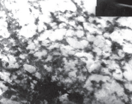
Hyperplasia and dilatation of the SER cisterns. EM χ 10 000
RER degranulation and fragmentation were also present (Figures 2, 3) that is, separation of ribosomes and their dilatation (Figure 4). The mitochondrias changed their size and shape, i.e. they became polymorphous with the thickening of their matrix (Figure 3). The swelling of mitochondrias and even the forming of giant mega-mitochondrias were not rare. The criste in them were shortened (Figure 4.) or fragmented followed by a simultaneous occurrence of crystal inclusion and myelinic figures.
FIGURE 2.

Chromatin condensation in the nuclear periphery, rare glycogen granules, dense mitochondrial matrix, reduced and partially degenerated RER. EM χ 10 000
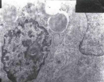
FIGURE 3.
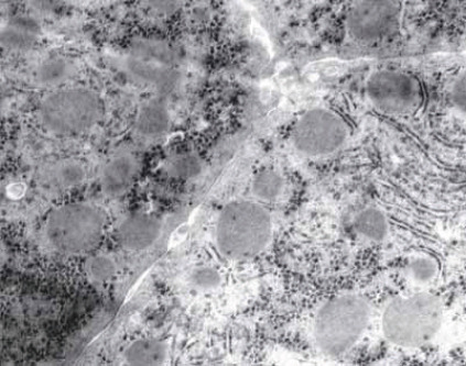
Narrowing of the intercellular space, villi protrusion and membrane “capillarization”. Mitochondrial polymorphism with matrix condensation. Focal degeneration (lack of ribosomes) and fragmentation of RER. Glycogen granules are dense in the hepatocyte periphery. Some of them contain crystalloid corpuscles. Matrix condensation is present in some of them. EM χ 10 000
FIGURE 4.
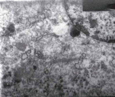
Lipid vesicles, megamitochondria, fragmented cysts, crystalloid corpuscles, vesicular RER dilatation, mitochondrial membrane thinning and multifocal fragmentation. EM χ 10 000
The lysosomes were not directly affected by drugs. The changes in them were of secondary nature and are the result of digestion of products of the damaged membranes of other organelles. The autophagal vacuoles, the increased number of lipofuscin pigment granule, and paracrystal inclusions-myelin figures were found in the cytoplasm of hepatocyte, and even in the cytoplasm of biliary epitheliums. (Figure 3.).
Changes in the cells membranes
The swelling and the thinning of microvilli on the vascular pole accompanied by the protrusion cytoplasm in Disse space were found. This phenomenon is known as shedding figure and represents an unspecific response to shedding (Figure 5). Much more important is the widening of intercellular space together with the fearing of desmosomes.
FIGURE 5.

The Kupfler cell’s dominant hypertrophy with narrowing of sinusoidal lumen. The Kupffer cell’s hyperactiv-ity i.e. hyperplasia of Golgi’s zone and RER. Thinning of the microvilli and peeling of the vascular site of hepatocytes. Marginalization and condensation of the hepatocyte nuclear chromatin is present in some of them. EM χ 26 000
Changes in the nucleus
The condensation of the chromatin and destruction of the nucleus was dominant (Figure 6).
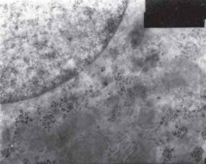
FIGURE 6.

Condensation and marginalization of nuclear chromatin. EM χ 10 WO
Degenerative changes
In the case of serious degeneration type lipid changes, depletion of glycogen in the cytoplasm of hepatocyte was noticeable (Figure 7).
FIGURE 7.
Evident depletion of glycogen or the glycogen’s absence in the hepatocyte cytoplasm. EM χ 13 000
Inflammatory changes
A mesenchymal inflammatory reaction was present in the lobules, portal and periportal areas. Kupffer’s and sinusoidal endothelial cells were increased in the lobules (Figure 5). In the cytoplasm of Kupffer’s cells there were various inclusions, corpuscles, above all, granules of the lipofuscin pigment, hemosiderin, ceroids and biliary inclusions. The expanded Disse space contained the cells of inflammation of the lymphocyte type and polymorphonuclears which are closely related with hepatocytes.
An important change was capillarization of the sinusoid (Figure 3). First, the thin amorphous basal membrane was created along the vascular pole. The basal membrane was followed by the increase of collagen fibres in Disse space, but also in the intercellular space. Those damages caused a disorder of microcirculation and further progress of cirrhosis.
Chronic active hepatitis
In chronic active hepatitis, lymphocyte-plasma cells infiltrate extended from the portal spaces nearly to the parenchyma (12, 13). Lymphocyte-plasma cells showed different stages of maturation and were stuck between hepatocytes. During this process, their cell membranes were in close contact. In the case of a limited lysis of the hepatocyte cytoplasmic membrane, the extensions of activated lymphocytes were invaginated into hepatocyte, which is known as emperipolesis (Figure 8). Plasma cells were in close connection with the hepatocyte. There were phagosomes in macrophages, and together with the fibroblasts they increased protein synthesis thereby enabling the multiplication of collagen. With the multiplication of fibrils, hepatocytes became isolated from the food source which led to irregular lobular organization. Collagen was especially prominent in Disse space near the vascular pole of hepatocyte. The lumen of sinusoids was reduced because of the hypertrophic sinusoidal macrophages (Figure 5), and multiplied lymphocytes and plasma cells. Condensation of the collagen fibres caused intralobular septa which connected the portal zones with the lobular centre damaging lobular structure even more. Besides fibrosis and mononuclear thick infiltrate, cholangiole were multiplied in the portal space, but had the shape of solid stripes without lumen.
FIGURE 8.
Activated lymphocyte with protrusion of its extensions in the hepatocyte cytoplasm - the process of emperiopolesis, the presence of paracrystaloid inclusions in the mitochondrial matrix. EM χ 10 000
Chronic persistent hepatitis
In port space, there were crowded lymphocytes, histiocytes, plasma cells and fibroblasts, accompanied by proliferation of bile channels. The border plates were intact and the lobular structure was preserved. Hepatocytes were very often without changes, although they sometimes contained HBsAg (the surface antigen of the Hepatitis-B-Virus). In the periphery, the stasis of the bilirubin pigment was found and it was the result of periportal inflammation and fibrosis. The multiplied biliary channels were surrounded by the collagen sheaf, but they had a lumen and basal membrane, as well as microvillus on apical poles. Mitochondria of the hepatocytes changed shape and size due to the paracrystaloid inclusions (Figure 8). Sometimes, a nucleoid corpuscle could be seen.
Cirrhosis
In cirrhosis, the nodules in Disse space showed different level of dilatation, as well as increased EM thickness (14). The presence of the continuous basal membrane with transformation of the sinus into capillaries was described (Figure 2). Bundles of collagen fibres around the basal membranes were polarized in parallel formations, thus creating thick or thin sheaths. The forming of basal membranes and sinusoidal capillarization (Figure 3) played an important role in the fibrogenesis of Disse spaces. Basal membranes surrounded the portal space and proliferating sinuses, while sinusoidal capillarization represented focus for the occurrence of cirrhotic fibrogenesis. Collagen fibres were formed even when the basal membrane was not present (15). The increase of the reticular and collagen fibres was found in both extended intercellular spaces and cirrhotic nodules which is known as pericellular fibrosis. It has been suggested that a changed Disse space represents a barrier for diffusion of substrates from the sinusoids. That is why the pathological microvillous borders in some occur as an adaptive response. Ito cells of the perisinusoidal spaces had the main role in the synthesis of collagen fibres. Mitochondrias had greater dimension compared to the megamitochondrias. In hepatocytes, there were Mallory corpuscles. Cholestasis was present in the hepatocytes and small canals and was accompanied by the hypertrophy of Golgi zone and promination of SER (16).
CONCLUSION
The most important ultrastructural findings in the liver of intravenous heroin addicts include:
-hyperplasia and hypertrophy of the smooth endoplasmic reticulum which is histologically proven vesicular degeneration of hepatocyte occurring as a result of the increased synthesis of enzymes of SER due to chronic IV heroin intake;
-the presence of a continuous basal membrane accompanied by the transformation of sinusoids into capillaries (in cases of chronic active hepatitis and cirrhosis), which causes disorders of microcirculation and further progress of cirrhosis.
List of Abbreviations
SER - smooth endoplasmic reticulum
RER - rough endoplasmic reticulum
IV - intravenous
EM - electron microscopic
HBsAg - the surface antigen of the Hepatitis-B-Virus
REFERENCES
- 1.Tennant F. Hepatitis C, B, D, and A: contrasting features and liver íunction abnormalities in heroin addicts. J. Addict. Dis. 2001;20(1):1–7. doi: 10.1300/J069v20n01_02. [DOI] [PubMed] [Google Scholar]
- 2.Passarino G, Ciccone G, Siragusa R, Tappero P, Mollo F. Histo-pathological findings in 851 autopsies of drug addicts, with toxicologic and virologic correlations. Am. J. Forensic Med. Pathol. 2005;26(2):106–116. [PubMed] [Google Scholar]
- 3.Skeie I, Brekke M, Lindbaek M, Waal H. Somatic health among heroin addicts before and during opioid maintenance treatment: a retrospective study. BMC Public Health. 2008;8:43. doi: 10.1186/1471-2458-8-43. [DOI] [PMC free article] [PubMed] [Google Scholar]
- 4.Steentoft A, Teige B, Holmgren P, et al. Fatal poisoning in Nordic drug addicts in 2002. For. Sci. Int. 2006;160:148–156. doi: 10.1016/j.forsciint.2005.09.004. [DOI] [PubMed] [Google Scholar]
- 5.Jezequel AM, Koch M, Galeazzi R, et al. Liver response to drugs in man. A correlation of morphometric and functional study. In: Leevy CM, editor. Disease of the liver and biliary tract. Basel: Karger; 1976. p. 153. [Google Scholar]
- 6.Bellei M, Battelli D, Fornieri C, et al. Changes in liver structure and function after short term and long term treatment of rats with dehydroepiandrosterone. J. Nutr. 1992;122(4):967–976. doi: 10.1093/jn/122.4.967. [DOI] [PubMed] [Google Scholar]
- 7.Naumova TA, Pauchenko LF, Pirozhkov SV, Baronets V, Aliabeva TV. Disorders in the immune status and hepatic pathologies in young heroin addicts. Vestn. Ross. Akad. Med. Nauk. 2003;3:32–36. [PubMed] [Google Scholar]
- 8.Stenger RJ. Organelle pathology of the liver. The endoplasmic reticulum. Gastroenterology. 1970;58(4):554–574. [PubMed] [Google Scholar]
- 9.Jezequel AM, Orlandi F, Tenconi LT. Changes of the smooth endoplasmic reticulum induced by rifampicin in human and guinea pig hepatocytes. Gut. 1971;12(12):984–987. doi: 10.1136/gut.12.12.984. [DOI] [PMC free article] [PubMed] [Google Scholar]
- 10.Jezequel AM, Koch M, Orlandi F. A morphometric study of the endoplasmic reticulum in human hepatocytes. Correlation between morphological and biochemical data in subjects under treatment with certain drugs. Gut. 1974;15(9):737–747. doi: 10.1136/gut.15.9.737. [DOI] [PMC free article] [PubMed] [Google Scholar]
- 11.Trigueiro de Araujo MS, Gerard F, Chossegros P, et al. Cellular and matrix changes and drug abusers liver sinusoids: A semiquantitative and morphometric ultrastructural study. Virchows Arch. A Pathol. Anat. Histopathol. 1993;422(2):145–152. doi: 10.1007/BF01607166. [DOI] [PubMed] [Google Scholar]
- 12.Pogromov AP, Mishnaevski AL, Mustain KK. Viral liver damage in heroin abusers. Viral liver damage in heroin abusers. Eksp. Klin. Gastroenterol. 2002;1:61–63. [PubMed] [Google Scholar]
- 13.Reimer J, Lorenzen J, Baetz B, et al. Multiple viral hepatitis in injection drug users and associated risk factors. J. Gastroenterol. Hepatol. 2007;22:80–84. doi: 10.1111/j.1440-1746.2006.04358.x. [DOI] [PubMed] [Google Scholar]
- 14.Toupalik P, Vanerkova H, Klir P, Bouska I. Morphologic findings in chronic abuse of heroin and pervitine. Soud. Lek. 2002;47(1):5–11. [PubMed] [Google Scholar]
- 15.Trigueiro de Araujo MS, Gerard F, Chossegros P, Guerret S, Grimand JA. Lack of hepatocyte involvement in the genesis of the sinusidal dilatation related to heroin addiction: A morphometric study. Virchows Arch. A Pathol Anat. Histopathol. 1992;420(2):149–153. doi: 10.1007/BF02358806. [DOI] [PubMed] [Google Scholar]
- 16.Lapis K. Normal Liver. In: Johannessen JV, editor. Electron Microscopy in Human Medicine. The Liver. New York, London, Tokyo: McGraw Hill International Book Company; 1989. pp. 3–19. [Google Scholar]