Abstract
Hair transplantation, a generally regarded as a safe surgical modality for the treatment of androgenetic alopecia, is not without its potential risks and complications. A case of an extensive keloid formation at donor site following follicular unit extraction is discussed. Hair transplant surgeons should be aware of this significant potential complication, especially in patients having previous keloidal tendencies to avoid such disastrous outcomes.
Keywords: Donor site, follicular unit extraction, hair transplantation, keloid, scar
INTRODUCTION
Hair transplantation techniques have gradually evolved from the use of large punch grafts, minigrafting, micrografting, and strip harvesting to most recent follicular unit extraction (FUE) method. FUE is minimally invasive as compared to strip harvesting method and expected to be less traumatic and least scarring or with keloidal tendency.[1] Inclination to form hypertrophic scar or keloid following any surgical procedure is individualistic and may depend on racial characteristics, site of trauma, previous personal or family history of keloid formation, and factors like tension at suture line, presence of secondary infection, etc.[2]
Only one published report of keloid formation at the donor site suggests that its exact risk is not predictable in an individual participant. A case with this unusual adverse effect of keloid formation in FUE is being reported. Preventive strategies for this extremely difficult to treat complication are discussed.
CASE REPORT
A 30-year-old male presented with increasing growth of reddish raised itchy areas over occiput for past 8 months, following a hair transplant session using FUE technique for androgenetic alopecia. The diseased area corresponded to the donor site for FUE. Postoperative period was uneventful, and healing over donor as well as recipient site was appropriate without any complications such as local edema, bleeding, or secondary infection. Six-week postoperative, patient experienced itching over donor site followed by formation of gradually increasing raised bumps.
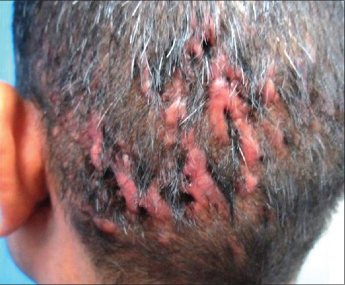
On examination, there were extensive erythematous irregular linear raised firm plaques forming irregular geographic areas devoid of hair with extending pseudopodia to normal skin over the occiput [Figure 1]. Recipient areas of frontal scalp were spared and had well taken up FUE grafts without cobblestoning. A diagnosis of FUE-induced keloids was thus made, and another keloid lesion over the chest was found. He was given 40 mg/ml triamcinolone acetonide intralesional injections on two occasions without much relief and was subsequently lost to follow up.
Figure 1.

Keloidal scar at donor site of follicular unit extraction
DISCUSSION
Keloids may form spontaneously or follow a traumatic and/or inflammatory event, extending to normal skin, no tendency toward spontaneous regression, whereas hypertrophic scars form after injury, do not extend beyond the initial site of injury, and regress over a period of few years.[2] Keloids are more commonly encountered in certain anatomic locations, such as the chest, upper back, earlobes, neck, and shoulders. There is a genetic predisposition to keloids as patients with keloids often report a positive family history. Acne keloidalis nuchae (AKN) a similar condition develops fibrotic plaques, papules, and alopecia on the occiput and/or nape of the neck following acute and chronic granulomatous inflammation surrounding hair shaft. The fibrotic plaques of AKN are, however, different from keloidal scar as being devoid of true keloidal collagen.[3]
FUE involves harvesting hair using small punches of 0.6–1.0 mm in size to individually extract the follicular units instead of removing a strip of hair-bearing skin in follicular unit transplantation to 4 mm punches in punch grafting, thereby resulting in quicker postoperative wound healing and less trauma for patients. FUE results in smaller scars, with less chances of hypertrophic scar or keloid formation.[1] A thorough search of literature found a single case report of keloid or hypertrophic scar formation at donor site following hair transplantation dating back to 1990.[4]
Selmanowitz and Orentreich recommended performing a test patch punch grafts, placing it adjacent to the fringe area of the potential recipient zone, and observing it for keloid formation for 3 months, especially in dark-skinned individuals.[5] However, both the positive and negative predictive value of this test patch is doubtful and cannot be relied on.
Although previous history of keloid formation is not an absolute contraindication for any surgical procedure including hair transplant, some precautions with a guarded prognosis to the patient is worthwhile in such a situation. The importance of interpretation of test patch should also be emphasized to the patient in no uncertain terms.
Further, the risk of keloid formation can be minimized by starting immediate postoperative prophylactic measures such as pressure therapy, silicone gel sheeting, silicone gel, and flavonoids.[6] Although the applicability, utility and efficacy of these preventive measures to scalp keloids following FUE in particular are unpredictable.
Early intervention in potentially developing lesions with intralesional steroids, 5-flurouracil, interferons, and cryotherapy forms the mainstay of treatment of keloid anywhere in the body.[6] Some of the recently introduced therapeutic strategies for the management of keloids methotrexate, retinoids, calcineurin inhibitors, calcium channel blockers, botulinum toxin, vascular endothelial growth factor inhibitors, basic fibroblast growth factor, interleukin-10, transforming growth factor beta, antihistamines, and prostaglandin E2 with variable efficacies may also be promising for FUE-induced keloids.[7]
A high index of suspicion directed toward potential keloid developing situations, informed explicit consent, explanation of the outcome to the patient, early prevention, and prompt early treatment may avoid such disastrous outcome of a cosmetic procedure.
To the best of our knowledge, this is first case report of extensive keloids following FUE hair transplant.
Financial support and sponsorship
Nil.
Conflicts of interest
There are no conflicts of interest.
REFERENCES
- 1.Dua A, Dua K. Follicular unit extraction hair transplant. J Cutan Aesthet Surg. 2010;3:76–81. doi: 10.4103/0974-2077.69015. [DOI] [PMC free article] [PubMed] [Google Scholar]
- 2.Gauglitz GG, Korting HC, Pavicic T, Ruzicka T, Jeschke MG. Hypertrophic scarring and keloids: Pathomechanisms and current and emerging treatment strategies. Mol Med. 2011;17:113–25. doi: 10.2119/molmed.2009.00153. [DOI] [PMC free article] [PubMed] [Google Scholar]
- 3.Shapero J, Shapero H. Acne keloidalis nuchae is scar and keloid formation secondary to mechanically induced folliculitis. J Cutan Med Surg. 2011;15:238–40. doi: 10.2310/7750.2011.10057. [DOI] [PubMed] [Google Scholar]
- 4.Brown MD, Johnson T, Swanson NA. Extensive keloids following hair transplantation. J Dermatol Surg Oncol. 1990;16:867–9. doi: 10.1111/j.1524-4725.1990.tb01575.x. [DOI] [PubMed] [Google Scholar]
- 5.Selmanowitz VJ, Orentreich N. Hair transplantation in blacks. J Natl Med Assoc. 1973;65:471–82. [PMC free article] [PubMed] [Google Scholar]
- 6.Gauglitz GG. Management of keloids and hypertrophic scars: Current and emerging options. Clin Cosmet Investig Dermatol. 2013;6:103–14. doi: 10.2147/CCID.S35252. [DOI] [PMC free article] [PubMed] [Google Scholar]
- 7.Viera MH, Amini S, Valins W, Berman B. Innovative therapies in the treatment of keloids and hypertrophic scars. J Clin Aesthet Dermatol. 2010;3:20–6. [PMC free article] [PubMed] [Google Scholar]


