

Introduction
Soft tissue calcification (calcinosis cutis) is a rare disorder with 5 different subtypes: dystrophic, metastatic, idiopathic, iatrogenic, and calciphylaxis.1 Dystrophic calcinosis refers to the deposition of calcium in areas of prior tissue injury in patients with normal serum calcium and phosphorus. Dystrophic calcification has been associated with various connective tissue disorders including dermatomyositis, overlap syndrome, diffuse cutaneous systemic sclerosis, and CREST (calcinosis, Raynaud phenomenon, esophageal dysfunction, sclerodactyly, and telangiectasia) syndrome; however, it is rarely seen in systemic lupus erythematosus (SLE).2 We report a unique case of bilateral preauricular platelike calcification in a 25-year-old woman with a 4-year history of SLE. We hypothesize that repeated tissue trauma secondary to photosensitivity was the cause of facial dystrophic calcification. To our knowledge, there are no documented cases of facial calcinosis in association with SLE.
Case report
A 25-year-old woman with treatment-resistant SLE complicated by multisystem organ involvement was admitted with a 2-week history of diffuse arthralgias and worsening pruritic rash involving the face, trunk, and upper extremities. SLE was diagnosed at the age of 21 after the patient presented with a malar rash, polyarthritis, headache, and positive serologies (anti–double-stranded DNA, antismith, anti–smooth muscle, antiribonucleoprotein, antinuclear, anti–β-2 microglobulin). Since diagnosis, she has had multiple complications including class V lupus nephritis, hemolytic anemia, myopericarditis, and recurrent lupus cerebritis. Cutaneous manifestations of SLE have included photodistributed erythematous papules on the face and arms, discoid lesions of the scalp with alopecia, hard palate ulcerations, and necrotic vasculitis of digits, nasal bridge, and superior left ear helix secondary to antiphospholipid antibody syndrome. There was no family history of SLE or other autoimmune conditions. Given the severity of her disease, she has been unable to work since finishing college.
Her SLE has been refractory to numerous treatments including azathioprine, bortezomib, and mycophenolic acid. A severe reaction to rituximab precluded its use. Her regimen at the time of presentation included abatacept, hydroxychloroquine, prednisone, and monthly cyclophosphamide infusions.
Recurrence of malar rash, often in the setting of poor compliance with photoprotection, has been a consistent feature of active SLE in this patient. Two weeks before admission, the patient was on vacation for 1.5 weeks, spending a significant amount of time in the sun. On presentation, she reported 1 week of headaches, back pain, and worsening rash on the face and upper extremities. Laboratory values were notable for decreased C3 (63 mg/dL)/C4 (9.0 mg/dL) complement and elevated double-stranded DNA IgG antibodies (102.7 IU/mL), supportive of an acute SLE flare. The dermatology department was consulted for evaluation of rock hard bilateral preauricular plaques.
The patient was unable to identify when the lesions first appeared because they were asymptomatic except for tenderness to touch. She denied recent dental work, surgery, or antecedent mechanical trauma to the area. On examination, approximately 6- × 5-cm hard, sheetlike subcutaneous plaques were present on the bilateral preauricular areas. Overlying the right subcutaneous plaque was an erythematous atrophic depression with scar (Fig 1). The left subcutaneous plaque had near identical overlying skin changes. Photodistributed red nonblanching scaly papules and plaques on the face and extremities were similar in quality to prior presentations. There were no palmar lesions, oral plaques, vesicles, bullae, or other subcutaneous nodules or plaques present elsewhere on the body.
Fig 1.
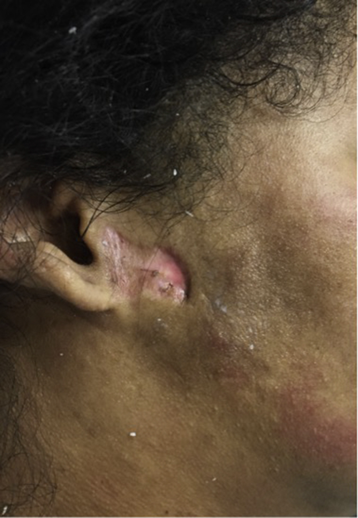
Right preauricular, subcutaneous, firm plaque with overlying atrophic depression and erythema.
Basic metabolic panel, serum calcium, phosphorus, parathyroid hormone levels, alkaline phosphatase, and hepatic function panel were normal. Complete blood count was remarkable only for stable leukopenia (3.13 × 10³/μL). Computed tomography scan of the paranasal sinuses showed calcification within the bilateral subcutaneous soft tissues lateral and anterior to the mandible (Fig 2). Computed tomography scans of the chest, abdomen, and pelvis performed over the course of her admission for various reasons were negative for aberrant calcification of soft tissue or deeper tissue planes. There was no history of lupus mastitis or lupus panniculitis.
Fig 2.

Axial (left) and coronal (right) images from noncontrast head CT showing calcifications (circled) within the bilateral subcutaneous soft tissues lateral and anterior to the mandible.
Biopsies of the preauricular lesions were not performed, as this would have provided little clinical or diagnostic benefit. Calcification represents the end stage of an inflammatory process; therefore, biopsy could not determine the underlying cause. In this case, biopsy would only confirm what was already observed on imaging and physical examination at the risk of impaired wound healing and cosmetic defect.
The patient was treated with pulse steroid doses and cyclophosphamide, which resulted in improvement of the maculopapular rash on her arms and face. The sheetlike calcific lesions remained unchanged.
Discussion
Calcinosis cutis refers to a group of disorders that are characterized by soft tissue calcification. Dystrophic calcinosis is the most common subtype and is associated with a variety of autoimmune disorders.1 It is rarely described in association with SLE, and there are only approximately 45 documented cases to date.2, 3, 4, 5, 6, 7, 8 Many of these patients were on systemic steroids or suffered mechanical trauma or tissue injury secondary to myopathy, skin ulcerations, and, rarely, osteonecrosis or bone infarction. The time of onset for dystrophic calcinosis is typically 20 years after the onset of SLE but is around 5 years in cases associated with lupus panniculitis.2, 3, 4, 5, 6, 7 Most cases involved the extremities and buttocks in women. Although most cases of calcinosis with SLE are dystrophic and localized in nature, diffuse calcification involving the muscle and fascia, known as calcinosis universals, has also been described.2 In this patient, there was no evidence of calcification other than the face.
Facial calcinosis cutis is an uncommon finding in all connective tissue disorders and is only described in a few cases of juvenile dermatomyositis and systemic scleroderma. There is one report of calcinosis cutis in an adolescent involving the mandible and submandibular area in association with linear cutaneous lupus erythematosus, a form of discoid lupus.5 Preauricular platelike calcification in SLE has not been previously reported.
Possible etiologies of dystrophic calcification in this case include preceding panniculitis, discoid lesions, or mechanical trauma. Lupus panniculitis, also known as lupus profundus, typically presents as subcutaneous indurated plaques or nodules and most frequently occur on the cheeks, shoulders, thighs, breast, and buttocks.9 Although lesions may heal with secondary calcification and scaring, this patient was unaware of any preceding panniculitislike lesion in the preauricular area. Furthermore, the extent of calcification and lesion symmetry would be an atypical presentation of lupus panniculitis. Although discoid lesions are common on the face, dystrophic calcinosis occurring in the setting of discoid lupus erythematosus is exceedingly rare, with only 5 cases reported in the literature.6 In such cases, it was hypothesized that calcification occurred as a result of trauma from scratching or mechanical irritation.5, 6
Preauricular and scalp calcification is characteristic of the dystrophic calcification reported in porphyria cutanea tarda, another photosensitivity disorder.10 It has been hypothesized that inflammation and local tissue damage alters the physiologic mechanisms that normally inhibit calcification. Calcium then preferentially deposits at an anatomic site where tissue injury exists. Photosensitivity with sun-induced solar elastosis and elastic tissue damage in both SLE and porphyria cutanea tarda may explain the preferential deposition and unusual distribution of dystrophic calcinosis in this case of SLE and the one report of dystrophic calcinosis on the scalp of a patient with SLE.4, 11
Treatment options for dystrophic calcinosis are limited, and there is no universally accepted treatment algorithm. Case reports have shown variable success in management with diltiazem, topical/intralesional sodium thiosulfate, and surgical excision; however, no randomized controlled trials have analyzed outcome measures.2, 12 Additional research to better understand the pathogenesis of this disorder should be pursued as more effective therapy is needed.
Footnotes
Funding sources: None.
Conflicts of interest: None declared.
References
- 1.Jimenez-Gallo D., Ossorio-Garcia L., Linares-Barrios M. Calcinosis cutis and calciphylaxis. Actas Dermosifiliogr. 2015;106(10):785–794. doi: 10.1016/j.ad.2015.09.001. [DOI] [PubMed] [Google Scholar]
- 2.Balin S.J., Wetter D.A., Andersen L.K., Davis M.P. Calcinosis cutis occurring in association with autoimmune connective tissue disease: The mayo clinic experience with 78 patients, 1996-2009. Arch Dermatol. 2012;148(4):455–462. doi: 10.1001/archdermatol.2011.2052. [DOI] [PubMed] [Google Scholar]
- 3.Kim M.S., Choi K.C., Kim H.S., Song I.G., Shin B.S. Calcinosis cutis in systemic lupus erythematosus: a case report and review of the published work. J Dermatol. 2010;37(9):815–818. doi: 10.1111/j.1346-8138.2010.00894.x. [DOI] [PubMed] [Google Scholar]
- 4.Eastham A., Velez N.F., Chesebro A.L., Townsend H.B., Vleugels R. Diffuse dystrophic calcinosis cutis of the scalp in a patient with scalp discoid lupus erythematosus and systemic lupus erythematosus. JAMA Dermatol. 2013;149(2):246–248. doi: 10.1001/jamadermatol.2013.1420. [DOI] [PubMed] [Google Scholar]
- 5.Ma H., Liao M., Qiu S., Lu R., Lu C. Linear cutaneous lupus erythematosus with calcinosis cutis and milia. Pediatr Dermatol. 2015;32(1):e33–e35. doi: 10.1111/pde.12496. [DOI] [PubMed] [Google Scholar]
- 6.Korekawa A., Nakajima K., Kaneko T., Nakano H., Sawamura D. Discoid lupus erythematosus with dystrophic calcinosis cutis. JAAD Case Rep. 2015;1(4):182–184. doi: 10.1016/j.jdcr.2015.01.006. [DOI] [PMC free article] [PubMed] [Google Scholar]
- 7.Rothe M.J., Grant-Kels J.M., Rothfield N.F. Extensive calcinosis cutis with systemic lupus erythematosus. Arch Dermatol. 1990;126(8):1060–1063. [PubMed] [Google Scholar]
- 8.Dima A., Berzea I., Baicus C. Impressive subcutaneous calcifications in systemic lupus erythematosus. Maedica (Buchar) 2015;10(1):55–57. [PMC free article] [PubMed] [Google Scholar]
- 9.Hansen C.B., Callen J.P. Connective tissue panniculitis: lupus panniculitis, dermatomyositis, morphea/scleroderma. Dermatol Ther. 2010;23(4):341–349. doi: 10.1111/j.1529-8019.2010.01334.x. [DOI] [PubMed] [Google Scholar]
- 10.Grossman M.E., Bickers D.R., Poh-Fitzpatrick M.B., Deleo V.A., Harber L.C. Porphyria cutanea tarda. Am J Med. 1979;67(2):277–286. doi: 10.1016/0002-9343(79)90403-0. [DOI] [PubMed] [Google Scholar]
- 11.Pugashetti R., Shinkai K., Ruben B.S., Grossman M.E., Maldonado J., Fox L.P. Calcium may preferentially deposit in areas of elastic tissue damage. J Am Acad Dermatol. 2011;64(2):296–301. doi: 10.1016/j.jaad.2010.01.046. [DOI] [PubMed] [Google Scholar]
- 12.Baumgartner-Nielsen J., Olesen A.B. Treatment of skin calcifications with intra-lesional injection of sodium thiosulphate: a case series. Acta Derm Venereol. 2016;96(2):257–258. doi: 10.2340/00015555-2206. [DOI] [PubMed] [Google Scholar]