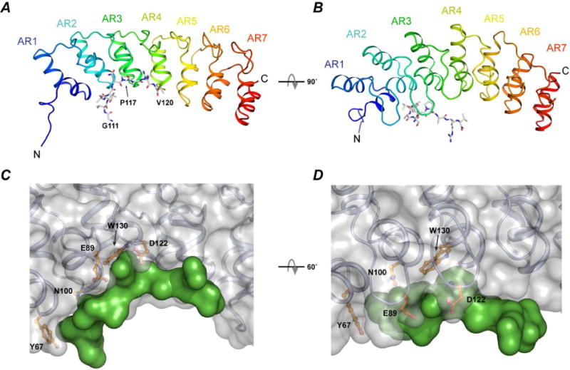
Figure 2. Crystal structure of the complex between ANK17 and Snap25111–120.

A–B) Cartoon and stick representation of the ANK17 and Snap25b peptide, respectively. C–D) Surface representation of the complex. The peptide (green surface) binds to the concave region of ANK17 between AR1–AR3. Some of the interacting residues in ANK17 are shown in orange.
