Cell Biology. In the article “Dictyostelium myosin II null mutant can still cap Con A receptors” by Carmen Aguado-Velasco and Mark S. Bretscher, which appeared in number 18, September 2, 1997, of Proc. Natl. Acad. Sci. USA (94, 9684–9686), the authors request that Figs. 1 and 2 be reprinted with higher contrast. The figures and their legends are shown below.
Figure 1.
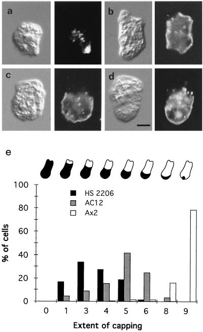
Con A distribution on the surfaces of amoebae after a 30-min incubation (Nomarski images on left, Con A fluorescence on right). (a) Wild-type Ax2 cell. (b–d) mhcA− (HS2206) cells showing ≈10% clearance from two regions at opposite ends of the cell (b), ≈30% clearance (c) and ≈50% clearance (d). (e) Histogram of the extent of capping Con A receptors in which all the cells (about 200) in a field were scored. The cartooned amoebae above the histogram indicate the extent of capping in each case: 0 signifies an uncapped amoeba, 1 ≈ 10% capped, 3 ≈ 30% capped, and so on. Note also that the cells in a–d all have some internalized fluorescent Con A. (Scale bar in d is 5 μm.)
Figure 2.
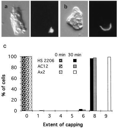
Con A is capped more efficiently when two additional layers of cross-linking are added. Micrographs are as in Fig. 1. (a) Wild-type Ax2 cell. (b) mhcA− (HS2206) cell. (c) Histogram showing the extent of capping Con A receptors (as defined in Fig. 1e; about 300 of each cell type were scored). Note that no substantial endocytosis of fluorescence occurs in these experiments.


