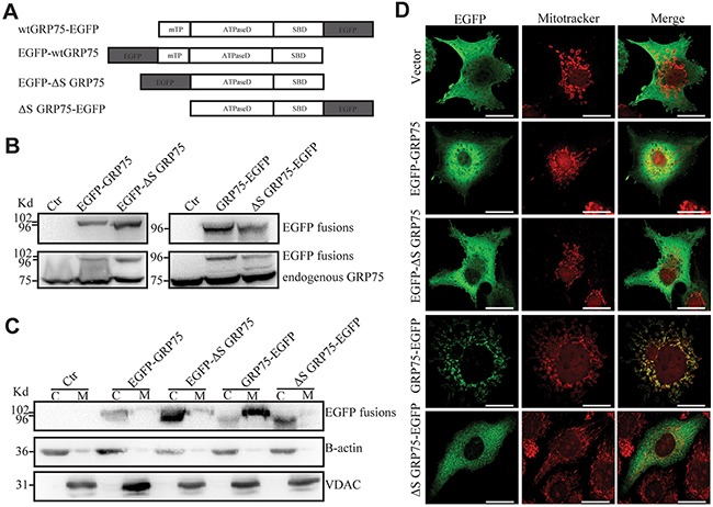
Figure 3. Signal peptide directed GRP75 expression-trafficked into mitochondria.

(A) Schematic representation of C-/N-terminal EGFP-fused GRP75 constructs with/without mitochondrial-targeting signal peptides. wt: wild type. mTP: mitochondrial-targeting signal peptide. ΔS: without signal peptide. ATPaseD: ATPase domain. SBD: substrate-binding domain. (B) HeLa cells were transfected with GRP75 constructs as listed above. Forty-eight hours post-transfection, the cell lysates were analyzed through SDS-PAGE followed by Western blotting with rabbit anti-GFP Ab and mouse anti-GRP75 Ab as described in Materials and Methods. (C) The cytoplasm (C) and mitochondria (M) fractions of transfected HeLa cells were analyzed by Western blotting with rabbit anti-GFP Ab (up row), anti-B-actin Ab (middle row), and anti-VDAC Ab (down row), respectively. (D) Transfected HeLa cells were first stained with mitoTrcker (300nM) for 10 min at 37°C for mitochondria labeling (red color). After fixation and permeabilization, cells were sequentially stained with rabbit anti-GFP Ab (1:500) and then with anti-rabbit AF488 (1:1000). The co-stained cells were viewed under a confocal microscope. Representative cell images show the contrast distribution of exogenous EGFP-fused proteins with mitochondria. Scale bar, 20 um.
