Abstract
Neurointerventional education relies on an apprenticeship model, with the trainee observing and participating in procedures with the guidance of a mentor. While educational videos are becoming prevalent in surgical cases, there is a dearth of comparable educational material for trainees in neurointerventional programs. We sought to create a high-quality, three-dimensional video of a routine diagnostic cerebral angiogram for use as an educational tool. A diagnostic cerebral angiogram was recorded using two GoPro HERO 3+ cameras with the Dual HERO System to capture the proceduralist’s hands during the case. This video was edited with recordings from the video monitors to create a real-time three-dimensional video of both the actions of the neurointerventionalist and the resulting wire/catheter movements. The final edited video, in either two or three dimensions, can serve as another instructional tool for the training of residents and/or fellows. Additional videos can be created in a similar fashion of more complicated neurointerventional cases. The GoPro HERO 3+ camera and Dual HERO System can be used to create educational videos of neurointerventional procedures.
Electronic supplementary material
The online version of this article (doi:10.1007/s10278-017-9948-7) contains supplementary material, which is available to authorized users.
Keywords: GoPro camera, Neurointerventional, Resident education, Training, Video
Background
From the earliest of times, medical education has relied on an apprenticeship model to pass on the skills and techniques necessary to perform procedures in a safe manner. During the course of their training, residents and fellows progress from observers to assistants to primary operators as their knowledge base and skills increase. Recently, restrictions to trainee work hours have placed new constraints on time and limited the potential number of cases in which they can participate [1, 2]. Increasingly, there is concern that residents/fellows who are training in this era of work-hour restrictions may not be comfortable or qualified to practice independently at the conclusion of their training [3].
To address this potential shortcoming, trainers and trainees have increasingly turned to technology to augment medical education. Video recordings of procedures have become commonplace and are available for viewing outside of the traditional, hospital-based teaching environment. High-quality, detailed instructions and videos from leading practitioners across the world are widely available throughout the internet (e.g., The Neurosurgical Atlas’s AANS Operative Grand Rounds, http://www.neurosurgicalatlas.com/grand-rounds/category/aans-operative-grand-rounds) to supplement a trainee’s understanding of the nuances and complexities of a particular procedure.
Initially, traditional two-dimensional videos were created through the use of the operative microscope or video cameras. With the rapid growth in video and camera technology, three-dimensional (3D) videos have become more prevalent [4]. These provide the viewer with a depth of field that could not be obtained from a single-lens camera; however, this added capability has increased the costs of the hardware needed to produce high-quality, 3D videos for educational purposes. We sought to create high-quality, 3D educational videos of neurointerventional procedures using technology that is readily available.
Methods
Hardware
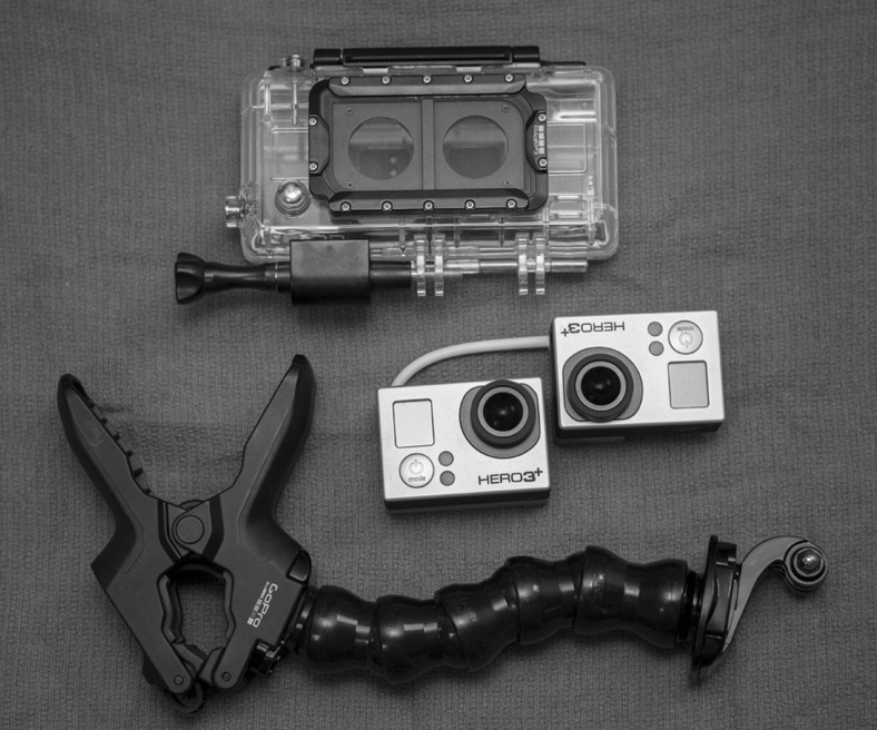
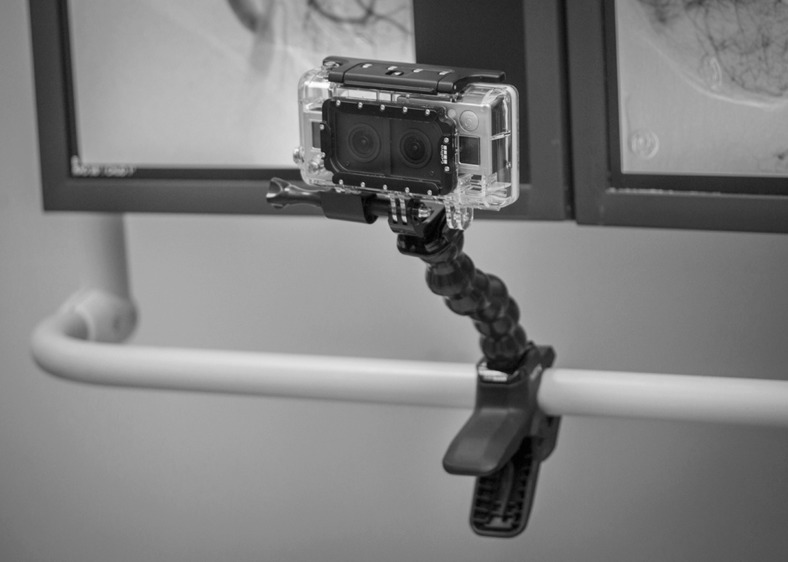
The GoPro HERO 3+ camera (GoPro, San Mateo, CA) is a commercially available product that was initially designed for use by practitioners of extreme sports. As popularity of these cameras has grown, they have infiltrated into many corners of everyday life. We used two GoPro HERO 3+ Black Edition cameras with the Dual HERO System to create 3D neuroangiography videos (Fig. 1). The cameras were connected by the HERO System USB sync and placed in the Dual HERO System housing, which was attached via a flexible mounting system (GoPro Jaws: Flex Clamp) to the video boom (Fig. 2).
Fig. 1.
Two GoPro HERO 3+ Black Edition cameras and the Dual HERO System housing
Fig. 2.
GoPro cameras mounted on the video boom with the GoPro Jaws Flex Clamp
The GoPro HERO 3+ Black Edition camera is a lightweight 12-megapixel camera with an ultra-sharp f/2.8–6 element aspherical glass lens with the capability of capturing ultra-wide angle videos with minimal distortion. This professional-grade camera allows for capturing 12-megapixel photographs in addition to videos. Additionally, the user may select ultra-wide to narrow field of views for the videos. Videos are recorded on microSD memory chips with up to 64 gigabytes of storage capacity. A wi-fi-enabled controller allows the videographer to remotely activate the recordings at prescribed moments, while maintaining sterile technique.
The Dual HERO System, which is only compatible with the HERO 3+ Black Edition cameras, contains the USB sync cord and the housing system. The USB sync cord electronically connects the two GoPro cameras and ensures simultaneous capture of videos. Starting recording on one camera automatically begins recording on the other. The cameras are mounted in the prescribed orientation within the housing system. Both cameras can be turned on with a single button on the housing system or via the sterile covered remote. The GoPro Jaws: Flex Clamp allows the Dual HERO housing system to be connected to a flexible arm and then clamped to any appropriately sized structure.
Additionally, a standard video camera was used to record the video monitors to create the simultaneous angiographic and operator/procedure videos. With currently available technology, however, the live feeds from the video monitor can be readily recorded and downloaded from the angiography suite.
Costs
The GoPro HERO 3+ Black edition camera retails between $350 and $400. The Dual HERO System, which is compatible with the HERO 3+ Black edition only, retails for $200. With the additional costs of the SD microchip and the flexible clamp, the total hardware cost of our system (two GoPro HERO 3+ Black edition cameras, Dual HERO System, SD microchip, and the flexible clamp) was approximately $1200.
Video Editing
Video editing was performed using Adobe Premiere Pro CS6 software on a PC. The Adobe Premiere Pro CS6 is a commercially available program targeted at video editing professionals and retails at over $500. Although the videos from the Dual HERO system can also be edited using the GoPro Studio program for Windows or Mac, which is available for free on GoPro’s website (www.gopro.com), the ability to have side-by-side images of the angiography monitors with the 3D videos of the angiography table and the operator’s hand manipulations required a more robust software editing program.
The paired GoPro cameras create left and right videos, each with 1920 × 1080 resolution. These files were imported into the video editing software to create a 3840 × 1080 sequence with two video layers, one for the left and one for the right. This allows the editor to place the videos side by side to create a stereoscopic video (Video 1). The video could also be formatted for standard 3D anaglyph viewing if desired. Additionally, by using only one of the videos, traditional two-dimensional videos can be created (Video 2).
Viewing
Once the educational database of the hands-on skills and an immersive 3D experience is acquired, the video can be reformatted to a variety of readily available viewing methods. Side-by-side virtual reality (VR) videos can be uploaded and viewed online using television-based stereoscopic displays with active or passive glasses, stereoscopic computer monitors with active or passive shutter glasses are available (Nvidia 3D Vision, Toronto, CA), or freely available Google (Google Inc., Menlo Park, California, USA) cardboard experiences can be used with modern smartphones. Anaglyph videos can be readily viewed on any monitor or screen with the use of the 3D anaglyph glasses.
As more immersive experiences are recorded, full-room VR experiences may be developed using multiple cameras in larger arrays. Stereoscopic videos produced with a simple two-camera setup can already be played within Oculus VR or Vive VR or Samsung Gear VR arrays. Although two cameras can provide an immersive “hands-on” experience, future acquisitions may be used to acquire full 360° videos, also freely playable online, in immersive viewers or VR environments.
Informed Consent
This study was determined to be exempt from Investigational Review Board approval.
Results
Diagnostic Cerebral Angiography
We were able to create a high-quality 3D instructional video on the performance of a routine diagnostic cerebral angiogram using the GoPro HERO 3+ cameras and the Dual HERO System. The GoPro cameras were focused on the operator’s hands during the procedure. In the final editing of the video, both the recordings from the video monitors and the neurointerventionalist’s hands could be viewed simultaneously using split screens. This allows the viewer to follow both the hand/digital manipulations and the resultant catheter/wire movements in real time.
Discussion
Many resident physicians and fellows currently in training are part of the “millennial generation” of individuals born between 1982 and 2004. These residents grew up with digital devices and are accustomed to using such devices in their everyday life. Given their familiarity with electronics, the way these residents learn may be different from that of physicians who trained previously, particularly in incorporating multimedia platforms [5]. Thus, the use of multimedia methods in medical education should be investigated further.
Video is widely used across multiple disciplines to enhance education and monitor compliance [6–8]. Beskind et al. [6] demonstrated that showing an educational video on cardiopulmonary resuscitation to high school students improved responsiveness, and the benefit was still seen at 2 months. Using video as both an educational tool and to provide feedback can assist in protocol adherence. When video was used to outline standard hospital protocol, which usually is only available in writing, the odds for failure of adherence were reduced [7]. Finally, video analysis can be an important tool to monitor protocol compliance and provide feedback when used in combination with direct observation [8].
Educational videos have already been included with success in many residency programs to enhance resident education. In a study by Phillips et al. [9], radiology residents were given an instructional video on how to perform a stereotactic core biopsy of the breast. Residents were asked to complete a set of pre- and post-test questions related to the procedure, and all residents showed improvement in their knowledge related to questions discussed in the video content. Visual media applications can also be expanded from passive observation to allow the viewer to become actively engaged using simulations. Alternatively, video can be used to objectively observe and assess resident skills. For instance, McGoldrick et al. [10] developed a scoring system based on motion analysis of expert microsurgeons. Resident performance was then recorded, and the motion components were assessed using the grading scale to provide objective surgical feedback.
Retention is increased when students are challenged and forced to repeatedly recall and evaluate skills [11, 12]. This approach can be seen in high-performance industries such as professional sports. In football, players routinely watch “coach’s tape,” which allows plays to be watched in slow motion and evaluated within the context of the entire game and the other players. Review of these tapes is considered crucial for players to improve as they learn which actions help or hurt performance. The same philosophy may be applied to medical education. By using video recordings of a trainee performing a procedure, that performance may be analyzed and reviewed as many times as necessary to instill proper technique and decision-making during procedures.
In practice, the trainee would be able to prepare for upcoming procedures by reading appropriate texts supplemented by watching high-quality videos of the procedures being performed by experienced practitioners. During the procedure, videos could be made of the trainee performing certain portions of the procedure. Following the case and after appropriate editing, the trainee and trainer would review the videos to identify areas of competence and areas requiring more practice. In this fashion, the trainee’s education is custom tailored to address current shortcomings and identify areas of focus for future cases.
Additionally, these videos could be used for online education by posting in a web-based repository such as The Neurosurgical Atlas’s AANS Operative Grand Rounds. In this fashion, medical education may transcend the boundaries of brick and mortar institutions and become available for a worldwide audience.
The utility of 3D versus 2D videos for neuroangiography education may, at first glance, be somewhat limited with this current recording configuration; however, with improvements in technology, the magnification and image resolution may improve to the point to allow for improved visualization of “micro-manipulations” of the catheter, wires, coils, and other elements that are used in more complicated neurointerventional procedures versus a more routine diagnostic cerebral angiogram (Videos 1 and 2). For instance, deployment of flow-diverting stents for treatment of intracranial aneurysms requires subtle millimeter movements of both of the operator’s hands to simultaneously move the microcatheter and delivery wire of the device. At the current resolution and magnification, these movements would be difficult to appreciate. Three-dimensional videos combined with improved video resolution may allow for better appreciation of the intricacies and nuances of neurointerventional techniques, which in turn may lead to a better educational product. These videos could be used to test a trainee’s efficacy before and after the immersive video “ride along.” Trainees, particularly those in visual/spatial skill-dependent specialties, may benefit from an immersive learning experience.
Limitations
One limitation to our system proved somewhat problematic at first use. The lack of an optical viewfinder or LCD screen to frame the field was an issue in our first attempt. The cameraperson had to make the best estimate of where to position the camera (both height and direction) to ensure the best possible video. Additionally, movements of the angiography table would lead to a significant change in the field of view, necessitating adjustments of the camera position. These problems could be minimized by use of a head-mounted system, which would preferentially focus the camera on the neurointerventionalist’s field of view; however, during most neurointerventional procedures, the operator is focused on the video monitors rather than their own hand movements.
Future Direction
Significant advances are being made with this camera platform on a frequent basis that transcend the capabilities of our modest GoPro Dual HERO system. The GoPro 3DH3Pro12, a 12-camera, snap-in holder, is designed to allow for the creation of 360-degree 3D video presentations. When its use is combined with the Oculus Rift virtual reality system (Oculus VR, Irvine, CA), viewers can experience the surgery as virtual participants in the operating room. Head-tracking technology on the Oculus Rift system would allow the user to scan across the entirety of the operating theater. Additionally, one could foresee the development of “real-time” video capabilities with this newer technology. In the training environment, this would allow an individual to virtually attend the procedure as if they were standing at the angiography table. With the Oculus Rift system’s head-tracking technology and the GoPro 3DH3Pro12 camera system, the viewer could alternately watch the operative site, the neurointerventionalist, the video monitors, the radiology technicians, and/or the circulating nurses in the room.
Virtual telepresence in the angiographic suite may have additional benefits. For example, if processed and streamed in real time, trainees in multiple global locations could immersively experience an unusual surgery or new technique or learn how to use a new device. The inexpensive acquisition of high-yield immersive content may better suit the next generation of learners, who grow up in 3D and virtual training environments. 3D content can increase learner engagement.
Conclusion
In an age where technology is becoming more integrated into every aspect of life, using video to enhance medical education is a natural progression in training. Video offers additional channels through which important concepts may be conveyed but also may engage active learning through simulation. The ability to review video post-procedurally greatly expands residents’ repertoire of educational material.
We were able to produce high-quality videos of routine diagnostic cerebral angiogram using the GoPro Hero 3+ Black edition camera with the Dual HERO System. This video system has the potential to allow for the widespread adoption of 3D video technology to more centers throughout the USA and the world for educational purposes.
Electronic Supplementary Material
Three-dimensional diagnostic cerebral angiography educational video. (MOV 73951 kb)
Two-dimensional diagnostic cerebral angiography educational video. (MOV 72984 kb)
Acknowledgments
The authors thank Kristin Kraus, MSc for her invaluable assistance with the preparation of this manuscript.
Compliance with Ethical Standards
This study was determined to be exempt from Investigational Review Board approval.
Footnotes
Electronic supplementary material
The online version of this article (doi:10.1007/s10278-017-9948-7) contains supplementary material, which is available to authorized users.
References
- 1.Connors RC, Doty JR, Bull DA, May HT, Fullerton DA, Robbins RC. Effect of work-hour restriction on operative experience in cardiothoracic surgical residency training. J Thorac Cardiovasc Surg. 2009;137:710–713. doi: 10.1016/j.jtcvs.2008.11.038. [DOI] [PubMed] [Google Scholar]
- 2.Picarella EA, Simmons JD, Borman KR, Replogle WH, Mitchell ME. “Do one, teach one”; the new paradigm in general surgery residency training. J Surg Ed. 2011;68:126–129. doi: 10.1016/j.jsurg.2010.09.012. [DOI] [PubMed] [Google Scholar]
- 3.Griner D, Menon RP, Kotwall CA, Clancy TV, Hope WW. The eighty-hour workweek: surgical attendings' perspectives. J Surg Ed. 2010;67:25–31. doi: 10.1016/j.jsurg.2009.12.003. [DOI] [PubMed] [Google Scholar]
- 4.Lee B, Chen BR, Chen BB, Lu JY, Giannotta SL. Recording stereoscopic 3D neurosurgery with a head-mounted 3D camera system. Br J Anesth. 2015;29:371–373. doi: 10.3109/02688697.2014.997664. [DOI] [PubMed] [Google Scholar]
- 5.Evans CH, Schenarts KD. Evolving educational techniques in surgical training. Surg Clin N Am. 2016;96:71–88. doi: 10.1016/j.suc.2015.09.005. [DOI] [PubMed] [Google Scholar]
- 6.Beskind DL, et al. Viewing a brief chest-compression-only CPR video improves bystander CPR performance and responsiveness in high school students: a cluster randomized trial. Resuscitation. 2016;104:28–33. doi: 10.1016/j.resuscitation.2016.03.022. [DOI] [PubMed] [Google Scholar]
- 7.Kandler L, Tscholl DW, Kolbe M, Seifert B, Spahn DR, Noethiger CB. Using educational video to enhance protocol adherence for medical procedures. Br J Anesth. 2016;116:662–669. doi: 10.1093/bja/aew030. [DOI] [PubMed] [Google Scholar]
- 8.Sánchez-Carrillo LA, et al.: Enhancement of hand hygiene compliance among health care workers from a hemodialysis unit using video-monitoring feedback. Am J Infect Control 44:868–872, 2016 [DOI] [PubMed]
- 9.Phillips J, et al. Educational videos: An effective tool to improve training in interventional breast procedures. J Am Coll Radiol. 2016;13:719–724. doi: 10.1016/j.jacr.2016.02.009. [DOI] [PubMed] [Google Scholar]
- 10.McGoldrick RB, Davis CR, Paro J, Hui K, Nguyen D, Lee GK. Motion analysis for microsurgical training: Objective measures of dexterity, economy of movement, and ability. Plas Reconst Surg. 2015;136:231e–240e. doi: 10.1097/PRS.0000000000001469. [DOI] [PubMed] [Google Scholar]
- 11.Karpicke J, Roediger H., III Repeated retrieval during learning is the key to long-term retention. J Mem Lang. 2007;57:151–162. doi: 10.1016/j.jml.2006.09.004. [DOI] [Google Scholar]
- 12.Wiklund Hörnqvist C, Jonsson B, Nyberg L. Strengthening concept learning by repeated testing. Scand J Psychol. 2014;55:10–16. doi: 10.1111/sjop.12093. [DOI] [PMC free article] [PubMed] [Google Scholar]
Associated Data
This section collects any data citations, data availability statements, or supplementary materials included in this article.
Supplementary Materials
Three-dimensional diagnostic cerebral angiography educational video. (MOV 73951 kb)
Two-dimensional diagnostic cerebral angiography educational video. (MOV 72984 kb)


