. 2017 Sep 19;6:e30647. doi: 10.7554/eLife.30647

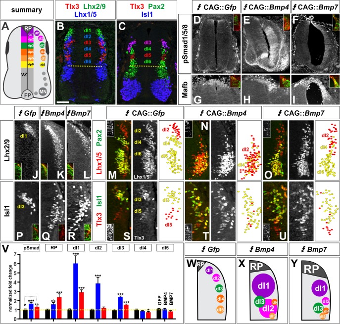
Figure 2. BMP4 and BMP7 direct distinct dorsal IN identities in vivo.

(A–C) Summary of dorsal progenitors (dP1-6) and post-mitotic neurons (dI1-6) in the developing spinal cord. The combinatorial use of antibodies against Lhx2/9 (green), Lhx1/5 (blue), Isl1 (blue), Pax2 (green) and Tlx3 (red) permit the unambiguous identification of dI1-dI5. (D–U) Chicken spinal cords were electroporated at HH stage 15 with Gfp (D, G, J, M, P, S), Bmp4 (E, H, K, N, Q, T) or Bmp7 (F, I, L, O, R, U), under control of the CAG enhancer (Miyazaki et al., 1989), and incubated until HH stage 25. Thoracic transverse sections were labeled with antibodies against Tlx3 (red, S–U), Lhx2/9 (red, J–L), Lhx1/5 (red,M–O), Isl1 (red P-R; green, S–U) or Pax2 (green M-O), pSmad1/5/8 (red, D–F) and Mafb (red, G–I). (D–F) Ectopic expression of Bmp4 (n = 52 sections from 5 embryos, p<0.0001) more effectively activates the R-Smads (Smad1/5/8) than Bmp7 (n = 30 sections 4 embryos, p<0.005), while the expression of Gfp has no effect (n = 37 sections from 4 embryos, p>0.72). (G–I) Mis-expression of Bmp4 or Bmp7 has dramatic, but distinct, effects on dorsal cell differentiation. Specifically, the ectopic expression of Bmp7 (I, n = 28 sections from 4 embryos, p<7.01×10⁻⁵) resulted in consistently more Mafb+ RP cells than Bmp4 (H, n = 37 sections from 5 embryos, p<0.12), and the Gfp control (G, n=36 sections from 2 embryos, p>0.67). (J–L, P–R) Mis-expression of Bmp4 however, most effectively directs cells towards the Lhx2/9+ dI1 (K, n = 28 sections from 3 embryos, p<0.0001) and Isl1+ dI3 fates (Q, 91 sections from 5 embryos, p<0.0001) compared to Bmp7 (dI1, L, n = 26 sections from 3 embryos, p<0.0001; dI3, R, n = 59 sections from 5 embryos, p<5.58×10⁻⁶). Mis-expression of Gfp has no effect (dI1, J, n = 46 sections from 3 embryos, p>0.47; dI3, P, n = 45 sections from 3 embryos, p>0.46). (P–R) Bmp4 (N, n = 47 sections from 3 embryos, p<0.0001) is the only BMP sufficient to direct cells toward an Lhx1/5+ dI2 identity, while Bmp7 can suppress Pax2+ dI4 fate (O, n = 24 sections from 3 embryos, p<0.01). The ectopic expression of Gfp has no effect (M, n = 35 sections from 3 embryos, p>0.77). Note that the presence of Bhlhlb5 (Skaggs et al., 2011), permitted the Pax2+ Lhx1/5- Bhlhb5- dI4s to be unambiguously distinguished from the Pax2- Lhx1/5+ Bhlhb5- dI2s and the Pax2+ Lhx1/5+ Bhlhb5+ dI6s (data not shown). (S–U) The mis-expression of the BMPs has no effect on the Tlx3+ dI5 fate (S, GFP: n = 38 sections from 3 embryos p>0.16; BMP4: T, n = 50 sections from 4 embryos, p>0.39; BMP7: U, n = 29 sections from 3 embryos, p>0.08) fates. (V) Quantification of the fold change in cell number normalized to Gfp control. The probability Bmp4 and Bmp7 misexpression result in the same distribution of cellular activities is p<0.0002 (Fisher test). (W–Y) Summary of the cellular changes directed by BMP4 and BMP7. We found that there are there are spatial organizational changes for Bmp4, but not Bmp7, misexpression. While the RP, dI1 and dI3 populations remain in the correct spatial order with respect to each other, the dI2s both expand and change location, such that they are intermingled with/ventral to the dI3 population. Probability of similarity between control and experimental groups, *=p<0.05, **p<0.005, ***p<0.0005. Student’s t-test or Mann-Whitney test. Scale bar: 65 µm.

Figure 2—source data 1. BMP4 and BMP7 direct distinct dorsal IN identities in vivo (experimental data).
DOI: 10.7554/eLife.30647.009

Figure 2.

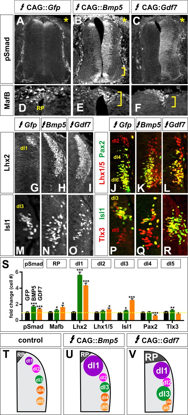
Figure 2—figure supplement 1. Other BMPs have unique activities specifying dorsal IN populations.

Figure 2—figure supplement 1.

(A–R) Chicken spinal cords were electroporated with Gfp (A, D, G, J, M, P), Bmp5 (B, E, H, K, N, Q) or Gdf7 (F, I, L, O, R) at HH stage 15 and incubated until HH stage 25. Thoracic transverse sections were labeled with antibodies against Lhx2/9 (G–I), Isl1 (M–R), Lhx1/5 (red, J–L), Pax2 (green, J–L) and Tlx3 (red, P–R). (A–C) Misexpression of Bmp5 (n = 44 sections from 3 embryos) and Gdf7 (n = 34 sections from 3 embryos) increases pSmad1/5/8 activity up to 2-fold, whereas misexpression of Gfp has no effect. In all experiments the Gfp control had no effect on cellular identity (n as for Figure 2). (D–F) Misexpression of Bmp5 (n = 35 sections from 3 embryos) and Gdf7 (n = 30 sections from 4 embryos) modestly increases the numbers of Mafb+ RP cells. (G–I) Ectopic expression of both Bmp5 (n = 34 sections from 3 embryos) and Gdf7 increase the number of Lhx2/9+ dI1s (n = 40 sections from 5 embryos). (J–L) Gdf7 modestly increases the number of dI2s (n = 24 sections from 3 embryos) and decreases the number of dI4s (n = 23 sections from 4 embryos) while neither Bmp5 (Lhx1/5: n = 43 sections from 3 embryos; Pax2: n = 27 sections from 3 embryos). (M–O) Gdf7 (n = 48 sections from 4 embryos) is much more effective than Bmp5 (n = 54 sections from 3 embryos) at increasing the number of dI3s. (P–R) Bmp5 can modestly increase the number of dI5s (n = 54 sections from 3 embryos), while neither Gdf7 (n = 29 sections from 3 embryos). (S) Quantification of the fold change in cell number normalized to the Gfp control. (T–V) Models for the different activities of BMP5 and BMP6. Probability of similarity between control and experimental groups, *p<0.05, **p<0.005,***p<0.0005, Student’s t-test. Scale bar: 70 µm.
Figure 2—figure supplement 2. BMP misexpression alters the expression of the electroporated BMP.
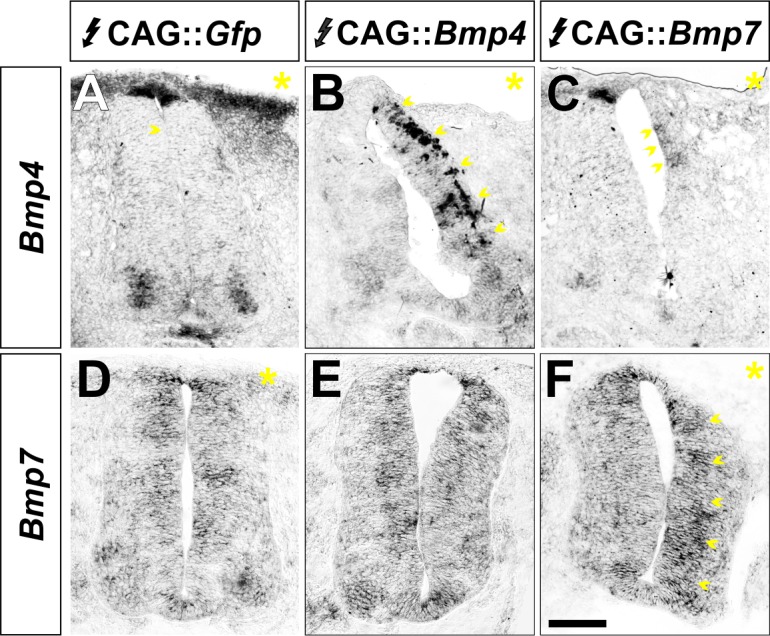
Figure 2—figure supplement 2.

(A–F) Chicken spinal cords were electroporated with CAG::Gfp (A, D), CAG::Bmp4 (B, E) or CAG::Bmp7 (C, F) at HH stage 15 and incubated until HH stage 25. Transverse sections were processed for in situ hybridization with probes for Bmp4 and Bmp7 expression. The yellow asterisk indicates the electroporated side. Increased Bmp4 expression was only observed on the embryos electroporated with CAG::Bmp4 (arrows, B). The levels of endogenous Bmp7 in the CAG::Bmp4 electroporated spinal cords are indistinguishable from control (E). Similarly, increased Bmp7 expression was only observed in embryos electroporated with CAG::Bmp7 (arrows, F). Endogenous Bmp4 expression is elongated and more diffuse in CAG::Bmp7 spinal cords (arrows, C), presumably as a result of the increase in RP cells.
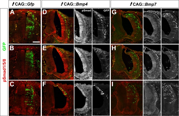
Figure 2—figure supplement 3. BMP misexpression results in consistent alterations in the levels of pSmad1/5/8.

Figure 2—figure supplement 3.

(A–I) Chicken spinal cords were electroporated at HH stage 15 with high concentrations (>500 ng/μl) of CAG::Gfp (A, D, G), CAG::Bmp4 (B, E, H) or CAG::Bmp7 (C, F, I) and incubated until HH stage 25. Transverse sections of brachial spinal cord were labeled with an antibody against pSmad1/5/8 (red, A–I). The presence of GFP (green) indicates the extent and pattern of the electroporated cells. (A–C) Electroporation of CAG::Gfp had no effect on endogenous pSmad1/5/8 activity. (D–F) In contrast, the electroporation of CAG::Bmp4 dramatically increased the level of pSmad1/5/8 staining throughout the spinal cord. Localized electroporation (F) increased pSmad levels to a similar extent as observed for broader patterns of electroporation (D, E). (G–I) Similarly, different patterns of CAG::Bmp7 electroporation result in the same lower levels of increased Smad1/5/8 activity throughout the spinal cord.
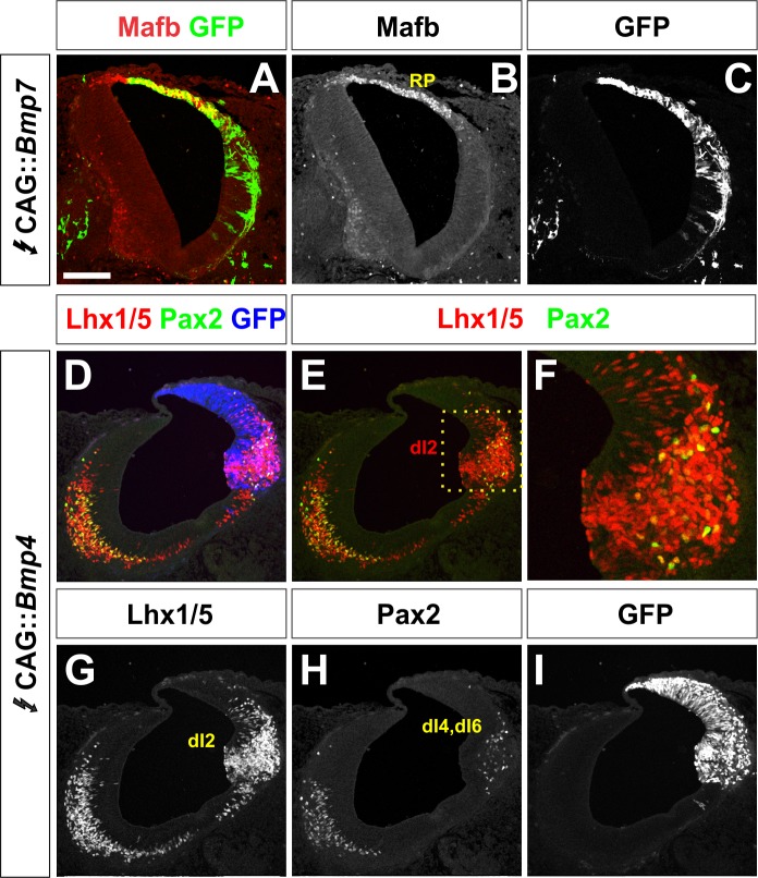
Figure 2—figure supplement 4. BMP misexpression can result in major morphological changes to the spinal cord.

Figure 2—figure supplement 4.

(A–I) Chicken spinal cords were efficiently electroporated at HH stage 15 with high concentrations (>500 ng/μl) of CAG::Bmp4 (D–I) or CAG::Bmp7 (A–C) and incubated until HH stage 25. Thoracic transverse sections were labeled with antibodies against Mafb (red, A–B), Lhx1/5 (red, D–G) or Pax2 (green, D–F, H). Gfp expression demonstrates electroporation efficiency. (A–C) The ectopic expression of Bmp7 results in the expansion of the Mafb+ RP. In this example, the expansion is profound, resulting in a widening of the ventricle as well as an elongation and thinning of the electroporated side of the spinal cord. (D–I) The ectopic expression of Bmp4 results in different morphological changes. In some embryos, a tumor-like ball of Lhx1/5+ cells has formed in the ventral spinal cord. These dI2-like cells are positioned where the motor column would normally be located. Scale bar: 100 µm.