Abstract
AIM
To determine peculiarities of tissue responses to manual and automated Ilizarov bone distraction in nerves and articular cartilage.
METHODS
Twenty-nine dogs were divided in two experimental groups: Group M - leg lengthening with manual distraction (1 mm/d in 4 steps), Group A - automated distraction (1 mm/d in 60 steps) and intact group. Animals were euthanized at the end of distraction, at 30th day of fixation in apparatus and 30 d after the fixator removal. M-responses in gastrocnemius and tibialis anterior muscles were recorded, numerical histology of peroneal and tibialis nerves and knee cartilage semi-thin sections, scanning electron microscopy and X-ray electron probe microanalysis were performed.
RESULTS
Better restoration of M-response amplitudes in leg muscles was noted in A-group. Fibrosis of epineurium with adipocytes loss in peroneal nerve, subperineurial edema and fibrosis of endoneurium in some fascicles of both nerves were noted only in M-group, shares of nerve fibers with atrophic and degenerative changes were bigger in M-group than in A-group. At the end of experiment morphometric parameters of nerve fibers in peroneal nerve were comparable with intact nerve only in A-group. Quantitative parameters of articular cartilage (thickness, volumetric densities of chondrocytes, percentages of isogenic clusters and empty cellular lacunas, contents of sulfur and calcium) were badly changed in M-group and less changed in A-group.
CONCLUSION
Automated Ilizarov distraction is more safe method of orthopedic leg lengthening than manual distraction in points of nervous fibers survival and articular cartilage arthrotic changes.
Keywords: Limb lengthening, Articular cartilage, Nerve, Histomorphometry, Dog
Core tip: Limb lengthening developed by Ilizarov is now well accepted method for correction of orthopedic problems but in some cases it is complicated with nerve and joint malfunctions or disturbances. In this animal study we present the comparative analysis of quantitative indices of nerves and articular cartilage structural reorganization during lengthening with manual and automatic Ilizarov bone distraction. Results of the study have indicated the benefits of automatic distraction.
INTRODUCTION
The technology of gradual limb lengthening developed by Ilizarov is now well accepted method for correction of limb length discrepancy, short stature or treatment of bone defects, resulting from trauma, congenital abnormality or oncologic resection. Distraction osteogenesis is an effective method for new bone tissue creation but in some cases is complicated with nonunion, neurovascular disturbances, muscle contractures and joint stiffness. Using manual circular distractor and specially developed automatic device, Ilizarov[1] investigated various rates and rhythms of distraction and have concluded that “rate of 1.0 mm/d led to the best results”. He also reported that “the greater the distraction frequency, the better the outcome” comparing the automatic distraction (1 mm/d in 60 steps) with manual (1 mm/d in 4 steps). This conclusion has been confirmed in clinical practice[2] but automatic distraction is not widely implemented method because of its technical difficulties and costs.
There are few research groups dealing with automatic distraction. Nakamura et al[3] have found less damage in knee cartilage in automatically distracted group (1 mm/d in 120 steps) compared with manual group (1 mm/d in two steps). In goats undergoing leg lengthening with automated distractor producing one, four or 720 increments per day it was found that “the intensity and dispersion of degenerative changes in muscles were in reverse proportion to the frequency of distraction”[4]. Distraction mode 1 mm/d in 1440 steps in comparison with 1 mm/d in 3 steps resulted in better range of motion and somatosensory evoked potentials, though muscle histology was the same[5]. Aarnes et al[6] reported that high frequency of distraction improved tissue adaptation during leg lengthening in humans. In recent research there was no difference in time to union or in the incidence of complications in comparison with manual low-frequent distraction[7].
Taking into account discrepancies in results it is important to revise experimental leg lengthening series from the points of quantitative histological and physiological methods. Such data is absent in global literature. The safety of automatic Ilizarov distraction for muscles, nerves and cartilage[8-11] was substantiated in Russian articles, but peculiarities of structural response to manual and automatic Ilizarov bone distraction in nerves and articular cartilage were not revealed.
The aim of our research - comparative analysis of structural changes in leg nerves and knee cartilage during experimental leg lengthening with manual (1 mm/d in four steps) and automatic (1 mm/d in 60 steps) distraction.
MATERIALS AND METHODS
Experiments were carried out in accordance with Principles of Laboratory Animal Care (NIH Publication No. 85-23, revised 1985). Twenty-nine mongrel adult dogs (weight 20-25 kg, 18-20 cm leg length) were used. Five animals formed the intact group and 24 were operated.
Surgery and experimental design
Anesthesia with intramuscular injections of atropine, dimedrol and xylazine was maintained with sodium pentobarbital (30 mg/kg i.v.) intravenously. Mid-diaphyseal osteoclasis and osteosynthesis by Ilizarov were performed. In M-group (n = 12) the lengthening protocol involved a 5-d latent period, and then manual movement of graded traction nodes at the rate of 1 mm/d in 4 steps was performed for 28 d. In A-group (n = 12) protocol was the same as in M-group, but distraction rate was 1 mm/d in 60 steps. The animals were euthanized at three time-points: D28 - the end of distraction (15% increase the initial leg length in both groups), F30 - 30 d of fixation in apparatus (bone regenerate consolidation in all animals of A-Group, but in M-Group consolidation was evident only in three animals), WA30 - 30 d without apparatus (full weight bearing of the operated limb in M-Group, but in A-Group it was noted immediately after the apparatus removal).
Neurophysiologic evaluation
Intramuscular EMG was performed after anesthesia also at D 28, F 30 and WA 30. M-responses in gastrocnemius and tibialis anterior muscles were recorded using a digital EMG-system DISA-1500 (DANTEC, Denmark). Bio potential leads were monopolar with modified needle electrodes. The active recording electrode was inserted percutaneously in muscle belly and the reference electrode - in its tendon. M-responses were recorded after supramaximal electrical stimulation of sciatic nerve through para neural needle electrodes using rectangular wave pulses of 1-ms duration. Muscle action potential amplitudes were measured from the top of the negative peak to the top of maximal positive peak.
Morphological evaluation
Histologic analysis was performed in 19 animals (all intact and seven from each experimental group). Animals were euthanized. Nerves and cartilage samples were fixed in mixture of 20 g/L solutions of glutaraldehyde and paraformaldehyde in phosphate buffer (рН 7.4) adding 1 g/L picric acid and then were partially embedded in paraffin. Paraffin sections were cut on “Reichert” microtome (Austria) and mounted in glasses with poly-L-lysin for reaction with ki-67 according protocols of producing company using the visualization system RE7140-К (Novocastra, Great Britain). Another parts were post-fixed in 1 g/100 mL tetraoxide osmium solution with 1.5 g/100 mL potassium ferricyanide and embedded in araldite. Transverse semi-thin sections (thickness 0.5-1.0 μm) were made with glass knives using the Nova ultratome LKB (Sweden), mounted on glass slides and then stained with toluidine blue and methylene blue-basic fuchsin. For numerical analysis the semi-thin epoxy sections of enlarged area (4-8 mm2 instead of standard 1 mm2) were used. Such technology provided the cellular details visualization in the light microscope and the sample representativeness[12]. Two calibrated experts conducted numerical analysis. True-color images were digitized using the stereomicroscope “AxioScope.A1”) with camera “AxioCam” (Carl Zeiss MicroImaging GmbH, Germany). Histomorphometry was performed with “VT-Master-Morphology” program (VideoTest, Russia, St. Petersburg). In 3 total sections (magnification 32 ×) nerves areas and summary areas of nerve fascicles with perineurium were determined. In 25 nonoverlapping fields of the endoneurial compartment (magnification 1250 ×), collected in a systematic random order, the numerical densities of endoneurial vessels were evaluated. About 400 samples of myelinated fibres for each nerve site were made, and morphometric parameters-diameters of myelinated nerve fibers, their axons and myelin sheath thickness-were measured. Per cent of degenerated myelinated nerve fibers in the samples were calculated.
Cartilage depth and volumetric chondrocytes density in 30 digital images were estimated, per cents of isogene groups and empty lacunas in random selection of 100 chondrocytes were calculated. Surfaces of epoxy blocks were exposed to silver deposition using Eico IB-6 ion coater and JEOL JEE-4X vacuum evaporator. The investigation of element composition was performed using scanning electron microscope JSM-840 (JEOL, Japan) equipped with energy dispersive X-ray analyzer (INCA 200, Oxford Instruments). Using accelerated voltage 20 kV concentrations of sulfur (ωS, weight %) and calcium (ωCa, weight %) were assessed. Results were obtained as smart maps, showing spatial distribution of elements and quantitative data in weight per cents. For SEM-micrographs cartilage samples were dehydrated in alcohols of ascending concentrations, permeated with camphene according to original method[13], air dried and coated with silver.
Statistical analysis
Statistical treatment of numerical data was performed in software package Attestat Program (version 9.3.1, developed by I. P. Gaidyshev, Certificate of Rospatent official registration No. 2002611109), the paired and unpaired Student t tests, Mann-Whitney U test and Fisher exact test were used at 0.05 significance level.
RESULTS
EMG changes
M-responses amplitudes were badly changed in all animals especially in m. tibialis anterior of M-group. At D28 and F30 the intramuscular EMG revealed fibrillations and sporadic positive sharp waves in both groups. At WA30 they disappeared. Table 1 shows that in m. gastrocnemius M-response amplitudes at D28 tended to be lower in A-group than in M-group, but at F30 and WA 30 the parameters of A-group became better than of M-group. In m. tibialis anterior they were better in A-group at all t-points.
Table 1.
M-response amplitudes in leg muscles
| t-points of EMG-testing |
M. gastrocnemius |
M. tibialis anterior |
||
| mean ± SE (mV) | Difference vs initial (%) | mean ± SE (mV) | Difference vs initial (%) | |
| Initial (before experiment) | 32.0 ± 2.9 | - | 22.6 ± 1.3 | - |
| M-group - manual distraction (1 mm/d in 4 steps) | ||||
| 28 d of distraction | 13.4 ± 3.1 | -58.1 | 8.8 ± 1.9 | -61.1 |
| 30 d of fixation | 10.6 ± 4.6 | -66.9 | 8.3 ± 1.3 | -63.3 |
| 30 d without apparatus | 16.3 ± 5.8 | -49.1 | 8.9 ± 0.1 | -60.6 |
| A-group - automatic distraction (1 mm/d in 60 steps) | ||||
| 28 d of distraction | 12.0 ± 1.7 | -62.5 | 12.7 ± 1.6a | -43.8 |
| 30 d of fixation | 11.9 ± 1.2a | -62.8 | 10.5 ± 0.5a | -53.5 |
| 30 d without apparatus | 18.3 ± 4.6a | -42.8 | 14.0 ± 2.0a | -38.1 |
P < 0.05 vs group M.
Changes of nerves sheaths
Pathologic conditions of epineurium, subperineurial space and endoneurium were more prominent in M-group. Table 2 shows that at F30 the thickening of Tn was noted in A-group and the thinning of Pn - in M-group. The first was determined by thickening and hypervascularity of epineurium, the second - by fibrotic changes of epineurium with marked loss of adipocytes. Laminated cellular structure of perineurium in lengthened nerves was maintained in both groups - presented in Figure 1A. In M-group the signs of subperineurial edema were noted - also visible in Figure 1A. Numerosity of perineurial cells nuclei was increased, fibrillar interlayers were thickened. In perineurial cells nuclei the high expression of ki-67 was noted - presented in Figure 1B.
Table 2.
Areas of nerves histologic transverse sections (An)
| Nerves/group and t-points |
Tibial nerve |
Peroneal nerve |
||
| (mean ± SD) (104 mKm2) | Difference vs contra-lateral (%) | (mean ± SD) (104 mKm2) | Difference vs contra-lateral (%) | |
| M-group - manual distraction (1 mm/d in 4 steps) | ||||
| 28 d of distraction | 353.7 ± 34.8 | 3 | 41.6 ± 9.4 | -2 |
| 30 d of fixation | 291.6 ± 26.2 | -4 | 63.6 ± 1.1 | -11a |
| 30 d without apparatus | 248.7 ± 29.2 | 3 | 53.3 ± 8.8 | -3 |
| A-group - automatic distraction (1 mm/d in 60 steps) | ||||
| 28 d of distraction | 287.3 ± 60.0 | -2 | 56.2 ± 3.4 | -1 |
| 30 d of fixation | 306.8 ± 76.7 | 15ac | 75.8 ± 16.3 | -1c |
| 30 d without apparatus | 224.8 ± 31.1 | 10c | 46.5 ± 1.1 | 1 |
P < 0.05 vs contralateral;
P < 0.05 vs group M.
Figure 1.

Peroneal nerve, F30, M-group. A: Perineurium, fragment of transverse semithin section, methylene blue - basic fuchsin stain; B: Proliferating cells in perineurium; C: Proliferating cells in endoneurium; fragments of paraffin-embedded sections stained using antibodies to Ki-67. Magnification 1250 ×.
Table 3 shows that at D28 the summary fascicular areas in transverse histologic sections of lengthened nerves were decreased compared to corresponding contralateral ones. Fascicular thinning was more prominent in Pn than in Tn. Extent of it in corresponding nerves in M and A-groups was approximately equal. At WA30 nerves restored their fascicular areas in A-group, in M-group nerve fascicles were thickened because of endoneural fibrosis.
Table 3.
Summary fascicular areas (Af) in transverse histologic sections of nerves
| Nerves/group and t-points |
Tibial nerve |
Peroneal nerve |
||
| (mean ± SD) (104 mKm2) | Difference vs contra-lateral (%) | (mean ± SD) (104 mKm2) | Difference vs contra-lateral (%) | |
| M-group - manual distraction (1 mm/d in 4 steps) | ||||
| 28 d of distraction | 68.7 ± 6.9 | -8 | 18.8 ± 8.1 | -15 |
| 30 d of fixation | 73.5 ± 5.8 | -7 | 40.1 ± 2.3 | -1 |
| 30 d without apparatus | 54.2 ± 4.8 | 9a | 27.8 ± 9.5 | 14a |
| A-group - automatic distraction (1 mm/d in 60 steps) | ||||
| 28 d of distraction | 83.7 ± 13.5 | -9a | 25.7 ± 2.5 | -16a |
| 30 d of fixation | 86.8 ± 13.5 | -4 | 29.6 ± 7.0 | 1 |
| 30 d without apparatus | 82.0 ± 6.4 | 5 | 25.9 ± 0.6 | 0c |
P < 0.05 vs contralateral;
P < 0.05 vs group M.
The level of ki-67 expression in endoneurial cells was increased especially at F30 - presented in Figure 1C. Table 4 shows that in M-group the numerical density of endoneural microvessels in lengthened Tn was increased only at F30, Pn nerve - at all t-points especially at D28 and WA30. In A-group the numerical density of endoneural microvessels in Tn were decreased at D28 compared with intact nerve, at F30 it was approximately equal to parameter of intact nerve and at WA 30 endoneurium of Tn was highly vascularized. The numerical densities of endoneural vessels in Pn in A-group were increased at all t-points.
Table 4.
Numerical densities of microvessels (NAmv) in endoneurium
| Nerves/group and t-points |
Tibial nerve |
Peroneal nerve |
||
| mean ± SE (mm-2) | Difference vs intact (%) | mean ± SE (mm-2) | Difference vs intact (%) | |
| Intact | 182 ± 22 | - | 141 ± 8 | - |
| M-group - manual distraction (1 mm/d in 4 steps) | ||||
| 28 d of distraction | 180 ± 11 | -1.1 | 192 ± 47a | 36.17 |
| 30 d of fixation | 203 ± 3a | 11.54 | 157 ± 36 | 11.35 |
| 30 d without apparatus | 187 ± 10 | 2.75 | 197 ± 52a | 39.72 |
| A-group - automatic distraction (1 mm/d in 60 steps) | ||||
| 28 d of distraction | 135 ± 21a | -25.8c | 164 ± 8a | 16.31c |
| 30 d of fixation | 181 ± 30 | -0.5c | 171 ± 29a | 21.28c |
| 30 d without apparatus | 219 ± 22a | 20.3c | 164 ± 28a | 16.31c |
P < 0.05 vs intact;
P < 0.05 vs group M.
Nerve fibers changes
Majority of nerve fibers in lengthened nerves survived but shares of nerve fibers with atrophic and degenerative changes were bigger in M-group.
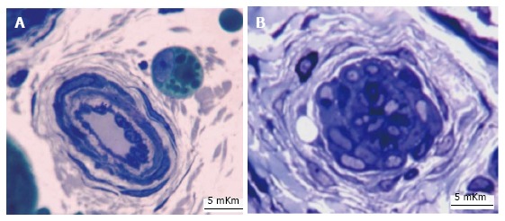
In two cases (one for each group) massive nerve fibers degeneration (more than 40% of nerve fibers with signs of demyelination, axonal or Wallerian degeneration) was revealed in Pn on the background of epineurial vessels obliteration or closing - presented in Figure 2. In all the rest animals of M-group the shares of degenerated myelin fibers overcame the corresponding indexes of intact nerves and of nerves in A-group at all t-points of experiment - presented in Tables 5 and 6. The shares of degenerated myelinated nerve fibers in Tn of A-group at F30 and WA30 were even smaller than in intact nerves.
Figure 2.

Fragments of canine Pn semithin sections. WA30, М-group. Toluidine blue stain. A: Normal condition of artery in epineurium; B: Epineural artery with closed lumen. Magnification 1250 ×.
Table 5.
Average morphometric parameters of tibial myelinated nerve fibers (mean ± SE)
| Parameters/group and t-points | Share of degenerated nerve fibers (%) | Nerve fibers diameters (mKm) | Axonal diameters (mKm) | Myelin sheath thickness (mKm) |
| Intact control | 1.6 ± 0.2 | 6.75 ± 0.01 | 4.63 ± 0.13 | 1.06 ± 0.02 |
| M-group - manual distraction (1 mm/d in 4 steps) | ||||
| 28 d of distraction | 5.0 ± 0.7a | 6.91 ± 0.17 | 4.57 ± 0.19 | 1.16a ± 0.01 |
| 30 d of fixation | 4.0 ± 0.9a | 6.77 ± 0.47 | 4.44 ± 0.28a | 1.17 ± 0.10 |
| 30 d without apparatus | 4.4 ± 2.4 | 6.61 ± 3.19 | 4.37 ± 2.10a | 1.12 ± 0.64 |
| A-group - automatic distraction (1 mm/d in 60 steps) | ||||
| 28 d of distraction | 2.4 ± 0.7b | 6.29 ± 0.13 | 4.63 ± 0.10 | 0.83 ± 0.02a |
| 30 d of fixation | 0.5 ± 0.2ac | 6.88 ± 0.11 | 4.99 ± 0.10a | 0.95 ± 0.12 |
| 30 d without apparatus | 0.9 ± 0.1ac | 7.26 ± 0.13a | 5.05 ± 0.09a | 1.11 ± 0.05 |
P < 0.05 vs intact;
P < 0.05 vs group M.
Table 6.
Average morphometric parameters of peroneal myelinated nerve fibers (mean ± SE)
| Parameters/group and t-points | Share of degenerated nerve fibers (%) | Nerve fibers diameters (mKm) | Axonal diameters (mKm) | Myelin sheath thickness (mKm) |
| Intact control | 1.9 ± 0.3 | 6.46 ± 0.07 | 4.39 ± 0.08 | 1.04 ± 0.04 |
| M-group - manual distraction (1 mm/d in 4 steps) | ||||
| 28 d of distraction | 6.0 ± 1.4a | 5.37 a ± 0.41 | 3.69 ± 0.29a | 0.84 ± 0.09a |
| 30 d of fixation | 4.3 ± 1.3a | 6.09 ± 0.63 | 4.44 ± 0.57 | 0.98 ± 0.06 |
| 30 d without apparatus | 4.2 ± 0.4a | 5.90 ± 0.43 | 4.10 ± 0.10a | 0.90 ± 0.17a |
| A-group - automatic distraction (1 mm/d in 60 steps) | ||||
| 28 d of distraction | 4.0 ± 0.8ac | 5.56 ± 0.26a | 3.70 ± 0.53a | 0.92 ± 0.13 |
| 30 d of fixation | 3.3 ± 0.1ac | 5.62 ± 0.07a | 3.91 ± 0.09a | 0.85 a ± 0.07 |
| 30 d without apparatus | 2.4 ± 0.6c | 6.17 ± 0.45 | 4.14 ± 0.16 | 1.01 ± 0.06 |
P < 0.05 vs intact;
P < 0.05 vs group M.
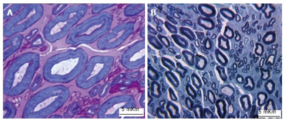
In comparison with intact nerves the average axonal diameter of Tn myelinated nerve fibers in M-group was decreased, the average myelin thickness increased. These changes are consistent with findings of myelinated fibers with signs of axonal atrophy and hypermyelination - presented in Figure 3A. In A-group the average axonal diameter in Tn at D28 was comparable with intact nerve, but the average myelin thickness was decreased. At subsequent t-points the average axonal diameter was bigger than in intact Tn nerve, the average myelin thickness was restored, although some nerve fibers were hypomyelinated at the end of experiment - presented in Figure 3B. In Pn the average axonal diameter was decreased in both groups at D28 and F30, but at the end of experiment this parameter didn’t significantly differed from intact nerve (Table 6). Restoration of all morphometric indices was evident only in Group A. In Group M the average diameter of nerve fibres and the average myelin thickness remained decreased.
Figure 3.

Fragments of canine Tn semithin sections, WA30. A: Some large myelinated nerve fibres has visibly thinned axons and thickened myelin sheaths, М-group, methylene blue - basic fuchsin stain, magnification 1250 ×; B: Two large nerve fibers (in the lower part of the image) are hypomyelinated; А-group, toluidine blue stain, magnification 500 ×.
Articular cartilage changes
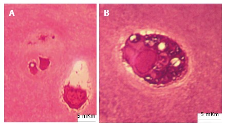
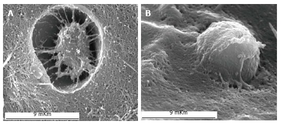
Alteration of morphometric parameters and mineral contents developed in both groups - more prominently in M-group. At D28 injuries of superficial cartilage zone were revealed - presented in Figure 4. More intensive separation of collagen network with usuras formation was noted in M-group. At F30 some chondrocytes of intermediate zone were with signs of apoptosis or necrotic death ranged mainly in M-group - presented in Figure 5A. In A-group many of chondrocytes were functionally active. They occupied the whole lacuna, had homogenic nuclei and well developed vacuolated cytoplasm - example of such cell presented in Figure 5B. Chondrocytes shrinkage and rounding of functionally active chondrocytes were also revealed by SEM - presented in Figure 6. At WA30 restoration of cellular cartilage architectonics was noted only in A-group. Cartilage thickness changes were divergent in studied groups - presented in Table 7. In M-group parameter increased at D28 and decreased at F30 and WA30 - compared with intact control. In A-group the thickness of cartilage decreased at D28 and at F30, but recovered at WA30. In both groups chondrocytes with signs of destruction were revealed mainly in superficial and deep cartilage zones. Maximal indices of empty lacunas and utmost decrease of chondrocytes volumetric densities were noted in M-group. Compensatory increase of isogenic clusters index was more prominent in A-group. In all experimental animals ωСа increase and ωS decrease were marked - more prominently in M-group presented in Table 8.
Figure 4.

Superficial zone of articular cartilage at D28 in semithin sections. A: М-group; B: A-group. Methylene blue - basic fuchsin stain. Magnification 500 ×.
Figure 5.

Chondrocytes of intermediate cartilage zone at F30. А: In the left lacuna there are two degenerated chondrocytes, in the right lacuna - chondrocyte shrinkage, chromatin condensed on periphery of caryolemma, М-group; B: Chondrocyte with well-developed functionally active vacuolated cytoplasm occupies the whole lacuna. A-group. Semithin sections. Methylene blue - basic fuchsin stain. Magnification 1250 ×.
Figure 6.

Chondrocytes in SEM at F30. А: Cellular shrinkage. М-group; B: Rounded cell. A-group. Magnification 5500.
Table 7.
Average morphometric parameters of articular cartilage (mean ± SD)
| Parameters/group and t-points | Cartilage thickness (mKm) | Chondrocytes volumetric density (%) |
Isogenic groups |
Empty lacunas |
| (% of chondrocyte sample) | ||||
| Intact control | 475.5 ± 1.31 | 9.03 ± 1.51 | 14.5 | 13.6 |
| M-group - manual distraction (1 mm/d in 4 steps) | ||||
| 28 d of distraction | 710.3 ± 7.16a | 4.13 ± 0.28a | 24.7 | 29.97 |
| 30 d of fixation | 421.1 ± 4.81a | 5.1 ± 0.27a | 20.18 | 20.85 |
| 30 d without apparatus | 416.9 ± 4.37a | 5.6 ± 0.19a | 16.1 | 21.12 |
| A-group - automatic distraction (1 mm/d in 60 steps) | ||||
| 28 d of distraction | 356.45 ± 1.55ac | 8.34 ± 0.48ac | 25.6 | 24.3 |
| 30 d of fixation | 392.7 ± 2.12ac | 6.8 ± 0.45ac | 28.8 | 16.3 |
| 30 d without apparatus | 464.6 ± 6.51c | 7.96 ± 0.37ac | 29.9 | 15.4 |
P < 0.05 vs intact;
P < 0.05 vs group M.
Table 8.
Content of sulfur and calcium in articular cartilage (mean ± SD, weight %)
| Parameters/group and t-points | ωS | ωCa |
| Intact control | 1.26 ± 0.02 | 0.15 ± 0.02 |
| M-group - manual distraction (1 mm/d in 4 steps) | ||
| 28 d of distraction | 0.71 ± 0.01a | 0.19 ± 0.02a |
| 30 d of fixation | 0.96 ± 0.02a | 0.39 ± 0.03a |
| 30 d without apparatus | 0.94 ± 0.02a | 0.27 ± 0.02a |
| A-group - automatic distraction (1 mm/d in 60 steps) | ||
| 28 d of distraction | 0.79 ± 0.01a | 0.16 ± 0.01c |
| 30 d of fixation | 1.09 ± 0.02ac | 0.18 ± 0.02c |
| 30 d without apparatus | 1.15 ± 0.02c | 0.20 ± 0.02ac |
P < 0.05 vs intact;
P < 0.05 vs group M.
DISCUSSION
So, better restoration of M-response amplitudes in leg muscles in group with automatic distraction was consistent with less structural alterations of nerves and articular cartilage. Adaptive growing processes in nerve sheaths were marked with ki-67-positive cells in both groups but probably the smaller incremental length (0, 017 mm in group with automatic distraction instead of 0, 25 mm in group with manual distraction) was associated with fewer disturbances in nerves sheaths. Subperineurial edema and fibrosis were evident only in group with manual distraction and restoration of nerve fascicles summary area was achieved only in group with automatic distraction. Per cents of degenerated nerve fibers were smaller in group with automatic distraction than in group with manual distraction at all time points of experiment. Being expressed in per cents this difference seems to be rather small, but in absolute figures it means that in group with automatic distraction thousands neurons don’t lose their connections with periphery and survive. Thousands neurons with degenerated axial cylinders in M-Group enter into regenerative status and create new outgrowths but such active changes may lead to death many of them. As for morphometric parameters of survived myelinated nerve fibers population, in peroneal nerve all of them were restored at the end of experiment only in group with automatic distraction. Automatic distraction prevented axonal atrophy and absolute myelin thickening in tibial nerve. Such changes were evident in group with manual distraction and even in conditions without distraction - after experimental shin bone fracture[14]. Increased axonal diameters in tibial nerves in group with automatic distraction were associated with better nerve fibers survival because shares of degenerated nerve fibers were smaller even in comparison with intact nerves. Limitation of our study - we have not study endoneurial circulation and axonal transport with special methods. But restoration of fascicular areas, smaller per cents of degenerated nerve fibers and bigger axonal diameters in group with automatic distraction bear indirect evidence that more discrete (high frequent) mode of automatic distraction resulted in fewer disturbances of endoneurial fluid and axoplasmic flow.
Articular cartilage alterations also depended on distraction frequency. All quantitative parameters (cartilage thickness, volumetric densities of chondrocytes, percentages of isogenic clusters and empty cellular lacunas, contents of sulfur and calcium) were less changed in group with automatic distraction.
And thus, automated distraction developed by Ilizarov (1 mm/d in 60 steps) is more advantageous because few alterations of nerves and cartilage structure than in manual mode (1 mm/d in 4 steps) provide better initial functional recovery and better functional prognosis.
ACKOWLEDGMENTS
The authors would like to thank the members of the engineering group for their technical support and Gaidyshev IP for his help in statistical analysis.
COMMENTS
Background
Comparative study of the effects of manual low-frequent and automated high-frequent distraction on the results of orthopedic limb lengthening at clinics is problematic. The safety of automatic Ilizarov distraction for nerve, cartilage and muscles changes in experimental animal leg lengthening was substantiated in Russian articles but careful multiparametric comparative analysis has not been done.
Research frontiers
It was stated that for a definite rate of distraction the higher frequent distraction improve bone formation and consolidation. Previous experimental researches have proved that automatic distraction is safer for nerves ultrastructures and cartilage relief than manual.
Innovations and breakthroughs
Special technology of tissue semi thin sections of enlarged shear provided representative histomorphometric data. This is the first study evaluating substantial difference of peroneal and tibial nerves adaptability to conditions of orthopedic leg lengthening. Complex research of articular cartilage histomorphometry, scanning electron microscopy and X-ray electron probe microanalysis revealed fewer alterations in group with automatic distraction.
Applications
Better nerve fibers survival and less arthrotic cartilage changes in conditions of automatic distraction are critically important for good functional prognosis of orthopedic leg lengthening. Development of axonal atrophy and degenerative myelin sheath thickening in tibial nerve fibers evident in group with manual distraction was effectively prevented in group with automatic distraction.
Terminology
Ilizarov method is one of the most clinically implemented distraction osteogenesis procedures. Ilizarov discovered that gradual tension stress maintain the regeneration and growth of living tissues. He was the first who developed the automated high frequent distraction. Different tissue responses on distraction with various frequencies may be revealed in standard experimental models, when groups differ in only one experimental condition - the frequency of distraction. Subtle differences in tissue structures may be of critical importance for functional prognosis.
Peer-review
The topic is interesting and the study may have a point.
Footnotes
Supported by Russian Foundation for Basic Research, No.14-4 4-00010.
Institutional review board statement: The article was reviewed by RISC “RTO” Review Board and statement including: (1) The manuscript is not simultaneously being considered by other journals or already published elsewhere; (2) The manuscript has no redundant publication, plagiarism, or data fabrication or falsification; (3) Experiments involving animal subjects were designed and performed in compliance with the relevant laws regarding the animal care and use of subjects; and (4) Material contained within the manuscript is original, with all information from other sources appropriately referenced.
Institutional animal care and use committee statement: All animal studies are approved by RISC “RTO” Ethical Committee - exerpts from the minutes #4 (50) under date of December 13, 2016.
Conflict-of-interest statement: The authors declare no conflict of interest.
Data sharing statement: No additional data are available.
Manuscript source: Invited manuscript
Specialty type: Orthopedics
Country of origin: Russia
Peer-review report classification
Grade A (Excellent): 0
Grade B (Very good): B
Grade C (Good): C
Grade D (Fair): D
Grade E (Poor): 0
Peer-review started: February 7, 2017
First decision: July 10, 2017
Article in press: August 3, 2017
P- Reviewer: Drampalos E, Fernandez-Fairen M, Mavrogenis AF S- Editor: Ji FF L- Editor: A E- Editor: Lu YJ
Contributor Information
Nathalia Shchudlo, Laboratory of Morphology FSBI, Russian Ilizarov Scientific Center “Restorative Traumatology and Orthopaedics”, 640014 Kurgan, Russia. nshchudlo@mail.ru.
Tatyana Varsegova, Laboratory of Morphology FSBI, Russian Ilizarov Scientific Center “Restorative Traumatology and Orthopaedics”, 640014 Kurgan, Russia.
Tatyana Stupina, Laboratory of Morphology FSBI, Russian Ilizarov Scientific Center “Restorative Traumatology and Orthopaedics”, 640014 Kurgan, Russia.
Michael Shchudlo, Laboratory of Morphology FSBI, Russian Ilizarov Scientific Center “Restorative Traumatology and Orthopaedics”, 640014 Kurgan, Russia.
Marat Saifutdinov, Laboratory of Morphology FSBI, Russian Ilizarov Scientific Center “Restorative Traumatology and Orthopaedics”, 640014 Kurgan, Russia.
Andrey Yemanov, Laboratory of Morphology FSBI, Russian Ilizarov Scientific Center “Restorative Traumatology and Orthopaedics”, 640014 Kurgan, Russia.
References
- 1.Ilizarov GA. The tension-stress effect on the genesis and growth of tissues: Part II. The influence of the rate and frequency of distraction. Clin Orthop Relat Res. 1989;239:263–285. [PubMed] [Google Scholar]
- 2.Shevtsov V, Popkov A, Popkov D, Prévot J. [Reduction of the period of treatment for leg lengthening. Technique and advantages] Rev Chir Orthop Reparatrice Appar Mot. 2001;87:248–256. [PubMed] [Google Scholar]
- 3.Nakamura E, Mizuta H, Takagi K. Knee cartilage injury after tibial lengthening. Radiographic and histological studies in rabbits after 3-6 months. Acta Orthop Scand. 1995;66:313–316. doi: 10.3109/17453679508995551. [DOI] [PubMed] [Google Scholar]
- 4.Makarov MR, Kochutina LN, Samchukov ML, Birch JG, Welch RD. Effect of rhythm and level of distraction on muscle structure: an animal study. Clin Orthop Relat Res. 2001;384:250–264. doi: 10.1097/00003086-200103000-00030. [DOI] [PubMed] [Google Scholar]
- 5.Shilt JS, Deeney VF, Quinn CO. The effect of increased distraction frequency on soft tissues during limb lengthening in an animal model. J Pediatr Orthop. 2000;20:146–150. [PubMed] [Google Scholar]
- 6.Aarnes GT, Steen H, Ludvigsen P, Kristiansen LP, Reikerås O. High frequency distraction improves tissue adaptation during leg lengthening in humans. J Orthop Res. 2002;20:789–792. doi: 10.1016/S0736-0266(01)00175-9. [DOI] [PubMed] [Google Scholar]
- 7.Bright AS, Herzenberg JE, Paley D, Weiner I, Burghardt RD. Preliminary experience with motorized distraction for tibial lengthening. Strategies Trauma Limb Reconstr. 2014;9:97–100. doi: 10.1007/s11751-014-0191-1. [DOI] [PMC free article] [PubMed] [Google Scholar]
- 8.Shevtsov VI, Shchudlo NA, Shchudlo MM, Filimonova GN. Structural adaptability and plasticity of skeletal muscles for limb lengthening. Genius of Orthopedics. 2009;4:39–47. [Google Scholar]
- 9.Shchudlo MM, Shchudlo NA, Varsegova TN, Borisova IV. Reaction of nerves to stretching and their structural adaptation to limb lengthening. Genius of Orthopedics. 2009;4:48–55. [Google Scholar]
- 10.Stupina TA, Shchudlo MM. Evaluation of articular cartilage regenerative potentials under different conditions of experimental lengthening of adjacent limb segment. Genius of Orthopedics. 2010;3:84–88. [Google Scholar]
- 11.Stupina TA, Shchudlo MM. Structural adaptability and articular cartilage reparative potentials depending on the conditions of limb adjacent segment lengthening. Traumatology and Orthopedics of Russia. 2011;4:62–68. [Google Scholar]
- 12.Stupina ТА, Shchudlo ММ. Method for preparing samples of non-decalcified articular cartilage with subjacent subchondral bone for multipurpose study. Russia patent RF 2466375. 2012
- 13.Silanteva ТА, Gorbach ЕN, Irjanov JМ, Stupina ТА, Varsegova ТN. Method of preparing biological tissue samples for analysis in scanning electron microscope. Russia patent RF 2397472. 2010
- 14.Varsegova TN, Shchudlo NA, Shchudlo MM, Saifutdinov MS, Stepanov MA. The effects of tibial fracture and Ilizarov osteosynthesis on the structural reorganization of sciatic and tibial nerves during the bone consolidation phase and after fixator removal. Strategies Trauma Limb Reconstr. 2015;10:87–94. doi: 10.1007/s11751-015-0227-1. [DOI] [PMC free article] [PubMed] [Google Scholar]


