Abstract
Sixty-eight cefotaxime-resistant Escherichia coli isolates were recovered from different water environments in Northern Thailand. Isolates were mostly resistant to ceftazidime and aztreonam (>90%). The most common extended-spectrum β-lactamase-encoding gene was blaCTX-M-group 1 (75%) followed by blaCTX-M-group 9 (13.2%). The co-existence of blaCTX-M and AmpC-type β-lactamase genes was detected in 4 isolates (5.9%). Two E. coli isolates carrying blaCTX-M from canal and river water samples belonged to the phylogenetic group B2-ST131, which is known to be pathogenic. This is the first study on blaCTX-M and blaCMY-2-carrying E. coli and the emergence of ST131 from water environments in Thailand.
Keywords: Escherichia coli, blaCTX-M, blaCMY-2, water, ST131
Escherichia coli is a normal inhabitant of the intestinal tract of humans and various animals. Although some strains appear to be harmless, several pathogenic strains are regarded as important causes of community-associated urinary tract, gastrointestinal, and systemic infections in humans (9). The presence of E. coli strains in water environments suggests fecal contamination and poses a public health risk. The dissemination of antibiotic-resistant E. coli in different water environments is a serious public health issue worldwide, particularly in the Indochina region, including Thailand, in which sanitary services may be inadequate (15). Individuals may be infected by resistant bacteria from water environments, resulting in serious infections. Similar genotypes between antibiotic-resistant E. coli from water environments and human clinical isolates suggest the spread of these organisms to humans (5).
Extended-spectrum β-lactamases (ESBL), which mediate resistance to third generation cephalosporins and monobactams, are widely disseminated among Gram-negative bacteria from hospital settings and communities. Several types of ESBL have been reported, such as TEM and SHV derivatives, which evolved from a mutation in parental β-lactamase TEM-1 and -2 and SHV-1, respectively. Another type of ESBL, CTX-M β-lactamase, has become the most common ESBL found in Enterobacteriaceae. ESBLs have extended their ability to hydrolyze cefotaxime, ceftazidime, ceftriaxone, and aztreonam and are inhibited by β-lactamase inhibitors (clavulanic acid, tazobactam, and sulbactam) (13). Resistance to third generation cephalosporins may be caused by the production of AmpC β-lactamase, with CMY-2 being the most prevalent (13). Cephalosporin resistance mediated by the AmpC enzyme includes mutations within regulatory regions, resulting in the permanently strong expression of AmpC and acquisition of a transferable plasmid-mediated AmpC β-lactamase (13).
To date, data on antibiotic-resistant E. coli, particularly those producing ESBL and AmpC, have frequently been obtained from various water environments, such as rivers, lakes, wastewater, tap water, and drinking water, in different regions across continents, but are more abundant from developing countries (1, 12). ESBL- and AmpC-carrying E. coli isolates have been widely disseminated in Thailand for many years (7, 14). Several ESBL- and AmpC-encoding genes have been identified; however, most studies have primarily focused on clinical isolates. Information on antibiotic-resistant E. coli from non-hospital sources, particularly water environments, is limited. A previous study in Thailand revealed the presence of ESBL-producing Enterobacteriaceae in different water environments; however, the resistant determinants and the genotypes of resistant isolates have not been reported (2). The carriage rate of these organisms among the healthy Thai population was reported to be high (2). Furthermore, approximately 22% of Swedish travelers returning from Thailand developed diarrhea caused by ESBL-producing E. coli (17). Hence, ESBL-producing E. coli may be present in various environments in Thailand. This study was conducted in order to clarify whether water environments in Thailand serve as reservoirs for antibiotic-resistant E. coli, particularly those producing ESBL and AmpC.
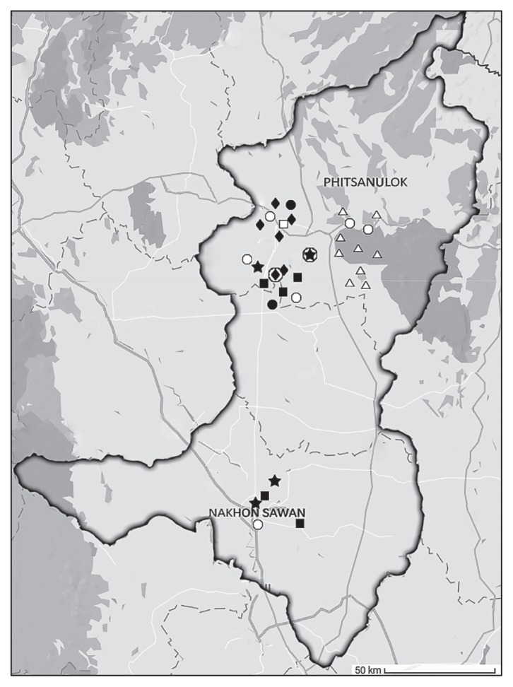
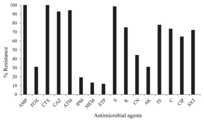
This study was conducted in the Phitsanulok and Nakhon Sawan provinces, two fast-growing cities in Northern Thailand. These areas have a relatively high population density and are regarded as active regions for livestock and aquaculture farming. Municipal water, mainly obtained from water resources within these cities, is used as a household water supply. Between August 2013 and January 2014, water samples were collected from 33 different water resources in the study area (river, 4; pond, 6; canal, 6; tap water, 8 and groundwater, 9). Sampling sites were shown in Fig. 1. Water samples were collected from 30 cm below the water surface using sterile bottles (500 mL bottle−1, 3 bottles sampling site−1). Groundwater was obtained by pumping water from drilling wells, while tap water was collected directly from a tap; approximately the first 500 mL was discarded in each case. All samples were kept on ice, transported to the laboratory, and processed within 24 h of collection. Ten milliliters of water samples were enriched in 90 mL EE broth (Becton, Dickinson and Company, NJ, USA) at 37°C for 24 h. Enrichment cultures were then 10-fold serially diluted, spread on MacConkey agar (Oxoid, Hampshire, UK) supplemented with 2 mg L−1 cefotaxime (Sigma-Aldrich, MO, USA), and incubated at 37°C for 24–48 h. Approximately 5–10 colonies with a typical E. coli morphology per sample were picked and sub-cultured on Tryptic Soy agar (Oxoid). In order to avoid working with clones of the same E. coli strain, an enterobacterial repetitive intergenic consensus polymerase chain reaction (ERIC-PCR) was performed as described previously (18). Isolates were considered to belong to the same clone if they shared the same ERIC-PCR pattern. Isolates representing distinct DNA patterns were selected for subsequent studies. Species identification was performed using standard biochemical tests (Gram staining, growth on EMB, oxidase test) and 16S rRNA gene sequencing (8). Among 33 water resources, cefotaxime-resistant E. coli isolates were found in samples from different rivers (n=4), ponds (n=5), and canals (n=6) (Fig. 1, Table 1). Two tap water samples yielded cefotaxime-resistant isolates, while no isolates were detected from groundwater samples. Sixty-eight isolates were recovered and antimicrobial susceptibility testing was performed using the disk diffusion method, as recommended by the Clinical and Laboratory Standards Institute (4). An isolate was defined as being multidrug resistance (MDR) if it was resistant to three or more classes of antimicrobial agents. Most isolates were resistant to ampicillin, cefotaxime, ceftazidime, aztreonam, and streptomycin (>90%) (Fig. 2, Table S1). High rates of resistance to kanamycin, tetracycline, chloramphenicol, ciprofloxacin, and trimethoprim-sulfamethoxazole (>60%) were also noted. It is a matter of concern that all 68 E. coli isolates showed the MDR phenotype and 58.8% of isolates exhibited resistance to up to ten or more of the antimicrobial agents tested. The Minimum Inhibitory Concentrations (MICs) of the selected antibiotics were assessed using the broth microdilution method according to CLSI guidelines (4). High-level resistance to cefotaxime (MIC90>128 mg L−1), ceftazidime (MIC90=128 mg L−1), and ciprofloxacin (MIC90=32 mg L−1) was noted. In contrast, low MIC90 values were observed for imipenem (4 mg L−1) and meropenem (2 mg L−1).
Fig. 1.
Location of water sampling. Black and white symbols represented sampling sites that were positive and negative for cefotaxime-resistant E. coli, respectively. Star, river; square, pond; diamond, canal; circle, tap water; triangle, groundwater. Circled diamond and star represent water environments that were positive for blaCTX-M-14- and blaCTX-M-27-carrying E. coli ST131, respectively.
Table 1.
Occurrence of cefotaxime-resistant E. coli from various water environments and the presence of β-lactamase genes.
| Different water environments (No. of sampling sites where E. coli were isolates) | No. of E. coli isolates (n=68) | β-lactamase genes | ||||||
|---|---|---|---|---|---|---|---|---|
|
| ||||||||
| none | blaCTX-M- 1 | blaCTX-M- 1+ blaTEM-1 | blaCTX-M- 1+ blaCMY-2 | blaCTX-M- 9 | blaCTX-M- 9+ blaTEM-1 | blaCTX-M- 9+ blaCMY-2 | ||
| River (n=4) | 20 | 2 | 3 | 9 | 0 | 3 | 1 | 2 |
| Pond (n=5) | 16 | 2 | 4 | 9 | 1 | 0 | 0 | 0 |
| Canal (n=6) | 26 | 4 | 1 | 18 | 1 | 2 | 0 | 0 |
| Tap water (n=2) | 6 | 0 | 1 | 4 | 0 | 1 | 0 | 0 |
| Total | 68 | 8 | 9 | 40 | 2 | 6 | 1 | 2 |
Fig. 2.
Occurrence ratio (%) of antimicrobial-resistant E. coli isolates. Antimicrobial resistance was assessed using the disk diffusion method according to the CLSI guidelines (4). AMP, ampicillin; FOX, cefoxitin; CTX, cefotaxime; CAZ, ceftazidime; ATM, aztreonam; IPM, imipenem; MEM, meropenem; ETP, ertapenem; S, streptomycin; K, kanamycin; CN, gentamicin; AK, amikacin; TE, tetracycline; C, chloramphenicol; CIP, ciprofloxacin, and SXT, trimethoprim-sulfamethoxazole.
The detection of genes coding for ESBL (blaTEM, blaSHV, and blaCTX-M series) and AmpC (MOX, CIT, DHA, ACC, EBC, and FOX types) was performed by multiplex PCR using specific primers and conditions as previously described (6, 10, 19). Nucleotide sequences were analyzed with software available from the National Center for Biotechnology Information website (http://www.ncbi.nlm.nih.gov) (Table S1). In this study, as many as 88.2% (60/68) of cefotaxime-resistant E. coli isolates carried genes encoding for ESBL and AmpC (Table 1). Fifty-one (75%, 51/68) and 9 isolates (13.2%, 9/68) carried blaCTX-M-group 1 and blaCTX-M-group 9, respectively. The results of the sequence analysis revealed that the most common blaCTX-M-group 1 was blaCTX-M-55 (74.5%, 38/51) followed by blaCTX-M-15 (25.5%, 13/51). blaCTX-M encoding for CTX-M-14 was found in all blaCTX-M-group 9-carrying isolates. These results were consistent with blaCTX-M-14, blaCTX-M-15, and blaCTX-M-55 being detected worldwide and commonly being found in Thai patients (7, 14). No blaTEM or blaSHV-related ESBL genes were detected. However, the broad-spectrum β-lactamase gene, blaTEM-1, was found in 41 blaCTX-M-carrying isolates. Although blaTEM-1 is considered to be a non-ESBL, the hyperproduction of TEM-1 with a change in outer membrane proteins may result in reduced susceptibility to cefotaxime (20). Furthermore, 4 cefotaxime-resistant E. coli isolates (4/68, 5.9%) were positive for CIT-type AmpC genes. The results of the sequence analysis showed that all were blaCMY-2, which was found in combination with blaCTX-M. The emergence of carbapenem-resistant E. coli from water environments was observed (Fig. 2). The detection of carbapenemase genes (blaIMP, blaVIM, blaNDM, blaKPC, and blaOXA) by multiplex PCR was performed (11); however, the results obtained were negative. Carbapenem resistance may be due to the production of other carbapenemases, alterations in outer membrane proteins, and efflux pump overexpression (13).
The 68 cefotaxime-resistant E. coli isolates were classified into phylogenetic groups A, B1, B2, or D by multiplex PCR of the genes chuA and yjaA and the DNA fragment TspE4C2, as previously described (3). These results showed that most isolates belonged to the commensal groups B1 (55.9%, 38/68) and A (36.8%, 25/68). A small number of isolates belonged to the pathogenic groups B2 (2.9%, 2/68) and D (4.4%, 3/68) (Table S1). The recent global spread of the multi-resistant and highly virulent extraintestinal E. coli B2-sequence type (ST) 131 has been observed (9). We performed the MLST analysis by the amplification and sequencing of 7 housekeeping genes (adk, fumC, gyrB, icd, mdh, purA, and recA) according to the protocols on the E. coli MLST website (http://mlst.warwick.ac.uk/mlst/dbs/Ecoli). The results obtained showed that 2 E. coli isolates belonging to group B2 were identified as E. coli ST131. Both isolates showed resistance to the third generation cephalosporins, aztreonam and ciprofloxacin, and carried blaCTX-M. The amplification and sequence analysis of full-length blaCTX-M revealed that one isolate carried blaCTX-M-14, while the other carried blaCTX-M-27.
The 2 E. coli ST131 isolates were typed by pulsed field gel electrophoresis (PFGE). The preparation of chromosomal DNA in agarose plugs and digestion with XbaI (Fermentas, NY, USA) were performed as described previously (21). DNA was electrophoresed through 1% Pulsed Field Certified agarose in 0.5×TBE (Tris–borate–EDTA) buffer (CHEF Mapper XA System, Bio-Rad Laboratories, CA, USA). Banding patterns were interpreted by visual inspection. We observed that both ST131 isolates were closely related (<3 band differences) according to the criteria defined by Tenover et al. (16) (Fig. 3). These results suggested that the 2 ST131 isolates originated from the same clone even though they were obtained from distantly related water resources (approximately 20 km) (Fig. 1). One ST131 isolate (blaCTX-M-14-positive) was recovered from a canal outside the city, near to which many agricultural and farming activities were being conducted, while the other (blaCTX-M-27-positive) was obtained from a river located in the residential area within the city. To date, the presence of E. coli ST131 in humans, animals, and food products has been documented; however, limited information is currently available on the prevalence of ST131 in water environments (9). Previous studies in European countries identified ST131 in water environments (5, 22). However, studies in Asian countries, such as India and Bangladesh, in which ESBL-producing E. coli isolates from water environments were studied, did not identify ST131 (1, 12). Our results suggested that water environments in Thailand contained virulent and MDR E. coli that have the potential to cause serious infections.
Fig. 3.

PFGE profiles of blaCTX-M-carrying E. coli ST131 isolated from different water environments in Phitsanulok province. M, Saccharomyces cerevisiae chromosomal DNA (Bio-Rad); lane 1, blaCTX-M-14-carrying E. coli ST131 isolated from a canal outside the city; lane 2, blaCTX-M-27-carrying E. coli ST131 isolated from a river within the city.
Our results of MDR E. coli carrying ESBL and AmpC from different water environments were similar to those reported from other regions, even in countries that have good water supply and sanitation services, such as Switzerland (22). These results were not unexpected because of the inappropriate use of antimicrobial agents for chemotherapy, livestock farming, and aquaculture (15). Moreover, antibiotics are easily obtained without a prescription in Thailand. Antimicrobial-resistant bacteria from human activities may easily be released into water environments and subsequently lead to the dissemination of these organisms within the community.
In conclusion, this is the first study on blaCTX-M- and blaCMY-2-carrying E. coli from different water environments in Thailand. The matter of most concern is the emergence of blaCTX-M-carrying E. coli ST131 due to its ability to cause severe infections. Our results have important implications for the health of individuals living in the community. Furthermore, these results highlight the need for the more extensive surveillance of water environments in Thailand.
The nucleotide sequences of blaCTX-M and blaCMY-2 reported in this study were deposited in the GenBank database under the accession numbers MF374734–MF374735, MF385034–MF385037, MF406113–MF406132, and MF422526.
Supplementary Material
Acknowledgements
This work was funded by Naresuan University (R2560B139). UT was supported by the Royal Golden Jubilee (RGJ)-PhD Program from the Thailand Research Fund (TRF) and Rajamangala University of Technology Lanna (PHD/0054/2555). AK was supported by the RGJ-Ph.D program from the TRF and Naresuan University (PHD/0181/2557). KA, PT, and PP were supported by the Faculty of Medical Science, Naresuan University.
References
- 1.Bajaj P., Singh N.S., Kanaujia P.K., Virdi J.S. Distribution and molecular characterization of genes encoding CTX-M and AmpC β-lactamases in Escherichia coli isolated from an Indian urban aquatic environment. Sci Total Environ. 2015;505:350–356. doi: 10.1016/j.scitotenv.2014.09.084. [DOI] [PubMed] [Google Scholar]
- 2.Boonyasiri A., Tangkoskul T., Seenama C., Saiyarin J., Tiengrim S., Thamlikitkul V. Prevalence of antibiotic resistant bacteria in healthy adults, foods, food animals, and the environment in selected areas in Thailand. Pathog Glob Health. 2014;108:235–245. doi: 10.1179/2047773214Y.0000000148. [DOI] [PMC free article] [PubMed] [Google Scholar]
- 3.Clermont O., Bonacorsi S., Bingen E. Rapid and simple determination of the Escherichia coli phylogenetic group. Appl Environ Microbiol. 2000;66:4555–4558. doi: 10.1128/aem.66.10.4555-4558.2000. [DOI] [PMC free article] [PubMed] [Google Scholar]
- 4.Cockerill F.R., Patel J.B., Alder J., et al. Performance standards for antimicrobial susceptibility testing; twenty-third informational supplement. Clinical and Laboratory Standards Institute; Wayne, PA: 2013. [Google Scholar]
- 5.Colomer-Lluch M., Mora A., López C., et al. Detection of quinolone-resistant Escherichia coli isolates belonging to clonal groups O25b:H4-B2-ST131 and O25b:H4-D-ST69 in raw sewage and river water in Barcelona, Spain. J Antimicrob Chemother. 2013;68:758–765. doi: 10.1093/jac/dks477. [DOI] [PubMed] [Google Scholar]
- 6.Dallenne C., Da Costa A., Decré D., Favier C., Arlet G. Development of a set of multiplex PCR assays for the detection of genes encoding important β-lactamases in Enterobacteriaceae. J Antimicrob Chemother. 2010;65:490–495. doi: 10.1093/jac/dkp498. [DOI] [PubMed] [Google Scholar]
- 7.Kiratisin P., Apisarnthanarak A., Laesripa C., Saifon P. Molecular characterization and epidemiology of extended-spectrum β-lactamase-producing Escherichia coli and Klebsiella pneumoniae isolates causing health care-associated infection in Thailand, where the CTX-M family is endemic. Antimicrob Agents Chemother. 2008;52:2818–2824. doi: 10.1128/AAC.00171-08. [DOI] [PMC free article] [PubMed] [Google Scholar]
- 8.Lane D.J. 16S/23S rRNA sequencing. In: Stackebrandt E., Goodfellow M., editors. Nucleic Acid Techniques in Bacterial Systematics. John Wiley and Sons; United Kingdom: 1991. pp. 115–175. [Google Scholar]
- 9.Nicolas-Chanoine M.H., Bertrand X., Madec J.Y. Escherichia coli ST131, an intriguing clonal group. Clin Microbiol Rev. 2014;27:543–574. doi: 10.1128/CMR.00125-13. [DOI] [PMC free article] [PubMed] [Google Scholar]
- 10.Pérez-Pérez F.J., Hanson N.D. Detection of plasmid-mediated AmpC β-lactamase genes in clinical isolates by using multiplex PCR. J Clin Microbiol. 2002;40:2153–2162. doi: 10.1128/JCM.40.6.2153-2162.2002. [DOI] [PMC free article] [PubMed] [Google Scholar]
- 11.Poirel L., Walsh T.R., Cuvillier V., Nordmann P. Multiplex PCR for detection of acquired carbapenemase genes. Diagn Microbiol Infect Dis. 2011;70:119–123. doi: 10.1016/j.diagmicrobio.2010.12.002. [DOI] [PubMed] [Google Scholar]
- 12.Rashid M., Rakib M.M., Hasan B. Antimicrobial-resistant and ESBL-producing Escherichia coli in different ecological niches in Bangladesh. Infect Ecol Epidemiol. 2015;5:26712. doi: 10.3402/iee.v5.26712. [DOI] [PMC free article] [PubMed] [Google Scholar]
- 13.Ruppé É, Woerther P.L., Barbier F. Mechanisms of antimicrobial resistance in Gram-negative bacilli. Ann Intensive Care. 2015;5:61. doi: 10.1186/s13613-015-0061-0. [DOI] [PMC free article] [PubMed] [Google Scholar]
- 14.Singtohin S., Chanawong A., Lulitanond A., Sribenjalux P., Auncharoen A., Kaewkes W., Songsri J., Pienthaweechai K. CMY-2, CMY-8b, and DHA-1 plasmid-mediated AmpC β-lactamases among clinical isolates of Escherichia coli and Klebsiella pneumoniae from a university hospital, Thailand. Diagn Microbiol Infect Dis. 2010;68:271–277. doi: 10.1016/j.diagmicrobio.2010.06.014. [DOI] [PubMed] [Google Scholar]
- 15.Suzuki S., Hoa P.T. Distribution of quinolones, sulfonamides, tetracyclines in aquatic environment and antibiotic resistance in indochina. Front Microbiol. 2012;3:67. doi: 10.3389/fmicb.2012.00067. [DOI] [PMC free article] [PubMed] [Google Scholar]
- 16.Tenover F.C., Arbeit R.D., Goering R.V., Mickelsen P.A., Murray B.E., Persing D.H., Swaminathan B. Interpreting chromosomal DNA restriction patterns produced by pulsed field gel electrophoresis: criteria for bacterial strain typing. J Clin Microbiol. 1995;33:2233–2239. doi: 10.1128/jcm.33.9.2233-2239.1995. [DOI] [PMC free article] [PubMed] [Google Scholar]
- 17.Tham J., Odenholt I., Walder M., Brolund A., Ahl J., Melander E. Extended-spectrum β-lactamase-producing Escherichia coli in patients with travellers’ diarrhoea. Scand J Infect Dis. 2010;42:275–280. doi: 10.3109/00365540903493715. [DOI] [PubMed] [Google Scholar]
- 18.Versalovic J., Koeuth T., Lupski J.R. Distribution of repetitive DNA sequences in eubacteria and application to fingerprinting of bacterial genomes. Nucleic Acids Res. 1991;19:6823–6831. doi: 10.1093/nar/19.24.6823. [DOI] [PMC free article] [PubMed] [Google Scholar]
- 19.Woodford N., Fagan E.J., Ellington M.J. Multiplex PCR for rapid detection of genes encoding CTX-M extended-spectrum β-lactamases. J Antimicrob Chemother. 2006;57:154–155. doi: 10.1093/jac/dki412. [DOI] [PubMed] [Google Scholar]
- 20.Wu T.L., Siu L.K., Su L.H., Lauderdale T.L., Lin F.M., Leu H.S., Lin T.Y., Ho M. Outer membrane protein change combined with co-existing TEM-1 and SHV-1 β-lactamases lead to false identification of ESBL-producing Klebsiella pneumoniae. J Antimicrob Chemother. 2001;47:755–761. doi: 10.1093/jac/47.6.755. [DOI] [PubMed] [Google Scholar]
- 21.Xiong Z., Zhu D., Wang F., Zhang Y., Okamoto R., Inoue M. Investigation of extended-spectrum β-lactamase in Klebsiellae pneumoniae and Escherichia coli from China. Diagn Microbiol Infect Dis. 2002;44:195–200. doi: 10.1016/s0732-8893(02)00441-8. [DOI] [PubMed] [Google Scholar]
- 22.Zurfluh K., Hächler H., Nüesch-Inderbinen M., Stephan R. Characteristics of extended-spectrum β-lactamase- and carbapenemaseproducing Enterobacteriaceae isolates from rivers and lakes in Switzerland. Appl Environ Microbiol. 2013;79:3021–3026. doi: 10.1128/AEM.00054-13. [DOI] [PMC free article] [PubMed] [Google Scholar]
Associated Data
This section collects any data citations, data availability statements, or supplementary materials included in this article.


