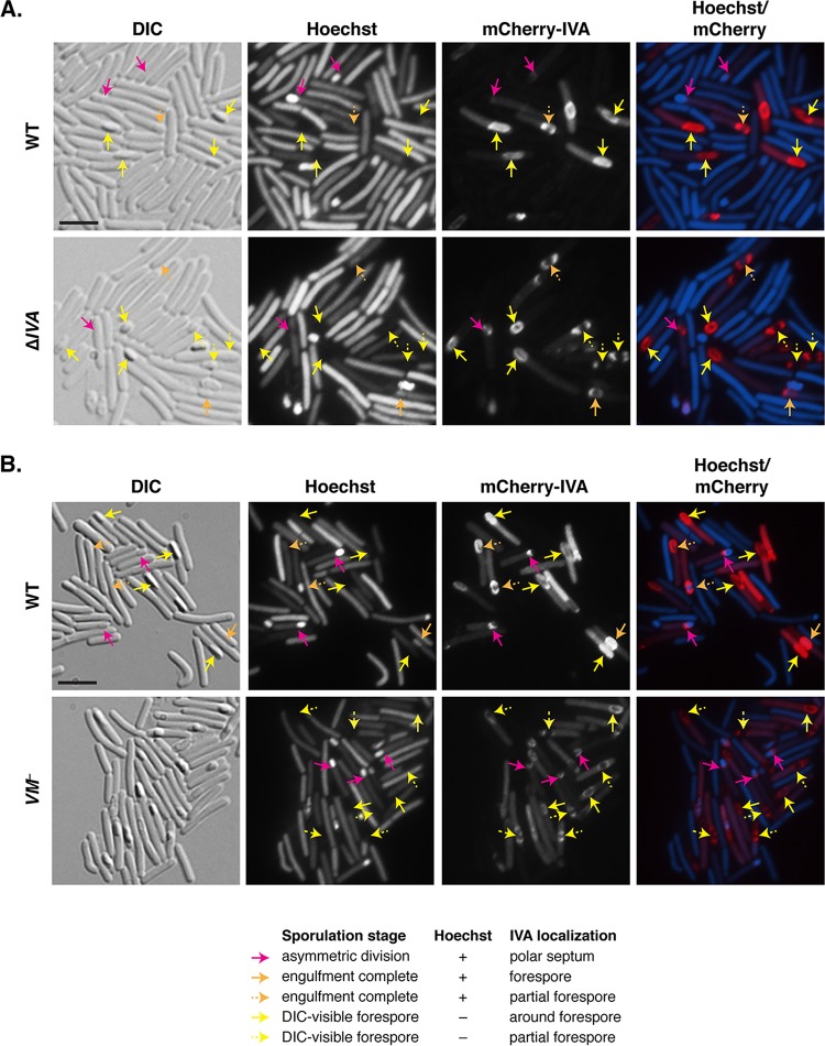
FIG 5 .
IVA can localize to and encase the forespore in the absence of VM. Fluorescence microscopy analyses of wild-type (WT) and ΔIVA sporulating cells (A) and wild-type and VM mutant sporulating cells (B) producing mCherry-IVA at 23 h post-sporulation induction. Differential interference contrast (DIC) microscopy was used to visualize forespores and free spores. The nucleoid was stained with Hoechst stain (blue), and mCherry-IVA fluorescence is shown in red. The merge of Hoechst stain and mCherry is also shown. Pink arrows highlight cells that have undergone asymmetric division but have not completed engulfment; these forespores stain with Hoechst stain (+), and mCherry-IVA localizes to the mother cell-forespore interface. Orange arrows highlight cells that appear to have completed engulfment but whose forespores retain the Hoechst dye (+). Yellow arrows mark forespores that are visible by DIC and no longer retain the Hoechst dye. Orange and yellow solid arrows indicate that mCherry-IVA fully encases the forespore, while dashed arrows indicate partial encasement of the forespore. Bars, 5 µm.

