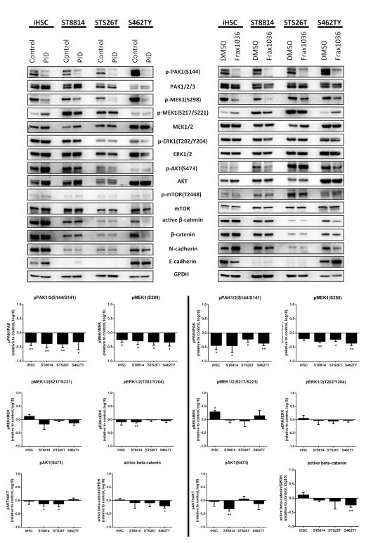
Figure 3. Inhibition of PAK1/2/3 affects MAPK, Akt, β-catenin pathways and cadherin expression.
Immunoblot analysis of MAPK, AKT/mTOR and β-catenin cascades signaling and N- and E-cadherin protein levels changes in iHSC, ST8814, STS26T and S462TY cells exposed to PAK1/2/3 inhibitors GST-PID and Frax1036. WB quantification of Pak, Mek/Erk, Akt/mTOR phosphorylation changes and changes of active β-catenin levels Error bars represent standard deviation (*= p<0.05, **= p<0.01).

