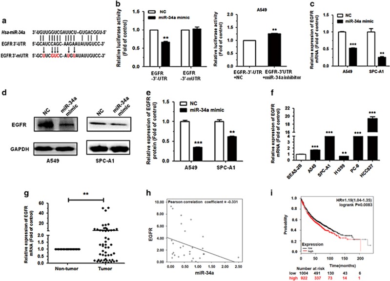
Figure 5.
EGFR is a direct target of miR-34a. (a) EGFR WT 3′-UTR contains predicted miR-34a-binding site. The data show alignment of miR-34a with EGFR WT 3′-UTR and arrows indicate mutagenesis nucleotides. (b) Dual luciferase reporter assay. pGL3-EGFR WT 3′-UTR (EGFR-3′-UTR) and pGL3-EGFR mut 3′-UTR (EGFR-3′-mUTR) were co-transfected with miR-34a mimic in HEK293T cells (left). EGFR-3′-UTR were co-transfected with miR-34a inhibitor in A549 cells (right). Data displayed are relative firefly luciferase expression, normalized to Renilla luciferase expression. (c, d) The mRNA levels of EGFR were detected by qRT–PCR and protein levels were detected by western blot in A549 and SPC-A1 cells transfected with miR-34a mimic. (e) Densitometry was used to quantify the relative expression of EGFR proteins in A549 and SPC-A1 cells transfected with miR-34a mimic, after normalization to GAPDH. (f) The expression of EGFR in the five human NSCLC cell lines. BEAS-2B cells were used as the normal control for comparison. (g) The relative expression of EGFR mRNA from corresponding non-tumor tissues and tumor tissues as measured by qRT–PCR. 18S was used as an internal control. (h) There was a negative correlation between miR-34a and EGFR, according to the Pearson correlation coefficient. (i) The effect of EGFR expression levels on the overall survival of 1926 lung cancer patients was analyzed. Kaplan–Meier plots were generated using a Kaplan–Meier Plotter. *P<0.05, **P<0.01 and ***P<0.001.

