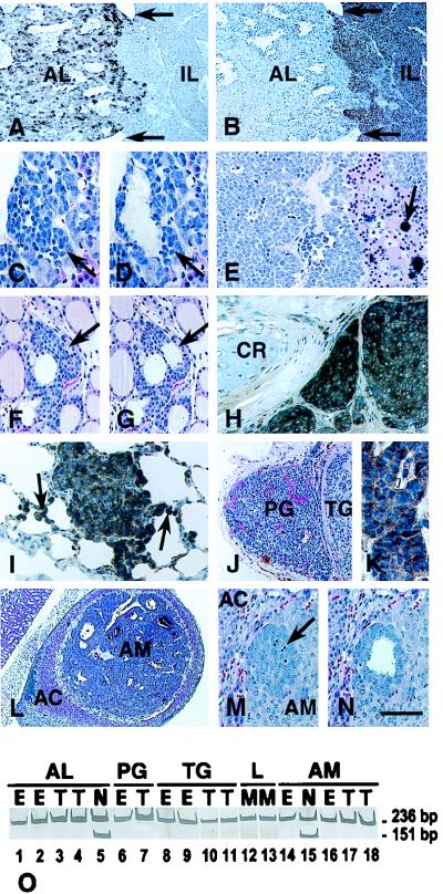
Medical Sciences. In the article “RB-mediated suppression of spontaneous multiple neuroendocrine neoplasia and lung metastases in Rb+/− mice” by Alexander Yu. Nikitin, María I. Juárez-Pérez, Song Li, Leaf Huang, and Wen-Hwa Lee, which appeared in number 7, March 30, 1999, of Proc. Natl. Acad. Sci. USA (96, 3916–3921), the following correction should be noted. The labeling of the gel in Fig. 1O was erroneously moved down and to the right. The corrected figure and its legend are shown below.
Figure 1.
Multiple neuroendocrine neoplasia in Rb+/− mice. (A and B) Gross compound pituitary tumor on P394. The tumor consists of two histologically distinct components that substitute for the pituitary anterior (AL) and intermediate (IL) lobes, which are demarcated by remnants of Rathke’s cleft (arrows). AL tumor cells contain α-GSU (A) and are positioned loosely around sinusoid-like vessels. IL tumor cells contain α-melanocyte-stimulating hormone (B) and form poorly vascularized epithelioid fields with central necrotic and hemorrhagic areas. (C and D) EAP in the anterior pituitary lobe on P90 before (C) and after (D) microdissection for genotype analysis by the PCR. (E) C-cell carcinoma on P340 showing the typical arrangement of polyhedral tumor cells in solid nests and rough hyalinized collagen with calcification (arrow). (F and G) C-cell EAP (arrow) on P60 before (F) and after (G) microdissection. The atypical cells show a parafollicular location. (H) Medullary thyroid carcinoma invading surrounding tissues on P469. Accumulation of calcitonin (brown color) is evident in the cytoplasm of tumor cells (arrow). CR, cartilage. (I) Calcitonin-containing metastatic cells in the lung on P463. The metastatic cells exhibit intraalveolar spreading (arrows). (J) A well-vascularized parathyroid tumor (PG) and a neighboring solid C-cell tumor of the thyroid gland (TG) on P370. (K) Parathyroid hormone expression in parathyroid tumor cells. (L) Pheochromocytoma of the adrenal medulla (AM) compressing the adrenal cortex (AC). (M and N) EAP in the adrenal medulla (AM) on P60 before (M) and after (N) microdissection. The arrow indicates multiple apoptotic figures. Staining: avidin-biotin-peroxidase immunostaining for α-GSU (A), α-melanocyte-stimulating hormone (B), calcitonin (H and I), or parathyroid hormone (K) with hematoxylin counterstaining; (C–G, J, and L–N) hematoxylin-eosin staining. [Bar: 160 μm (A and B), 40 μm (C, D, and I), 110 μm (E), 60 μm (F and G), 50 μm (H), 150 μm (J), and 20 μm (K), 390 μm (L), and 70 μm (M and N).] (O) Absence of the wild-type Rb allele (151-bp PCR product) in gross tumors (T; lanes 3, 4, 7, 10, 11, 17, and 18) and EAPs (E; lanes 1, 2, 6, 8, 9, 14, and 16) of the pituitary anterior lobe (AL, lanes 1–4), the parathyroid gland (PG, lanes 6 and 7), thyroid C cells (TG, lanes 8–11), lung metastases (L, lanes 12 and 13), or adrenal medulla (lanes 14 and 16–18). N, Rb+/− normal tissue (lanes 5 and 15). Nondenaturing 12% polyacrylamide gel stained with silver. The 236-bp band corresponds to the mutant Rb allele (11).


