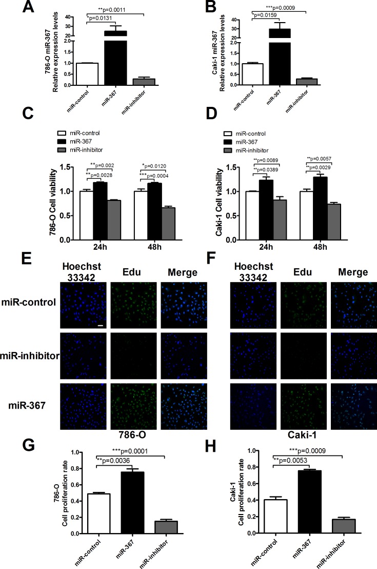
Figure 2. MiR-367 regulated cell proliferation in ccRCC cells.
(A and B) MiR-367 level was measured by qRT-PCR after transfection with miR-control (negative control), miR-367 mimics (miR-367) or miR-inhibitor in 786-O and Caki-1 cells. (C and D) Caki-1 and 786-O viabilities were detected using MTT assay. ccRCC cells were transfected with miR-367 or miR-367 inhibitor for 24 h and 48 h. (E and F) Representative images of Edu staining showing proliferation cells (stained in green). Nuclei that double labeled with EdU (green) and Hoechst 33342 (blue) were considered to be new proliferative cells. Scale bar indicates 100 μm. (G and H) Quantitative analysis of double labeled nuclei was performed. The number of double labeled nuclei was significantly increased in miR-367-treated group compared with miR-control-treated group. Data are expressed as mean ± SEM. **p<0.01 or *p<0.05 vs. miR-control.

