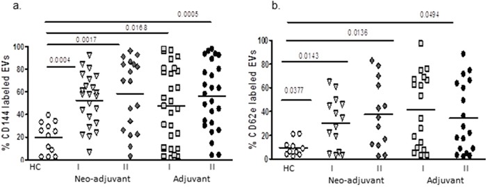
Figure 3. EVs endothelial markers.

EVs were isolated by a series of centrifugations. Antigen levels of endothelial markers VE-cadherin (CD144) and E-selectin (CD62E) were measured using specific fluorescent antibodies on EVs obtained from healthy controls and on EVs obtained from patients before chemotherapy (time point I) and at the last chemotherapy treatment (time point II). The percentage of labelled EVs was calculated from the total number EVs using FACS analysis.
