
Figure 2. Characterization and physicochemical properties of Bufalin-BSA-NP.
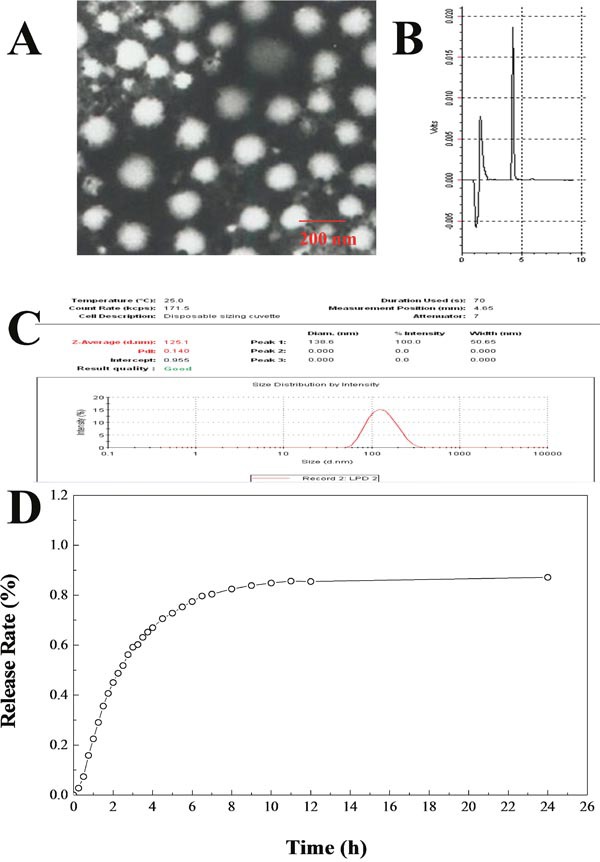
(A) The transmission electron microscope photograph of Bufalin-BSA-NP; (B) HPLC results of Bufalin-BSA-NP; (C) size distribution by intensity of Bufalin-BSA-NP; (D) release curve of Bufalin-BSA-NP in vitro.