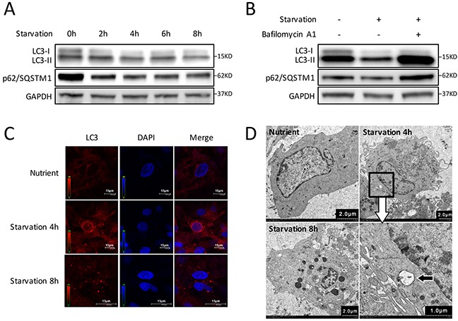
Figure 1. Autophagy is induced by starvation with EBSS in CMs.

(A) Time course of autophagy activation stimulated by starvation. Western blot shows the LC3-I decreased while LC3-II increased during the first 4 hours and gradually recovered after starvation for 6 h. The degradation of p62/SQSTM1 increased gradually in the first 4 hours, then decreased gradually. (B) CMs were treated with Bafilomycin A1 and starved for 4 h. LC3-II accumulation was markedly increased, and p62/SQSTM1 degradation was blocked in Bafilomycin A1-treated groups. GAPDH was used as endogenous control. (C) The intracellular autophagosome and autolysosomes at 4 h starvation were probed by LC3 dots (red) for cell immunofluorescence. Nuclei were stained with DAPI (blue). (D) Transmission electron microscopy showed double-membraned autophagosomes formed in CMs at 4 h starvation (black arrow).
