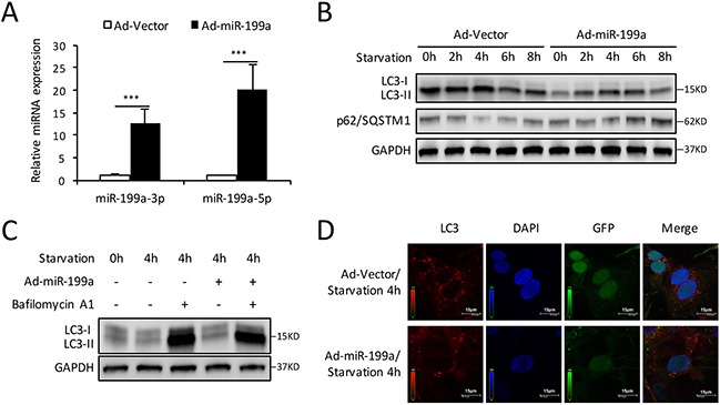
Figure 3. Overexpression of miR-199a blocks the autophagy activity in CMs.

(A) After transfection with miR-199a overexpression adenovirus (Ad-miR-199a), the miR-199a-3p and miR-199a-5p expression levels were markedly up-regulated compared with cells infected with Ad-vector. The differences between two groups were analyzed using t-tests. (***P<0.001 versus Ad-vector, n=3) (B) Western blot showed that overexpression of miR-199a significantly suppressed LC3-II accumulation and p62/SQSTM1 degradation at starvation for 2-8 h. (C) After 48 h transfection, CMs were treated with Bafilomycin A1 and exposed to starvation for 4 h. Western blot shows reduced LC3-II levels in miR-199a overexpression CMs treated with or without Bafilomycin A1. GAPDH was used as endogenous control. (D) MiR-199a blocked starvation-induced autophagosomes formation. At 4 h starvation, confocal microscopy showed that the number of autophagosomes as represented by LC3 dots (red) was reduced in Ad-miR-199a group compared with Ad-vector group. Nuclei were stained with DAPI (blue). GFP: green fluorescent protein fused in adenovirus.
