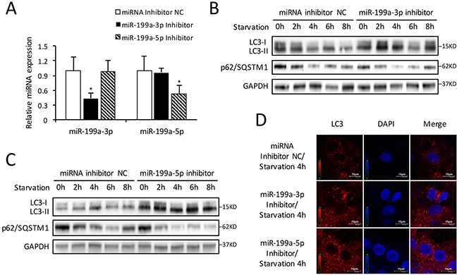
Figure 4. Inhibition of endogenous miR-199a stimulates autophagic activity.

(A) After transfection with miR-199a-3p inhibitor, the miR-199a-3p expression was suppressed while miR-199a-5p showed no significant change. Conversely, after transfection with miR-199a-5p inhibitor, the miR-199a-5p expression was decreased while miR-199a-3p showed no significant change. The analysis of variance for multi-group comparison was using ANOVA (*P<0.05 versus miRNA inhibitor NC, n=3). (B and C) Knockdown of either miR-199a-3p or miR-199a-5p stimulated LC3-II accumulation and p62/SQSTM1 degradation in CMs. GAPDH was used as endogenous control. (D) The results of immunofluorescence suggest that at 4 h starvation, knockdown of either miR-199a-3p or miR-199a-5p could increase the number of LC3 fluorescent dots (red) which indicated autophagosomes formation. Nuclei were stained with DAPI (blue).
