Abstract
The integrity of taste buds is intimately dependent on an intact gustatory innervation, yet the molecular nature of this dependency is unknown. Here, we show that differentiation of new taste bud cells, but not progenitor proliferation, is interrupted in mice treated with a hedgehog (Hh) pathway inhibitor (HPI), and that gustatory nerves are a source of sonic hedgehog (Shh) for taste bud renewal. Additionally, epithelial taste precursor cells express Shh transiently, and provide a local supply of Hh ligand that supports taste cell renewal. Taste buds are minimally affected when Shh is lost from either tissue source. However, when both the epithelial and neural supply of Shh are removed, taste buds largely disappear. We conclude Shh supplied by taste nerves and local taste epithelium act in concert to support continued taste bud differentiation. However, although neurally derived Shh is in part responsible for the dependence of taste cell renewal on gustatory innervation, neurotrophic support of taste buds likely involves a complex set of factors.
KEY WORDS: Shh, Cell renewal, Innervation, Lingual epithelium, Taste receptor cells
Summary: The production and maintenance of taste bud cells requires Shh from both local epithelium and gustatory innervation, and this dual supply mechanism may be relevant for progenitor populations of other renewing epithelia.
INTRODUCTION
The ability to taste allows us to discriminate between foods rich in nutrients (sweet, salty and savory/umami tastes) and potentially toxic substances (bitter and sour tastes). These stimuli are detected by taste buds, the functional receptor units of the taste system, located on the tongue within specialized epithelial appendages called taste papillae. The dependence of taste buds on the gustatory nerve supply has been recognized since the late 1800s (Von Vintschgau and Hönigschmied, 1877). Interruption of the gustatory innervation results in loss of taste buds within a few days of nerve lesion. Even simple disruption of axonal transport in gustatory nerves is sufficient to cause loss of taste buds (Sloan et al., 1983), yet the necessary trophic substances supplied by the nerves have yet to be identified. The canonical neurotrophin BDNF serves the inverse role in this system, being secreted by taste buds and providing trophic support to the innervating gustatory nerve fibers. What the nature of the reciprocal signal might be − from nerve to taste epithelium − is unclear.
Unlike receptor cells in many other sensory endorgans, the ∼100 taste cells comprising each taste bud are transient, being continually renewed with an average lifespan of 10-14 days, albeit with significantly shorter- and longer-lived populations (Beidler and Smallman, 1965; Farbman, 1980; Hamamichi et al., 2006; Perea-Martinez et al., 2013). Mitotically active progenitor cells adjacent to taste buds generate post-mitotic cells, which enter taste buds as taste precursor (type IV) cells; these, in turn, differentiate into mature taste cells (Barlow and Klein, 2015; Delay et al., 1986; Hirota et al., 2001). Proliferating progenitors express the epithelial markers keratin (K) 14 and K5, and by genetic lineage tracing, have been shown to give rise to both taste bud cells and non-taste cells in adjacent epithelium (Okubo et al., 2009). Previous studies have focused primarily on cellular dynamics of this turnover, while analyses of molecular regulation of taste cell renewal and differentiation are limited.
The sonic hedgehog (Shh) signaling pathway regulates homeostasis of numerous epithelia (Ingham and McMahon, 2001; Petrova and Joyner, 2014). In lingual epithelium, Shh is expressed by post-mitotic taste precursor cells (Miura et al., 2001, 2003, 2004), while the Hh receptor patched 1 (Ptch1) and target gene Gli1 are expressed by K5+ progenitor cells surrounding each bud (Miura et al., 2001), suggesting that Shh+ cells within taste buds signal to adjacent progenitors to regulate cell renewal. Consistent with this hypothesis, broad misexpression of Shh in lingual progenitors triggers formation of ectopic taste buds, suggesting Shh promotes taste bud differentiation (Castillo et al., 2014).
The importance of Hh signaling in taste bud maintenance is evidenced by disruption of taste function in cancer patients given chemotherapeutics that inhibit the Hh pathway (HPIs) (Basset-Seguin et al., 2015; LoRusso et al., 2011); and in mice, HPIs cause loss of taste buds and taste nerve responses (Kumari et al., 2015; Yang et al., 2015). However, the cellular mechanisms of Shh support of taste bud maintenance are unidentified.
RESULTS
Inhibition of Hh signaling reduces addition of new taste cells to fungiform taste buds with minimal impact on progenitor proliferation
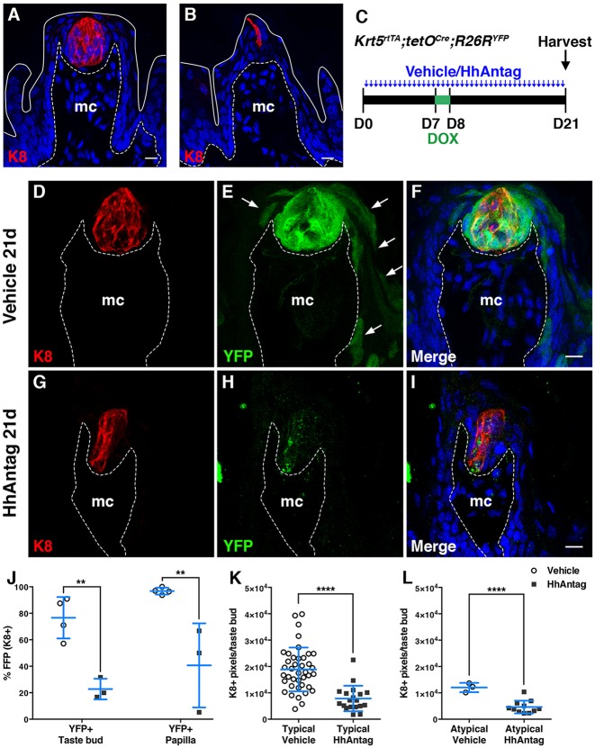
In this study, we have focused on taste buds housed in fungiform taste papillae (FFP) on the anterior tongue. Mice were treated for 21 days with HhAntag, a HPI that binds Smo and inhibits activation of Hh target genes (Yauch et al., 2008). Using Keratin (K) 8 immunofluorescence to mark mature taste cells (Knapp et al., 1995), we found the number of FFP and taste buds (‘typical FFP’; see example in Fig. 1A) was significantly decreased (Fig. S1A). Conversely, significantly more atypical, conical FFP that house slender taste buds (see example in Fig. 1B), a morphology indicative of degenerating FFP (Nagato et al., 1995; Oakley et al., 1990), were evident in HhAntag-treated mice compared with controls (Fig. S1B). These findings are consistent with a previous report using another HPI, LDE225 (Kumari et al., 2015).
Fig. 1.
Mice treated with HhAntag for 21 days have reduced renewal of taste bud and FF papilla epithelium. (C) Krt5rtTA;tetOCre;R26RYFP mice were treated with vehicle or HhAntag twice daily (blue arrows) for 21 days, and fed dox chow overnight on day 7. Typical (A) and atypical (B) FFP with K8+ taste buds (red) are present in controls and mutants. (D-F) Control FFP taste buds have robust levels of K8+ cells (red), and K5-YFP+ progeny are evident (green) in taste buds and FFP epithelium (arrows). (G-I) HhAntag-treated mice have fewer K8+ taste cells (red) and K5-YFP+ lineage-traced cells (green), and distorted morphology. (J) HhAntag treatment results in significantly fewer taste buds and FFP with K5-YFP lineage-traced cells. (K,L) Taste bud size, i.e. the number of K8+ pixels, is reduced in typical (K) and atypical (L) FFP in HhAntag-treated mice. Nuclei are counterstained with Draq5 (blue); white dashed lines indicate basement membrane; solid line indicates tongue surface; mc, mesenchymal core. Images are compressed confocal z-stacks. Scale bars: 10 μm. n=3 or 4 mice per condition. Data are represented as mean±s.d. analyzed using two-way ANOVA (J) or Student's t-test (K,L). **P<0.01, ****P<0.0001.
To test whether HhAntag blocks differentiation of taste cells, we used lineage tracing to track input of new cells into buds from K5+ progenitors. Krt5rtTA;tetOCre;R26RYFP mice were given HhAntag or vehicle for 7 days, fed doxycycline (dox)-chow overnight on day 7 and treated with HhAntag or vehicle for another 14 days (Fig. 1A). Mice receiving vehicle and dox chow had normal FFP (Fig. 1D-F), with robust YFP expression in taste buds and FFP epithelium (Fig. 1E, arrows). Mice treated with HhAntag had significantly fewer taste buds and FFP with YFP+ cells (Fig. 1G-J), suggesting HhAntag blocks differentiation of new taste cells. Consistent with a reduced inflow of new cells, taste buds in both typical and atypical FFP were smaller in drug-treated mice (Fig. 1K,L).
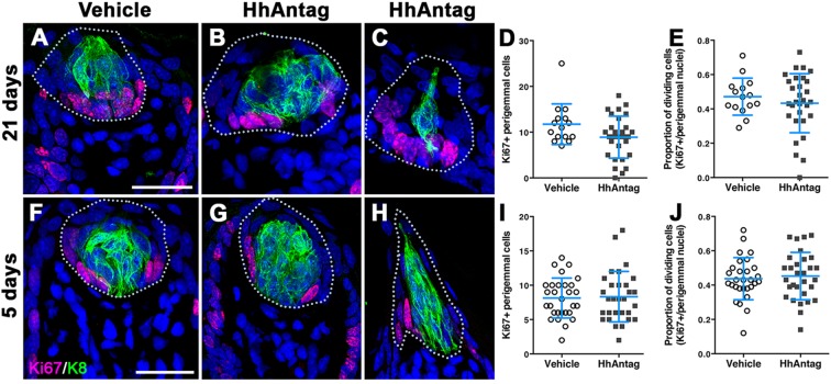
Although HhAntag led to smaller taste buds, this decrease in size could also be attributable to reduced progenitor proliferation. In controls, Ki67+ progenitors reside at the basement membrane of the lingual epithelium, as well as adjacent to taste buds (Fig. S2A). HhAntag treatment for 21 days did not grossly disturb this pattern (Fig. S2B), and quantification of epithelial Ki67+ cells (Fig. S2C,D,D′) revealed no difference in proliferating lingual progenitors due to drug treatment (Fig. S2E-H). We next focused on epithelial cells adjacent to FFP taste buds (perigemmal cells), as conditional epithelial deletion of a Shh effector, Gli2, or overexpression of a dominant-negative form of Gli2 impacted proliferation of these presumed taste progenitors (Ermilov et al., 2016). In contrast to results reported for Gli2 manipulations, neither the number nor the proportion of Ki67+ perigemmal cells differed significantly between HhAntag- and vehicle-treated mice at 21 days (Fig. 2A-E), although some taste buds were associated with fewer dividing cells in drug-treated animals. We also tested whether inhibition of Hh signaling had an immediate effect on perigemmal cell proliferation. However, proliferation was unaltered by 5 days of HhAntag treatment (Fig. 2F-J). In fact, even taste buds in FFP that appeared to be degenerating and assuming an atypical FFP morphology were associated with perigemmal Ki67+ cells (Fig. 2C,H). In summary, our new data suggest that blocking Hh signaling has minimal impact on progenitor proliferation but more likely disrupts taste bud renewal by decreasing differentiation of new taste cells, consistent with our previous findings (Castillo et al., 2014).
Fig. 2.
HhAntag does not alter proliferation of perigemmal taste progenitor cells in FFP. (A-C) Proliferating epithelial cells (Ki67+ red) are situated perigemmally around taste buds (K8+, green) in mice treated with vehicle (A) or HhAntag (B,C) for 21 days. (D,E) Although fewer taste buds are present in HhAntag mice at 21 days (see Fig. S1), the number (D) and proportion (E) of Ki67+ perigemmal cells in the remaining FFP with buds do not differ from controls. (F-H) Similarly, Ki67+ perigemmal cells are detected in vehicle (F) and HhAntag-treated mice (G,H) after 5 days. (I,J) Neither the number of Ki67+ perigemmal cells (I) nor proportion of Ki67+ perigemmal cells (J) differs with treatment. Images are compressed confocal z-stacks. Scale bars: 25 µm. n=3-6 mice per condition. Data are mean±s.d. analyzed using Student's t-test with Welch's correction.
Genetic deletion of Shh in K5+ progenitors and their progeny does not impact taste bud renewal
K5+ cells generate daughter cells that enter taste buds 12-24 h after mitosis and become type IV cells, which are immediate precursors of mature taste cells (Miura et al., 2006, 2014). Type IV cells are Shh+ (Miura et al., 2006, 2014; Nguyen and Barlow, 2010) and are a proposed source of the Shh necessary for taste bud cell renewal (Miura et al., 2001, 2014). Thus, we explored the requirement for type IV cell-supplied Shh by genetically deleting Shh in K5+ progenitors.
Feeding dox chow to Krt5rtTA;tetOCre;Shhflox/flox (K5-ShhcKO) mice for 14, 21 or 42 days (Fig. 3A,B) should result in a majority deletion of Shh in the K5+ lineage. In genetic controls (Krt5+/+;tetOCre;Shhflox/flox fed dox), 60% of taste bud profiles were Shh+, whereas in mutants, only 9% of taste bud profiles were Shh+ at 42 days (Fig. 3C-E). Additionally, Shh expression in lingual epithelium was reduced in mutants compared with controls (Fig. 3F), but neither the number nor size of typical FFP taste buds was altered compared with controls, even by 42 days (Fig. 3G,I). Atypical FFP were more common in mutants, but this was not significant, nor was the difference in size of resident taste buds (Fig. 3H,J). Interestingly, Gli1 expression was not reduced in K5-ShhcKO mutant epithelium (Fig. 3F), suggesting Hh signaling to taste progenitors persisted despite knockdown of epithelial Shh. One interpretation of these data was that the efficacy of our Cre driver system was low; persistent epithelial Hh ligand supported taste bud renewal, explaining why long-term epithelial deletion of Shh did not phenocopy inhibition by HhAntag. Alternatively, we reasoned a previously unappreciated Hh source(s) may support taste bud renewal.
Fig. 3.
Genetic deletion of Shh in K5+ progenitor cells does not alter taste bud renewal. (A) Krt5rtTA;tetOCre;Shhflox/flox (K5-ShhcKO) mice were fed dox for 14, 21 or 42 days. (B) Deleting Shh in K5 progenitors results in Shhneg taste precursors (marked with crosses). (C-E) Shh-expressing precursor cells are evident in most control taste bud profiles using Nomarksi optics (C) but absent in most mutant profiles (D,E). (F) Shh mRNA expression is reduced in K5-ShhcKO epithelium, although because of high variability in control values, this difference is not significant. Gli1 expression does not differ between mutants and controls. (G,H) The number of K8+ typical (G) and atypical (H) FFP does not differ between control and K5-ShhcKO mice after 14, 21 or 42 days. (I,J) K5-ShhcKO does not affect taste bud size (K8+ pixels) within typical (I) and atypical (J) FFP. Black dashed lines indicate basement membrane; black circles indicate taste buds; mc, mesenchymal core. Scale bars: 25 μm. n=3 or 4 mice per condition. Data are presented as mean±s.d. analyzed using Student's t-test (E) or two-way ANOVA (G-J). ***P<0.001.
Sensory neurons that innervate fungiform taste buds express Shh
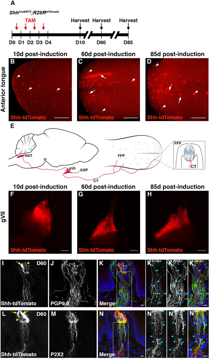
We next used ShhCreERT2/+;R26RtdTomato (Shh-tdTomato) lineage tracing to identify other sources of Shh relevant to taste buds. Mice received four doses of tamoxifen and tongues were harvested at 10, 60 or 85 days (Fig. 4A). At 10 days, tdTomato was restricted to taste buds (Fig. 4B, arrowheads), as Shh+ cells differentiate into taste receptor cells in this time frame (Miura et al., 2014). At 60 and 85 days, however, although tdTomato was still apparent within FFP (Fig. 4C,D, arrowheads), dimmer thread-like tdTomato signal emanated from FFP (Fig. 4C,D, arrows), a pattern suggestive of innervation (Lopez and Krimm, 2006).
Fig. 4.
Gustatory ganglion cells that innervate taste buds express Shh-tdTomato. (A) ShhCreERT2;R26RtdTomato (Shh-tdTomato) mice were given tamoxifen daily for 4 days and harvested at 10, 60 or 85 days. (B-D) In Shh-tdTomato+ tongues, punctate signal (red) is evident in FFP at all times (arrowheads); at 60 and 85 days (C,D) gustatory innervation (arrows) associated with FFP (arrowheads) is also labeled. (E) Gustatory neurons in the geniculate ganglion (gVII) innervate FFP via the chorda tympani (CT) nerve, and project to the nucleus of the solitary tract (NST) in the brainstem. The greater superficial petrosal (GSP) nerve innervates soft palate taste buds (not shown). gVII neurons project centrally to the NST. (F-H) Cells in gVII express tdTomato at all time points. (I-K‴) At 60 days, PGP9.5+ nerve fibers (white in J,K″; green in K,K‴) innervate taste buds and adjacent FF epithelium, whereas Shh-tdTomato+ neurites (white in I,K′; red in K,K‴), which are also PGP9.5+, innervate taste buds exclusively (K′-K‴, cyan arrowheads). (L-N‴) P2X2+ taste fibers (white in M,N′; green in N,N‴) express Shh-tdTomato (white in L,N′; red in N,N‴; cyan arrowheads). Boxes in K and N are shown at higher magnification in K′-K‴ and N′-N‴, respectively. Sparse bright lineage-labeled taste cells are detected at later times (I,L, arrows), and are distinguishable from dimmer Shh-tdTomato+ neurites (I,L, arrowheads within taste buds). Nuclei are counterstained with Draq5 (blue). B-D and F-H are images of whole tongue and ganglia. I-N‴ are compressed confocal z-stacks. Scale bars: 1 mm in B-D; 150 μm in F-H; 10 μm in I-N‴.
FFP taste buds are innervated by the chorda tympani nerve (CT) of the geniculate ganglion (Fig. 4E, gVII). Gustatory gVII neurons project fibers centrally to the brainstem, specifically to the nucleus of the solitary tract (Fig. 4E, NST) (Contreras et al., 1982; Corson et al., 2012; Finger et al., 2000; Krimm and Barlow, 2007; Sugimoto et al., 1997). Shh-tdTomato+ cells were detected in gVII at 10, 60 and 85 days (Fig. 4F-H). Additionally, Shh-tdTomato+ neurons were detected in the trigeminal ganglia (gV) (Fig. S3), which provide somatosensory innervation to the anterior tongue and FFP epithelium surrounding taste buds.
Although both gV and gVII possess Shh lineage-traced cells, Shh-tdTomato+ fibers innervated only taste buds and not the surrounding FFP epithelium (Fig. 4I,L), consistent with gustatory innervation by gVII and not somatosensory innervation by gV (Castillo et al., 2014; Finger and Simon, 2000; Finger et al., 2005; Kanazawa and Yoshie, 1996). Shh-tdTomato+ neurites within FFP express PGP9.5 (UCHL1), a general marker of nerve fibers (Kanazawa and Yoshie, 1996) (Fig. 4J-K‴ cyan arrowheads), but not all PGP9.5+ nerves were Shh-tdTomato+. Immunostaining for the purinergic receptor P2X2 (P2RX2), present in taste but not in somatosensory fibers (Finger et al., 2005) (Fig. 4M) revealed extensive colocalization of P2X2 in Shh-tdTomato+ nerve fibers (Fig. 4N-N‴, cyan arrowheads).
We also examined Shh-tdTomato expression in brainstem termini of gVII neurons. tdTomato+/P2X2+ gustatory afferents were readily evident in the NST (Fig. S4, white dashed circles), whereas tdTomato+/P2X2neg somatosensory fibers of gV projected to the spinal trigeminal tract (Fig. S4, arrowheads). Shh-tdTomato+ cell bodies were not detected in this region of the brainstem (data not shown). Thus, Shh is expressed by gustatory neurons of gVII, suggesting nerve-derived Shh may maintain taste buds.
Given that (1) epithelial deletion of Shh had no effect on taste buds and (2) gustatory sensory neurons express Shh, we hypothesized that Shh supplied by the gustatory innervation supports taste bud renewal. This hypothesis was particularly attractive, as taste buds are maintained by an intact innervation (Cheal and Oakley, 1977; Guagliardo and Hill, 2007; Sloan et al., 1983). Importantly, HPIs would block receipt of Shh, regardless of tissue source, explaining the discrepancy between our K5-ShhcKO and HhAntag results (Figs 1,2, Fig. S1) (Kumari et al., 2015; Yang et al., 2015). To test our hypothesis, ShhCreERT2/flox;R26RtdTomato mice were given tamoxifen (Shh-ShhcKO) and tissues were examined after another 31 days (Fig. 5A). In this model, tdTomato expression is induced and Shh is permanently deleted in long-lived cells expressing Shh, e.g. sensory neurons, but Shh loss should be transient in taste buds, as new Shh+ precursor cells are continually generated from unaffected K5+/Shhneg progenitors after cessation of tamoxifen (Fig. 5B).
Fig. 5.
Genetic deletion of Shh in Shh+ cells, including gustatory neurons, minimally affects taste buds. (A) ShhCreERT2/flox;R26RtdTomato (Shh-ShhcKO) and genetic control mice were given tamoxifen for 4 days and harvested at 35 days. (B) In tamoxifen-treated Shh-ShhcKO mice, Shh is deleted permanently from ganglion cells and nerves (indicated by the cross), but transiently from taste buds (see text). (C,D) tdTomato reports Shh deletion in tongue (C) and gVII (D) of mutant mice. (E) Quantitative PCR reveals that neither Shh nor Gli1 expression is significantly reduced in mutant tongue epithelium. (F) Expression of Shh, but not Gli1, is significantly reduced in mutant gVII. (G,H) Typical FFP number does not differ between mutants and controls (G), although atypical FFP increase in mutants (H). (I,J) The size of taste buds in typical (I) and atypical (J) FFP in mutants does not differ from controls. (K) Shh-tdTomato+ neurites (red) innervate a taste bud (K8+, green) in an atypical FFP in a Shh-ShhcKO mouse (Shh-descendent taste cell, arrow). (L) The proportion of FFP innervated by P2X2+ fibers is not affected by Shh-ShhcKO. (M,N) P2X2+ innervation density of taste buds in typical (M) and atypical (N) FFP does not differ between controls and Shh-ShhcKO mice. Nuclei counterstained with Draq5 (blue); white dashed lines delimit basement membrane; white solid lines delimit epithelial surface. C and D are images of whole tongue and gVII. K is a compressed confocal z-stack. Scale bars: 1 mm in C; 150 μm in D; 10 μm in K. n=3-5 mice per condition. Data are mean±s.d., except E and F, which are mean±s.e.m; I and N represent the median with interquartile range. Results were analyzed using Student's t-test (E-H,J,L,M) or Mann–Whitney U-test (I,N). ***P<0.001, **P=0.05.
Expression of tdTomato in Shh-ShhcKO-Tom mice confirmed CreER activation in gVII neurons and associated fibers (Fig. 5C,D), whereas tdTomato was generally absent in taste cells after 35 days, reflecting the continual replacement of Shh+ cells from K5+ progenitors in which Shh had not been deleted. qPCR confirmed Shh was reduced in mutant gVII, but not in lingual epithelium (Fig. 5E,F). Nonetheless, we did encounter sparse persistent Shh-descendent cells in some taste buds. We suspect the presence of lineage-labeled taste cells is due to reiterative Cre activation by ingestion of tamoxifen-containing feces (Brake et al., 2004; Fromson et al., 1973; Robinson et al., 1991). Even so, these long-persistent labeled cells represent a small fraction of the total new taste cells continually generated (see Miura et al., 2014). As mature taste cells do not express Shh, they do not impact the level of Shh mRNA expressed by the epithelium at 30 days (Fig. 5E). Thus, in Shh-ShhcKO mice, Shh is permanently deleted in sensory neurons but not in taste epithelium. Consistent with the hypothesis that Shh+ gustatory neurons signal to taste progenitor cells in lingual epithelium, Gli1 expression was reduced in lingual epithelium, although not significantly (Fig. 5E).
Nonetheless, despite reduction of Shh in Shh-expressing cells, the number and size of typical FFP taste buds did not differ between mutants and controls (Fig. 5G,I). Although the number of atypical FFP buds increased significantly in mutants (Fig. 5H), taste bud size in these degenerating FFP was unaffected (Fig. 5J). Because we observed a mild increase in atypical FFP number, we investigated whether this effect was indirect, as autocrine Shh within gVII might be required to maintain taste bud innervation and thereby only secondarily affect taste buds. However, the proportion of taste buds innervated by P2X2+ fibers (Fig. 5K,L) and P2X2+ innervation density within taste buds (Fig. 5M,N) did not differ between mutants and controls. This outcome is consistent with qPCR data showing Gli1 levels in Shh-ShhcKO ganglia do not differ from controls (Fig. 5F), and further suggests that intraganglionic Hh signaling is not affected by loss of Shh in Shh+ gustatory neurons. Thus, significant reduction of Shh in post-mitotic and non-renewing cells, including gVII neurons, has minimal impact on FFP, taste buds and their innervation.
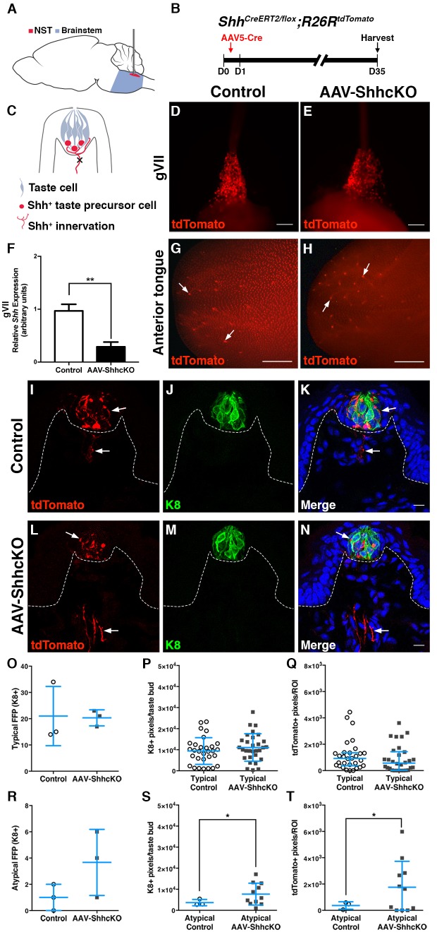
We next aimed to restrict Shh deletion solely to the gustatory innervation by developing a method to deliver virally encoded Cre recombinase into the NST (Fig. 6A); virus is taken up by gustatory afferents and transported retrogradely to neuronal cell bodies in gVII for Cre excision of loxp-flanked sequence. Adeno-associated virus 5 Cre (AAV5-Cre) was injected into the NST of R26RtdTomato mice and brainstems, and cranial ganglia and tongues were then examined at 14, 35 and 60 days. tdTomato expression was evident in the region of the brainstem housing the NST at all time points (Fig. S5A,B and data not shown), including in the gustatory rostral NST identified via P2X2+ immunostaining (Fig. S5C-E, green). Cell bodies in gVII and gustatory nerve fibers innervating taste buds were also tdTomato+ at all times post-injection (Fig. 6D,G and data not shown).
Fig. 6.
Viral deletion of Shh in gustatory neurons has little effect on taste bud number, size or innervation density. (A) Sagittal schematic of the brain depicting stereotaxic injection of the NST (red) in the brainstem (blue). (B) Experimental design for AAV5-Cre injection of ShhCreERT2/flox;R26RtdTomato (AAV-ShhcKO) mice. (C) AAV5-Cre deletes Shh in gustatory neurons that project to NST and innervate FFP (indicated by the cross). (D-H) AAV5-Cre injection into the NST activates tdTomato+ expression in gVII neurons of control (D) and mutant (E) mice, reduces Shh expression in gVII in mutants (F), and reveals tdTomato+ nerve fibers (arrows) innervating the anterior tongue of controls (G) and mutants (H). (I-N) FFP taste buds (K8+, green) in both control (I-K) and AAV-ShhcKO (L-N) mice are innervated by AAV5-tdTomato+ neurites (arrows). (O,R) Typical FFP number is not affected by AAV5-ShhcKO (O), whereas atypical FFP numbers increase but not significantly (R). (P-T) Taste bud size (P) and density of tdTomato+ neurites (Q) of typical FFP are similar in controls and AAV-ShhcKO mice, whereas taste buds of atypical FFP (S) are larger in mutants, with increased density of tdTomato+ neurites (T). Nuclei are counterstained with Draq5 (blue); white dashed lines delimit basement membrane. (D,E,G,H) Images of whole ganglia and tongues. Scale bars: 150 μm in D,E; 1 mm in G,H. n=3 mice per condition. Data are mean±s.d., except Q, which is the median with interquartile range. Results were analyzed using Student's t-test (F,O,P,R-T) or Mann–Whitney U-test (Q). *P≤0.05, **P<0.01.
To test specifically the role of neurally supplied Shh, the NST of ShhCreERT2/flox;R26RtdTomato mice was injected with AAV5-Cre (AAV-ShhcKO) (Fig. 6A,B). Neural deletion of Shh was accomplished by viral Cre delivery; mice were not treated with tamoxifen and ShhCreERT2 functioned as a null allele (Harfe et al., 2004). After 35 days, gVII and nerve fibers of AAV-injected control and AAV-ShhcKO mice were tdTomato+ (Fig. 6D,E,G-N). Importantly, NST injection of AAV5-Cre did not label taste cells (Fig. 6K,N); neither Cre-recombinase nor tdTomato protein is transferred from sensory fibers to taste cells, nor does centrally injected AAV5-Cre virus infect taste cells innervated by gustatory fibers. Importantly, Shh was significantly reduced in mutant ganglia (Fig. 6F).
Similar to genetic deletion of Shh in Shh+ cells (see Fig. 5), typical FFP taste bud number and size did not differ between control and mutant mice (Fig. 6O,P), nor was innervation density altered in typical FFP, as assessed by tdTomato+ fibers (Fig. 6Q). However, ganglionic reduction of Shh tended to increase the number and size of atypical FF taste buds (Fig. 6R,S), and the density of tdTomato+ fibers innervating these degenerating taste buds (Fig. 6T). The discrepancy between the large effect of HhAntag (Fig. 1, Fig. S1) versus the minimal impact of Shh deletion from tongue epithelium (Fig. 3) or gustatory ganglia (Figs 5 and 6) suggested that both epithelial and nerve-derived Shh maintain taste bud renewal, and that coincident abolition of Shh from both tissues would lead to taste bud loss.
Simultaneous Shh deletion from lingual epithelium and gustatory neurons mirrors the impact of HhAntag on taste buds
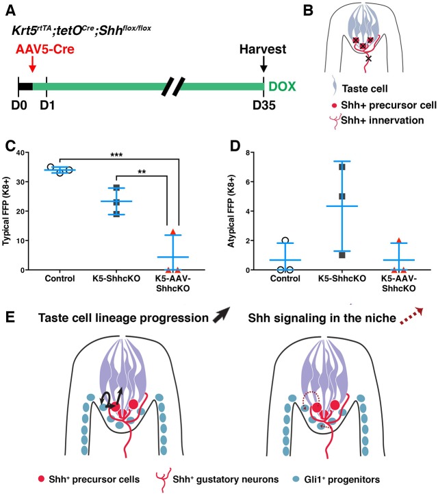
To test whether Shh+ precursor cells and gVII neurons together are required for taste bud renewal, we combined inducible, tissue-specific genetics and Cre delivery via AAV5 stereotaxic injection. The NST of Krt5rtTA;tetOCre;Shhflox/flox (K5-ShhcKO; as in Fig. 3) was injected with AAV5-Cre (as in Fig. 6) to generate dual deletion animals (K5-AAV-ShhcKO), and beginning on the day of injection, K5-AAV-ShhcKO, control (Krt5rtTA;tetOCre) and epithelial deletion only (K5-ShhcKO) mice were fed dox chow for 35 days (Fig. 7A,B).
Fig. 7.
Simultaneous deletion of Shh from tongue epithelium and sensory neurons abolishes taste buds. (A) Experimental design for concurrent neural and epithelial deletion of Shh. The NST of Krt5rtTA;tetOCre;Shhflox/flox (K5-ShhcKO) mice is injected with AAV5-Cre on day 0, and mice are fed dox for 35 days. (B) In K5-AAV-ShhcKO mice, Shh is deleted in both lingual epithelium and gustatory neurons (marked by crosses). (C) Typical FFP number is significantly decreased in K5-AAV-ShhcKOs compared with control and K5-ShhcKO mice. (D) Atypical FFP number does not vary significantly across conditions, although more are evident in K5-ShhcKO mice. n=3 mice per condition. Data are mean±s.d. One-way ANOVA (C,D); **P<0.01, ***P<0.001. (E) Model for the role of Shh in taste cell renewal. Taste cell lineage progression (black arrows, left panel): New taste cells are generated from self-renewing Gli1+ progenitors (blue), which exit the cell cycle, enter buds as Shh+ precursor cells (red) and differentiate as taste cells (gray). Shh signaling in the taste progenitor niche (red dashed arrows, right panel). The niche for Gli1+ progenitors comprise both gustatory neurons and taste precursor cells expressing Shh (red). Shh ligand promotes taste cell differentiation from these Shh-responsive progenitors.
In K5-ShhcKO mice, the number of K8+ typical FFP was slightly but not significantly reduced, and the number of atypical degenerating FFP was highly variable, albeit trending higher at 35 days (Fig. 7C,D), mirroring prior results (see Fig. 3G,H). In contrast, K5-AAV-ShhcKO mice had dramatically reduced typical and atypical FFP (Fig. 7C,D). In fact, in two of three dual-deleted mice, both typical and atypical taste buds were completely absent. Thus, concurrent deletion of Shh from both the gustatory innervation and lingual epithelium reveals that these two tissue sources in concert maintain FFP taste buds.
DISCUSSION
Shh signaling is implicated in stem cell proliferation, maintenance and differentiation in multiple systems; here, we report that Shh is required for maintenance of adult taste buds. Shh is expressed by taste cell precursors within each bud, while progenitors express Shh target genes, suggesting Shh signals to progenitor cells to regulate taste cell renewal (Gaillard et al., 2015; Liu et al., 2013; Miura et al., 2001, 2014; Okubo et al., 2009) (Fig. 7E).
A role for Shh in maintaining taste buds is further indicated by loss of taste buds in mice and taste dysfunction in cancer patients receiving HPI drugs. The most parsimonious explanation for taste loss is that, as is the case for tumor cells, progenitor proliferation is blocked by HPIs, which disrupts supply of new taste cells. However, we had posited previously that Shh functions to promote taste cell differentiation because broad misexpression of Shh drives differentiation of taste buds in ectopic locations throughout the lingual epithelium (Castillo et al., 2014). Here, we show that inhibition of Shh signaling by HhAntag has no impact on lingual epithelial progenitor proliferation in general, and, more importantly, the proliferation of perigemmal progenitors adjacent to FFP taste buds is not altered by HhAntag. Combined with our findings that input of newly differentiated taste cells into buds is significantly reduced, our data support a model where Shh promotes taste cell differentiation rather than progenitor proliferation in taste epithelium (Fig. 7E).
Our data contrast with recent findings where proliferation was reduced by vismodegib (a different Smo antagonist), or by genetic manipulation of Gli2, a Shh pathway effector, in lingual progenitors. In mice treated with vismodegib for 15 weeks, Ki67+ cells associated with taste buds in the circumvallate papilla of the posterior tongue were reduced, but only by 5% compared with controls (Yang et al., 2015). Our results are difficult to compare with this study because: (1) we examined anterior FFP taste buds, which are known to have differential molecular regulation from posterior buds (see Barlow and Klein, 2015); and (2) our study was short term (5 days and 21 days) rather than 15 weeks, although we do see reduced proliferation associated with a subset of taste buds at our longer time point (see Fig. 2D,E).
In another recent study, Mistretta and colleagues explored the role of Gli2, a transcription factor that regulates gene expression downstream of Hedgehog signaling. Deletion or overexpression of a dominant-negative form of Gli2 (dnGli2) in K5+ lingual progenitors leads to gradual (16 days) or rapid (5 days) loss, respectively, of most taste buds (Ermilov et al., 2016). Additionally, dnGli2 reduces proliferation of keratinocytes in the basal and perigemmal compartments of FFP, while loss of Gli2 impacts solely perigemmal cell proliferation (Ermilov et al., 2016); the latter is the set of cells within which we found no significant difference in proliferation with HhAntag treatment. What might explain this discrepancy? In addition to regulating expression of target genes in response to Shh, Gli2 functions downstream of other signaling pathways involved in proliferation, including TGFβ and Wnt (Gu and Xie, 2015; Javelaud et al., 2011); both pathways are present and function in taste epithelium (Gaillard et al., 2015; Nakamura et al., 2010). Thus, altering Gli2 expression in lingual epithelium likely impacts several molecular regulators, in addition to Shh, accounting for the discrepancy with our results where we inhibited Smo pharmacologically.
Shh+ cells within taste buds have been considered the sole source of Hh ligand relevant to taste cell renewal (Miura et al., 2001, 2006, 2014), and therefore it was surprising that significant reduction of Shh in tongue epithelium did not phenocopy taste bud loss from HhAntag treatment. Using lineage tracing, we identified Shh-expressing gustatory sensory neurons in gVII, suggesting this neural source might maintain taste buds. However, reduction of Shh in gustatory neurons via conditional genetics or viral deletion only minimally affected taste buds. These results may partly be attributable to our inability to completely knockout Shh using each of these Cre driver systems. Nonetheless, our results indicate that a combination of epithelial and neural Shh maintain taste buds, as coincident deletion of epithelial and neural Shh abolishes taste buds, a result comparable with the effect of HhAntag. Thus, in the anterior tongue, epithelial and neural sources of Shh function redundantly to support taste bud differentiation.
Shh plays an important role in homeostasis of many epithelial and neuroepithelial tissues. In ectodermally derived mouse incisors, HhAntag decreases differentiation of enamel-producing ameloblasts from incisor stem cells, but only minimally affects stem cell proliferation (Seidel et al., 2010). Likewise, for ectodermally derived fungiform taste buds (Stone et al., 1995), we show here and in Castillo et al. (2014), that the primary function of Shh is to signal to K5+ progenitors to direct their differentiation into taste cells, but this does not impact proliferation.
Our study adds to recent reports of neurally supplied Hh in several renewing epithelia. In adult hair follicles, Gli1+ bulge stem cells experience Shh from sensory neurons of the dorsal root ganglia (Brownell et al., 2011). In touch domes, mechanosensory epidermal structures associated with guard hair follicles, Merkel cells are maintained by Hh ligand from the innervation (Peterson et al., 2015; Xiao et al., 2015). Additionally, mouse incisor mesenchymal stem cells are supplied with Shh ligand via a neurovascular bundle (Zhao et al., 2014). Thus, neural delivery of Hh ligand to progenitor populations of renewing epithelia appears to be a common cellular mechanism.
Shh is crucial for development of the central nervous system and functions in axon guidance in the developing brain (Charron et al., 2003). Because Shh is expressed in gVII neurons, we postulated loss of Shh might regulate gustatory innervation, indirectly impacting taste buds. However, innervation of taste buds is not affected by deletion of Shh in gVII (see Figs 5 and 6). Moreover, gustatory innervation persists when taste buds are lost in mice with genetic alteration of epithelial Gli2 or treated with HPI for several weeks (Ermilov et al., 2016; Kumari et al., 2015). Thus, loss of taste buds in the absence of neural and epithelial Shh is likely due to a direct requirement for this pathway at the level of the epithelium and not to loss of innervation.
For over a century, innervation, and hence nerve-dependent factors, have been shown to maintain taste buds in mammals (Von Vintschgau and Hönigschmied, 1877). In rodents, denervation, by crush or transection of the chorda tympani nerve, causes FFP taste bud regression and transformation of FFP into atypical filiform-like papillae (Guagliardo and Hill, 2007; Nagato et al., 1995; Oakley et al., 1990). In the Mongolian gerbil, impairing axonal transport in the lingual-chorda tympani nerve with a colchicine nerve cuff causes rapid FFP taste bud degeneration (Sloan et al., 1983), further suggesting a nerve-derived factor(s) delivered by gustatory ganglion cells maintains taste buds. Here, we hypothesized that Shh ligand was a key nerve-derived trophic factor that maintains mammalian adult taste buds; yet loss of Shh in gustatory neurons only minimally affects taste buds in the anterior tongue. Thus, although we demonstrate Shh is necessary for taste bud maintenance, Shh cannot be the only neurally supplied trophic factor. Deletion of Shh from gustatory nerves alone does not cause loss of taste buds as epithelially supplied Shh can support ongoing taste cell renewal. Yet nerve transection does result in taste bud loss, indicating that epithelial Shh is insufficient to rescue taste cell genesis in the absence of all neural factors. In fact, when taste buds of the circumvallate papilla of the posterior tongue are denervated, Shh expression is lost almost immediately, before taste buds degenerate (Miura et al., 2001); the degree to which Shh expression in FFP taste buds is neurally maintained remains to be tested. Nonetheless, these studies suggest that gustatory nerves likely provide additional factors to act in concert with Shh to maintain taste buds.
MATERIALS AND METHODS
Animals
Male and female mice were on a mixed background. Mouse lines used were: Krt5rtTA, a gift from Sarah E. Miller, University of Pennsylvania, PA, USA (Diamond et al., 2000); tetOCre (Jax 006224) (Perl et al., 2002); Shhflox (Jax 004293) (Lewis et al., 2001); ShhCreERT2 (Jax 005623) (Harfe et al., 2004); R26RYFP (Jax 006148) (Srinivas et al., 2001); and R26RtdTomato (Jax 007914) (Madisen et al., 2010). Mice were 6-12 weeks of age at the start of each experiment and data were gathered from three or more mice per time point. Mice were genotyped as described (Gaillard et al., 2015). Selection of experimental and control mice was based on genotype and age. Once selected, no mice were excluded from the analysis. All mouse work was carried out in accordance with protocols approved by the Institutional Animal Care and Use Committee, University of Colorado Anschutz Medical Campus, USA.
HhAntag administration
HhAntag (a generous gift from Genentech) was prepared and administered as described previously (Yauch et al., 2008) via oral gavage for 21 days. Mice were sacrificed at day 22.
Doxycycline and tamoxifen induction of Cre
K5-ShhcKO: Krt5rtTA;tetOCre;Shhflox/flox mice were fed doxycycline (dox) chow (1 g doxycycline/kg; S3949, Bio-Serv) continuously until sacrificed at 14, 21, 35 or 42 days. Lineage-labeling experiments with HhAntag were carried out as follows: Krt5rtTA;tetOCre;R26RYFP mice were fed dox chow overnight on day 7 of drug or vehicle treatment, and sacrificed following 14 more days of drug or vehicle. Shh lineage tracing and Shh-ShhcKO were carried out as follows: ShhCreERT2;R26RtdTomato and ShhCreERT2/flox;R26RtdTomato mice were gavaged with 100 mg/kg tamoxifen (T5648, Sigma) dissolved in corn oil once every morning for 4 days; mice were sacrificed 10, 32-35 (‘35 day time point’), 60 or 85 days. As mice ingest feces and tamoxifen is excreted in feces (Brake et al., 2004; Fromson et al., 1973; Robinson et al., 1991), this paradigm results in progressive tamoxifen dosing over the span of 2 weeks or more, until cages are changed; furthermore, this results in persistent labeling of new Shh+ precursor cells (Lauren Gross, Jennifer Scott, D.C.-A., Dany Gaillard and L.A.B., unpublished).
Stereotaxic brain injections of viral vector encoding Cre
Adeno-associated virus 5 (AAV5.CMV.PI.Cre.rBG; ‘AAV5-Cre’) was obtained from the Viral Vector Core, University of Pennsylvania, USA (https://www.med.upenn.edu/gtp/vectorcore/). Virus injections were performed by stereotaxic targeting of the nucleus of the solitary tract (NST) in the brainstem. Each mouse received six injections (three per left and right side) to span the volume of the NST (see supplementary Materials and Methods for details). Mice were sacrificed after 35-36 days (‘35 days’).
Tissue preparation
Harvested tongues were perfusion fixed or fresh frozen. For perfusion fixation, animals were anesthetized by intraperitoneal (i.p.) injection of 250 mg/kg of body weight avertin (2,2,2-tribromoethanol) and transcardially perfused with periodate-lysine-paraformaldehyde (PLP; 75 mM L-lysine monohydrochloride, 1.6% paraformaldehyde, 10 mM sodium periodate) (Pieri et al., 2002). Dissected tongues were post-fixed in PLP for 3 h at 4°C and cryoprotected in 20% sucrose in 1× phosphate buffer (PB) overnight at 4°C. Tissue was embedded in Tissue-Tek OCT compound (4583, Sakura), frozen and stored at −80°C. Fresh dissected tongues were rinsed in sterile ice-cold 1× phosphate-buffered saline (PBS), embedded in OCT compound, frozen and stored at −80°C. Processing of tongues was restricted to the anterior 1.5 mm of the tongue. cryosections (12 μm) were collected on Superfrost Plus slides (12-550-15, Fisher Scientific). Brains and cranial ganglia were harvested from perfusion-fixed mice. Whole heads with exposed brains from PLP-perfused animals were immersed in PLP overnight at 4°C. Dissected brains and ganglia V and VII were cryoprotected in 20% sucrose in 1× PBS overnight at 4°C and embedded, frozen and stored as above. Hindbrains were cryosectioned at 40 μm, processed as free-floating sections (Stratford and Thompson, 2016), and ganglia were cryosectioned at 12 μm serial sections, collected on Superfrost Plus slides.
RNA extraction and qPCR
Freshly harvested tongues were injected between epithelium and muscle with Ringer's solution containing 2 mg/ml collagenase (LS 004174, Worthington) using a 30G needle, incubated at room temperature for 30 min in Ringer's solution, and then lingual epithelium freed from underlying mesenchyme by manual dissection (Castillo-Azofeifa and Barlow, 2017). Isolated epithelia were lysed in Qiazol reagent (79306, Qiagen) and RNA purified using the RNeasy Mini Kit (74104, Qiagen) according to the manufacturer's instructions. Geniculate ganglia (gVII) isolated from ShhCreERT2/flox;R26RtdTomato mice and non-transgenic controls were collected in Qiazol reagent (Qiagen) and RNA purified using the RNeasy Micro Kit (Qiagen). GVII RNA from AAV-Cre treated and control Krt5rtTA; tetOCre; Shhflox/flox mice was purified using the PicoPure RNA Isolation Kit (KIT0204, Thermo Fisher Scientific). Purified mRNA was reverse transcribed into cDNA with iScript cDNA synthesis kit (170-8891, Bio-Rad). SYBR Green-based qPCR was performed using Power SYBR Green Master Mix reagent (4367659, Applied Biosystems) and gene-specific primers for Shh, Gli1 and the ribosomal protein Rpl19, used as an endogenous reference gene (Shh F, AAGTACGGCATGCTGGCTCGC; Shh R, GCCACGGAGTTCTCTGCTTTCACAG; Gli1 F, GGAAGTCCTATTCACGCCTTGA; Gli1 R, CAACCTTCTTGCTCACACATGTAAG; Rpl19 F, GGTCTGGTTGGATCCCAATG; Rpl19 R, CCC GGG AAT GGA CAG TCA). qPCRs were carried out in triplicate using a StepOne Plus Real-Time PCR System (4376600, Applied Biosystems) and relative gene expression analyzed using the ΔΔCT method (Schmittgen and Livak, 2008).
Immunofluorescence and in situ hybridization
Immunofluorescence was performed on perfusion-fixed 12 or 40 μm cryosections as described previously (Nguyen and Barlow, 2010; Stratford and Thompson, 2016). The most anterior 1.5 mm of each tongue was collected as eight serial sets of 16 cryosections. Primary antibodies used were: rat anti-K8 (Troma) (1:250; DSHB, University of Iowa; RRID AB_531826), rabbit anti-P2X2 (1:500; APR-003, Alomone Labs; RRID:AB_2040054), rabbit anti-PGP9.5 (1:1000; 7863-0504, AbD Serotec; RRID AB_2210505), rabbit anti-Ki67 (1:200; RM-9106-S, Thermo Fisher Scientific; RRID:AB_2341197) and chicken anti-GFP to detect YFP (1:1000; GFP-1020, Aves Labs; RRID:AB_10000240). Secondary antibodies used were from Thermo Fisher Scientific (A11006, A11081, A21247, A11008, A11010, A21245, A11039, A21208, A21206, A31573, S11225), Jackson ImmunoResearch (712-165-153, 712-605-150, 703-545-155) and Vector Laboratories (PK-6101), used at 1:1000 (host: goat), 1:800 (host: donkey) and 1:500 (rabbit IgG biotinylated). Sections were counterstained with Draq5 (1:8000; 108410, AbCam) or Nissl (1:100; NeuroTrace 640/660, N-21483, Thermo Fisher Scientific) and coverslipped with Fluormount G (0100-01, SouthernBiotech) or ProLong Gold Antifade (P36930, Thermo Fisher Scientific). Detection of Shh mRNA by in situ was performed as described previously (Gaillard et al., 2015), with hybridization and stringency wash (0.2× SCC: 30 mM NaCl, 3 mM sodium citrate dihydrate) performed at 65°C. The Shh antisense RNA probe has been described previously (340-2668: Genbank AKO77688) (Echelard et al., 1993).
Image acquisition
All images were acquired by investigators blinded to condition. Bright-field and fluorescence images were acquired using a Zeiss Axioplan II microscope or Olympus SZX12 stereo microscope, Axiocam CCD camera with Axiovision software or Retiga 4000R camera with Q-Capture Pro-7 software. Confocal images were obtained as z-stacks of 0.76 μm optical sections acquired sequentially using a Leica TCS SP5 II confocal microscope with LASAF software. Whole slide multisection scans were acquired sequentially using a Leica DFC 365FX camera on a Leica DM6000B microscope with Surveyor by Objective Imaging software. A series of 10× or 20× images was obtained for each color channel (Texas Red, Fitc, Cy-5), aligned and stitched together using the Best Focus option in Surveyor. The final rendering is an RGB montage of each physical section.
Analysis
All analyses were completed by investigators blinded to condition. FFP taste bud counts: The following criteria to identify and tally taste buds: K8+ cells are located in FFP, and at least 1 K8+ cell with a nuclear profile is present. FFP taste buds were also categorized by papilla morphology: (1) Typical FFP taste bud: papilla with a flat apex and broad mesenchymal core bounded by papilla epithelium. K8+ taste bud has a characteristic onion shape (Fig. 1B); (2) Atypical FFP taste bud: papilla and mesenchymal core are narrow; the papilla apex is conical; and K8+ taste buds are narrow with a stretched appearance (Fig. 1C) (Nagato et al., 1995; Oakley et al., 1990). All taste buds of either FFP type were tallied and numbered. All atypical FFP taste buds were analyzed while a subset of 10 typical FFP taste buds per mouse was selected randomly (using random.org) from the total per mouse tongue. Z-stack confocal images of each taste bud were acquired sequentially in the red, green and far red channels (Castillo et al., 2014). Taste bud size was quantified by summing the total number of K8+ pixels per taste bud in Z-stack files using an imstack toolbox we developed in MATLAB (Mathworks, Natick, MA) (see supplementary Materials and methods). In K5-YFP+ lineage tracing experiments with HhAntag and vehicle, YFP contribution to FFP (typical and atypical were pooled) was determined by calculating the percentage of taste buds and FFP with at least one YFP+ cell. To quantify taste bud innervation, the total number of P2X2+ or tdTomato+ pixels was acquired using the imstack toolbox and pixel density was quantified within the standard ROI used to determine K8+ taste bud size (see supplementary Materials and Methods).
For analysis of cell proliferation in transverse tongue sections from HhAntag- and vehicle-treated mice, ImageJ (NIH) was used to measure the area of lingual epithelium occupied by Ki67+ cells after 21 days (see supplementary Materials and Methods). For analysis of perigemmal proliferation in HhAntag- and vehicle-treated mice, taste buds were tallied and enumerated, and for analysis at 5 days, 10 taste buds from mouse tongues were randomly selected for imaging. At 21 days, five taste buds from mouse tongues were randomly selected. Perigemmal nuclei (counterstained with Draq5) and Ki67+ nuclei were tallied in confocal z-stack images. Perigemmal cells were defined as those located at the basement membrane in the apical FFP and adjacent to K8+ taste buds.
Statistical analysis
Normally distributed data were analyzed using parametric tests including two-tailed Student's t-test with Welch's correction, one-way ANOVA with Tukey–Kramer post hoc test or two-way ANOVA with Sidak's multiple comparisons test. The non-parametric Mann–Whitney U-test was used if the data did not fit a normal distribution. Significance was taken as P<0.05 with a confidence interval of 95%. Data are presented as mean±s.d. for parametric data or as median with interquartile range for non-parametric data.
Acknowledgements
We thank Jennifer L. Scott, Lauren Gross, Dany Gaillard, Kelly Zaccone, Jennifer Stratford, Mei Li and Brooke Baxter for excellent technical assistance; Diego Restrepo and Abigail Person for discussions of experimental design for stereotaxic injections; and Fred deSauvage and Genentech for HhAntag.
Footnotes
Competing interests
The authors declare no competing or financial interests.
Author contributions
Conceptualization: D.C.-A., T.E.F., L.A.B.; Methodology: D.C.-A., J.T.L., E.S., E.J.G., L.A.B.; Software: E.S.; Validation: E.S.; Formal analysis: D.C.-A., E.J.G., L.A.B.; Investigation: D.C.-A., J.T.L., L.A.B.; Writing - original draft: D.C.-A., L.A.B.; Writing - review & editing: D.C.-A., J.T.L., E.S., E.J.G., T.E.F., L.A.B.; Visualization: D.C.-A., L.A.B.; Supervision: L.A.B.; Project administration: L.A.B.; Funding acquisition: L.A.B.
Funding
This work was supported by the National Institutes of Health/National Institute on Deafness and Other Communication Disorders (R01 DC012383 and R01 DC012675 to L.A.B.; P30 DC004567 and R01 DC000566 to Diego Restrepo). Deposited in PMC for release after 12 months.
Supplementary information
Supplementary information available online at http://dev.biologists.org/lookup/doi/10.1242/dev.150342.supplemental
References
- Barlow L. A. and Klein O. D. (2015). Developing and regenerating a sense of taste. Curr. Top. Dev. Biol. 111, 401-419. 10.1016/bs.ctdb.2014.11.012 [DOI] [PMC free article] [PubMed] [Google Scholar]
- Basset-Seguin N., Hauschild A., Grob J.-J., Kunstfeld R., Dréno B., Mortier L., Ascierto P. A., Licitra L., Dutriaux C., Thomas L. et al. (2015). Vismodegib in patients with advanced basal cell carcinoma (STEVIE): a pre-planned interim analysis of an international, open-label trial. Lancet Oncol. 16, 729-736. 10.1016/S1470-2045(15)70198-1 [DOI] [PubMed] [Google Scholar]
- Beidler L. M. and Smallman R. L. (1965). Renewal of cells within taste buds. J. Cell Biol. 27, 263-272. 10.1083/jcb.27.2.263 [DOI] [PMC free article] [PubMed] [Google Scholar]
- Brake R. L., Simmons P. J. and Begley C. G. (2004). Cross-contamination with tamoxifen induces transgene expression in non-exposed inducible transgenic mice. Genet. Mol. Res. 3, 456-462. [PubMed] [Google Scholar]
- Brownell I., Guevara E., Bai C. B., Loomis C. A. and Joyner A. L. (2011). Nerve-derived sonic hedgehog defines a niche for hair follicle stem cells capable of becoming epidermal stem cells. Stem Cell 8, 552-565. 10.1016/j.stem.2011.02.021 [DOI] [PMC free article] [PubMed] [Google Scholar]
- Castillo D., Seidel K., Salcedo E., Ahn C., de Sauvage F. J., Klein O. D. and Barlow L. A. (2014). Induction of ectopic taste buds by SHH reveals the competency and plasticity of adult lingual epithelium. Development 141, 2993-3002. 10.1242/dev.107631 [DOI] [PMC free article] [PubMed] [Google Scholar]
- Castillo-Azofeifa D. and Barlow L. A. (2017). Disassembling taste: peeling and dissociation of adult lingual epithelium to achieve single cell suspensions for use in cell sorting and high-throughput analysis. In Methods in Molecular Biology, Craniofacial Biology (ed. Michon F.) (in press). [Google Scholar]
- Charron F., Stein E., Jeong J., McMahon A. P. and Tessier-Lavigne M. (2003). The morphogen sonic hedgehog is an axonal chemoattractant that collaborates with netrin-1 in midline axon guidance. Cell 113, 11-23. 10.1016/S0092-8674(03)00199-5 [DOI] [PubMed] [Google Scholar]
- Cheal M. and Oakley B. (1977). Regeneration of fungiform taste buds: temporal and spatial characteristics. J. Comp. Neurol. 172, 609-625. 10.1002/cne.901720405 [DOI] [PubMed] [Google Scholar]
- Contreras R. J., Beckstead R. M. and Norgren R. (1982). The central projections of the trigeminal, facial, glossopharyngeal and vagus nerves: an autoradiographic study in the rat. J. Auton. Nerv. Syst. 6, 303-322. 10.1016/0165-1838(82)90003-0 [DOI] [PubMed] [Google Scholar]
- Corson J., Aldridge A., Wilmoth K. and Erisir A. (2012). A survey of oral cavity afferents to the rat nucleus tractus solitarii. J. Comp. Neurol. 520, 495-527. 10.1002/cne.22715 [DOI] [PMC free article] [PubMed] [Google Scholar]
- Delay R. J., Roper S. D. and Kinnamon J. C. (1986). Ultrastructure of mouse vallate taste buds: II. Cell types and cell lineage. J. Comp. Neurol. 253, 242-252. 10.1002/cne.902530210 [DOI] [PubMed] [Google Scholar]
- Diamond I., Owolabi T., Marco M., Lam C. and Glick A. (2000). Conditional gene expression in the epidermis of transgenic mice using the tetracycline-regulated transactivators tTA and rTA linked to the keratin 5 promoter. J. Investig. Dermatol. 115, 788-794. 10.1046/j.1523-1747.2000.00144.x [DOI] [PubMed] [Google Scholar]
- Echelard Y., Epstein D. J., St-Jacques B., Shen L., Mohler J., McMahon J. A. and McMahon A. P. (1993). Sonic hedgehog, a member of a family of putative signaling molecules, is implicated in the regulation of CNS polarity. Cell 75, 1417-1430. 10.1016/0092-8674(93)90627-3 [DOI] [PubMed] [Google Scholar]
- Ermilov A. N., Kumari A., Li L., Joiner A. M., Grachtchouk M. A., Allen B. L., Dlugosz A. A. and Mistretta C. M. (2016). Maintenance of taste organs is strictly dependent on epithelial hedgehog/GLI signaling. PLoS Genet. 12, e1006442 10.1371/journal.pgen.1006442 [DOI] [PMC free article] [PubMed] [Google Scholar]
- Farbman A. I. (1980). Renewal of taste bud cells in rat circumvallate papillae. Cell Tissue Kinet 13, 349-357. [DOI] [PubMed] [Google Scholar]
- Finger T. E. and Simon S. A. (2000). Cell biology of taste epithelium. In The Neurobiology of Taste and Smell (ed. Finger T. E., Silver W. L., and Restrepo D.) pp. 287-314. New York: Wiley-Liss. [Google Scholar]
- Finger T. E., Silver W. L. and Restrepo D. (eds) (2000). The Neurobiology of Taste and Smell. 2nd edn Chicester, UK: Wiley. [Google Scholar]
- Finger T. E., Danilova V., Barrows J., Bartel D. L., Vigers A. J., Stone L., Hellekant G. and Kinnamon S. C. (2005). ATP signaling is crucial for communication from taste buds to gustatory nerves. Science 310, 1495-1499. 10.1126/science.1118435 [DOI] [PubMed] [Google Scholar]
- Fromson J. M., Pearson S. and Bramah S. (1973). The metabolism of tamoxifen (I.C.I. 46,474). I. In laboratory animals. Xenobiotica 3, 693-709. 10.3109/00498257309151594 [DOI] [PubMed] [Google Scholar]
- Gaillard D., Xu M., Liu F., Millar S. E. and Barlow L. A. (2015). β-catenin signaling biases multipotent lingual epithelial progenitors to differentiate and acquire specific taste cell fates. PLoS Genet. 11, e1005208 10.1371/journal.pgen.1005208 [DOI] [PMC free article] [PubMed] [Google Scholar]
- Gu D. and Xie J. (2015). Non-canonical Hh signaling in cancer-current understanding and future directions. Cancers (Basel) 7, 1684-1698. 10.3390/cancers7030857 [DOI] [PMC free article] [PubMed] [Google Scholar]
- Guagliardo N. A. and Hill D. L. (2007). Fungiform taste bud degeneration in C57BL/6J mice following chorda-lingual nerve transection. J. Comp. Neurol. 504, 206-216. 10.1002/cne.21436 [DOI] [PMC free article] [PubMed] [Google Scholar]
- Hamamichi R., Asano-Miyoshi M. and Emori Y. (2006). Taste bud contains both short-lived and long-lived cell populations. Neuroscience 141, 2129-2138. 10.1016/j.neuroscience.2006.05.061 [DOI] [PubMed] [Google Scholar]
- Harfe B. D., Scherz P. J., Nissim S., Tian H., McMahon A. P. and Tabin C. J. (2004). Evidence for an expansion-based temporal Shh gradient in specifying vertebrate digit identities. Cell 118, 517-528. 10.1016/j.cell.2004.07.024 [DOI] [PubMed] [Google Scholar]
- Hirota M., Ito T., Okudela K., Kawabe R., Hayashi H., Yazawa T., Fujita K. and Kitamura H. (2001). Expression of cyclin-dependent kinase inhibitors in taste buds of mouse and hamster. Tissue Cell 33, 25-32. 10.1054/tice.2000.0146 [DOI] [PubMed] [Google Scholar]
- Ingham P. W. and McMahon A. P. (2001). Hedgehog signaling in animal development: paradigms and principles. Genes Dev. 15, 3059-3087. 10.1101/gad.938601 [DOI] [PubMed] [Google Scholar]
- Javelaud D., Alexaki V. I., Dennler S., Mohammad K. S., Guise T. A. and Mauviel A. (2011). TGF-β/SMAD/GLI2 signaling axis in cancer progression and metastasis. Cancer Res. 71, 5606-5610. 10.1158/0008-5472.CAN-11-1194 [DOI] [PMC free article] [PubMed] [Google Scholar]
- Kanazawa H. and Yoshie S. (1996). The taste bud and its innervation in the rat as studied by immunohistochemistry for PGP 9.5. Arch. Histol. Cytol. 59, 357-367. 10.1679/aohc.59.357 [DOI] [PubMed] [Google Scholar]
- Knapp L., Lawton A., Oakley B., Wong L. and Zhang C. (1995). Keratins as markers of differentiated taste cells of the rat. Differentiation 58, 341-349. 10.1046/j.1432-0436.1995.5850341.x [DOI] [PubMed] [Google Scholar]
- Krimm R. F. and Barlow L. A. (2007). Development of the taste system. Olfaction and Taste (ed. Smith D. V., Firestein S., and Beauchamp G. K.), pp. 1-31. San Diego: Academic Press. [Google Scholar]
- Kumari A., Ermilov A. N., Allen B. L., Bradley R. M., Dlugosz A. A. and Mistretta C. M. (2015). Hedgehog pathway blockade with the cancer drug LDE225 disrupts taste organs and taste sensation. J. Neurophysiol. 113, 1034-1040. 10.1152/jn.00822.2014 [DOI] [PMC free article] [PubMed] [Google Scholar]
- Lewis P. M., Dunn M. P., McMahon J. A., Logan M., Martin J. F., St-Jacques B. and McMahon A. P. (2001). Cholesterol modification of sonic hedgehog is required for long-range signaling activity and effective modulation of signaling by Ptc1. Cell 105, 599-612. 10.1016/S0092-8674(01)00369-5 [DOI] [PubMed] [Google Scholar]
- Liu H. X., Ermilov A., Grachtchouk M., Li L., Gumucio D. L., Dlugosz A. A. and Mistretta C. M. (2013). Multiple Shh signaling centers participate in fungiform papilla and taste bud formation and maintenance. Dev. Biol. 382, 82-97. 10.1016/j.ydbio.2013.07.022 [DOI] [PMC free article] [PubMed] [Google Scholar]
- Lopez G. F. and Krimm R. F. (2006). Refinement of innervation accuracy following initial targeting of peripheral gustatory fibers. J. Neurobiol. 66, 1033-1043. 10.1002/neu.20289 [DOI] [PMC free article] [PubMed] [Google Scholar]
- LoRusso P. M., Rudin C. M., Reddy J. C., Tibes R., Weiss G. J., Borad M. J., Hann C. L., Brahmer J. R., Chang I., Darbonne W. C. et al. (2011). Phase I trial of hedgehog pathway inhibitor vismodegib (GDC-0449) in patients with refractory, locally advanced or metastatic solid tumors. Clin. Cancer Res. 17, 2502-2511. 10.1158/1078-0432.CCR-10-2745 [DOI] [PMC free article] [PubMed] [Google Scholar]
- Madisen L., Zwingman T. A., Sunkin S. M., Oh S. W., Zariwala H. A., Gu H., Ng L. L., Palmiter R. D., Hawrylycz M. J., Jones A. R. et al. (2010). A robust and high-throughput Cre reporting and characterization system for the whole mouse brain. Nat. Neurosci. 13, 133-140. 10.1038/nn.2467 [DOI] [PMC free article] [PubMed] [Google Scholar]
- Miura H., Kusakabe Y., Sugiyama C., Kawamatsu M., Ninomiya Y., Motoyama J. and Hino A. (2001). Shh and Ptc are associated with taste bud maintenance in the adult mouse. Mech. Dev. 106, 143-145. 10.1016/S0925-4773(01)00414-2 [DOI] [PubMed] [Google Scholar]
- Miura H., Kusakabe Y., Kato H., Miura-Ohnuma J., Tagami M., Ninomiya Y. and Hino A. (2003). Co-expression pattern of Shh with Prox1 and that of Nkx2.2 with Mash1 in mouse taste bud. Gene Expr. Patterns 3, 427-430. 10.1016/S1567-133X(03)00081-4 [DOI] [PubMed] [Google Scholar]
- Miura H., Kato H., Kusakabe Y., Tagami M., Miura-Ohnuma J., Ninomiya Y. and Hino A. (2004). A strong nerve dependence of sonic hedgehog expression in basal cells in mouse taste bud and an autonomous transcriptional control of genes in differentiated taste cells. Chem. Senses 29, 823-831. 10.1093/chemse/bjh248 [DOI] [PubMed] [Google Scholar]
- Miura H., Kusakabe Y. and Harada S. (2006). Cell lineage and differentiation in taste buds. Arch. Histol. Cytol. 69, 209-225. 10.1679/aohc.69.209 [DOI] [PubMed] [Google Scholar]
- Miura H., Scott J. K., Harada S. and Barlow L. A. (2014). Sonic hedgehog-expressing basal cells are general post-mitotic precursors of functional taste receptor cells. Dev. Dyn. 243, 1286-1297. 10.1002/dvdy.24121 [DOI] [PMC free article] [PubMed] [Google Scholar]
- Nagato T., Matsumoto K., Tanioka H., Kodama J. and Toh H. (1995). Effect of denervation on morphogenesis of the rat fungiform papilla. Acta Anat (Basel) 153, 301-309. 10.1159/000147739 [DOI] [PubMed] [Google Scholar]
- Nakamura S.-I., Kawai T., Kamakura T. and Ookura T. (2010). TGF-beta3 is expressed in taste buds and inhibits proliferation of primary cultured taste epithelial cells. In Vitro Cell. Dev. Biol.-Anim. 46, 36-44. 10.1007/s11626-009-9239-9 [DOI] [PubMed] [Google Scholar]
- Nguyen H. M. and Barlow L. A. (2010). Differential expression of a BMP4 reporter allele in anterior fungiform versus posterior circumvallate taste buds of mice. BMC Neurosci. 11, 129 10.1186/1471-2202-11-129 [DOI] [PMC free article] [PubMed] [Google Scholar]
- Oakley B., Wu L.-H., Lawton A. and Desibour C. (1990). Neural control of ectopic filiform spines in adult tongue. Neuroscience 36, 831-838. 10.1016/0306-4522(90)90026-Z [DOI] [PubMed] [Google Scholar]
- Okubo T., Clark C. and Hogan B. L. M. (2009). Cell lineage mapping of taste bud cells and keratinocytes in the mouse tongue and soft palate. Stem Cells 27, 442-450. 10.1634/stemcells.2008-0611 [DOI] [PMC free article] [PubMed] [Google Scholar]
- Perea-Martinez I., Nagai T. and Chaudhari N. (2013). Functional cell types in taste buds have distinct longevities. PLoS ONE 8, e53399 10.1371/journal.pone.0053399 [DOI] [PMC free article] [PubMed] [Google Scholar]
- Perl A.-K. T., Wert S. E., Nagy A., Lobe C. G. and Whitsett J. A. (2002). Early restriction of peripheral and proximal cell lineages during formation of the lung. Proc. Natl. Acad. Sci. USA 99, 10482-10487. 10.1073/pnas.152238499 [DOI] [PMC free article] [PubMed] [Google Scholar]
- Peterson S. C., Eberl M., Vagnozzi A. N., Belkadi A., Veniaminova N. A., Verhaegen M. E., Bichakjian C. K., Ward N. L., Dlugosz A. A. and Wong S. Y. (2015). Basal cell carcinoma preferentially arises from stem cells within hair follicle and mechanosensory niches. Stem Cell 16, 400-412. 10.1016/j.stem.2015.02.006 [DOI] [PMC free article] [PubMed] [Google Scholar]
- Petrova R. and Joyner A. L. (2014). Roles for Hedgehog signaling in adult organ homeostasis and repair. Development 141, 3445-3457. 10.1242/dev.083691 [DOI] [PMC free article] [PubMed] [Google Scholar]
- Pieri L., Sassoli C., Romagnoli P. and Domenici L. (2002). Use of periodate-lysine-paraformaldehyde for the fixation of multiple antigens in human skin biopsies. Eur. J. Histochem. 46, 365-375. 10.4081/1749 [DOI] [PubMed] [Google Scholar]
- Robinson S. P., Langan-Fahey S. M., Johnson D. A. and Jordan V. C. (1991). Metabolites, pharmacodynamics, and pharmacokinetics of tamoxifen in rats and mice compared to the breast cancer patient. Drug Metab. Dispos. 19, 36-43. [PubMed] [Google Scholar]
- Schmittgen T. D. and Livak K. J. (2008). Analyzing real-time PCR data by the comparative CT method. Nat. Protoc. 3, 1101-1108. 10.1038/nprot.2008.73 [DOI] [PubMed] [Google Scholar]
- Seidel K., Ahn C. P., Lyons D., Nee A., Ting K., Brownell I., Cao T., Carano R. A. D., Curran T., Schober M. et al. (2010). Hedgehog signaling regulates the generation of ameloblast progenitors in the continuously growing mouse incisor. Development 137, 3753-3761. 10.1242/dev.056358 [DOI] [PMC free article] [PubMed] [Google Scholar]
- Sloan H. E., Hughes S. E. and Oakley B. (1983). Chronic impairment of axonal transport eliminates taste responses and taste buds. J. Neurosci. 3, 117-123. [DOI] [PMC free article] [PubMed] [Google Scholar]
- Srinivas S., Watanabe T., Lin C.-S., William C. M., Tanabe Y., Jessell T. M. and Costantini F. (2001). Cre reporter strains produced by targeted insertion of EYFP and ECFP into the ROSA26 locus. BMC Dev. Biol. 1, 4 10.1186/1471-213X-1-4 [DOI] [PMC free article] [PubMed] [Google Scholar]
- Stone L. M., Finger T. E., Tam P. P. and Tan S. S. (1995). Taste receptor cells arise from local epithelium, not neurogenic ectoderm. Proc. Natl Acad. Sci. USA 92, 1916-1920. 10.1073/pnas.92.6.1916 [DOI] [PMC free article] [PubMed] [Google Scholar]
- Stratford J. M. and Thompson J. A. (2016). MSG-Evoked c-Fos activity in the nucleus of the solitary tract is dependent upon fluid delivery and stimulation parameters. Chem. Senses 41, 211-220. 10.1093/chemse/bjv082 [DOI] [PMC free article] [PubMed] [Google Scholar]
- Sugimoto T., Fujiyoshi Y., Xiao C., He Y.-F. and Ichikawa H. (1997). Central projection of calcitonin gene-related peptide (CGRP)- and substance P (SP)-immunoreactive trigeminal primary neurons in the rat. J. Comp. Neurol. 378, 425-442. [DOI] [PubMed] [Google Scholar]
- Von Vintschgau M. and Hönigschmied J. (1877). Nervus Glossopharyngeus und Schmeckbecher. Pflügers Arch. ur. J. Physiol. 14, 443-448. 10.1007/BF01635485 [DOI] [Google Scholar]
- Xiao Y., Thoresen D. T., Williams J. S., Wang C., Perna J., Petrova R. and Brownell I. (2015). Neural Hedgehog signaling maintains stem cell renewal in the sensory touch dome epithelium. Proc. Natl. Acad. Sci. USA 112, 7195-7200. 10.1073/pnas.1504177112 [DOI] [PMC free article] [PubMed] [Google Scholar]
- Yang H., Cong W.-n., Yoon J. S. and Egan J. M. (2015). Vismodegib, an antagonist of hedgehog signaling, directly alters taste molecular signaling in taste buds. Cancer Med. 4, 245-252. 10.1002/cam4.350 [DOI] [PMC free article] [PubMed] [Google Scholar]
- Yauch R. L., Gould S. E., Scales S. J., Tang T., Tian H., Ahn C. P., Marshall D., Fu L., Januario T., Kallop D. et al. (2008). A paracrine requirement for hedgehog signalling in cancer. Nature 455, 406-410. 10.1038/nature07275 [DOI] [PubMed] [Google Scholar]
- Zhao H., Feng J., Seidel K., Shi S., Klein O., Sharpe P. and Chai Y. (2014). Secretion of shh by a neurovascular bundle niche supports mesenchymal stem cell homeostasis in the adult mouse incisor. Cell Stem Cell 14, 160-173. 10.1016/j.stem.2013.12.013 [DOI] [PMC free article] [PubMed] [Google Scholar]


