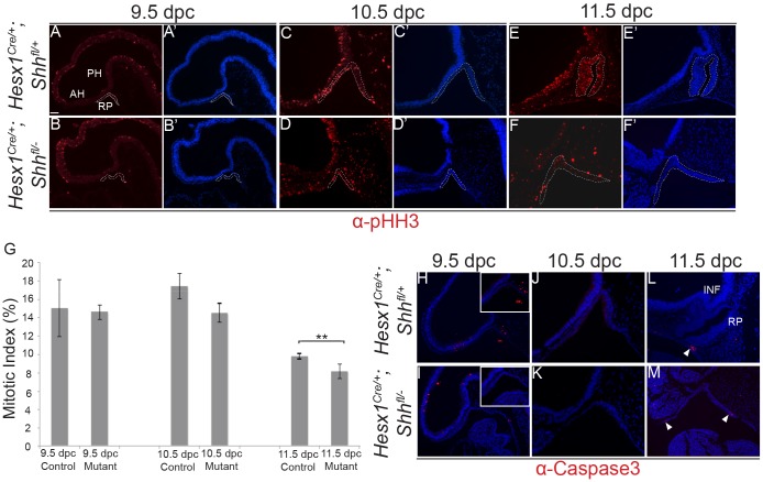
Fig. 7.
Reduced proliferation of RP epithelium in Hesx1Cre/+;Shhfl/− mutant embryos. (A-G) Immunofluorescent staining against phospho-Histone H3 (pHH3) on mid-sagittal sections of control and Hesx1Cre/+;Shhfl/− embryos from 9.5 to 11.5 dpc. Note the significant decrease in the mitotic index in the RP epithelium in Hesx1Cre/+;Shhfl/− mutant compared with control embryos at 11.5 dpc. The mitotic index is the ratio of pHH3+ cells out of the total DAPI+ nuclei. Student's t-test: 9.5 dpc, P=0.92; 10.5 dpc, P=0.0645; 11.5 dpc, P<0.001 (n=6 embryos per group). (H-M) Immunofluorescence against activated cleaved caspase 3 revealing the absence of positive cells in RP epithelium in both genotypes from 9.5 and 10.5 dpc. Apoptotic cells are observed only in the ‘feet’ of the evaginating RP at 11.5 dpc (arrowheads). Mesenchymal cells around RP show reduced staining in the mutant embryo. Positive signal in the cavities is artefactual. Dashed lines in A-F′ delineate the area that was quantified. RP, Rathke's pouch; PH, posterior hypothalamus; AH, anterior hypothalamus. Scale bar: 100 μm.

