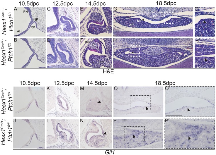
Fig. 8.
Conditional deletion of Ptch1 in Hesx1Cre/+;Ptch1fl/fl mutants results in pituitary hyperplasia. (A-H) Haematoxylin and Eosin staining on mid-sagittal (A-F) and coronal (G,H) sections of control and Hesx1Cre/+;Ptch1fl/fl embryos throughout pituitary organogenesis. At 10.5 dpc (A,B), the morphology of the RP is comparable between genotypes, but looks slightly expanded in the mutant compared with the control embryo at 12.5 dpc (C,D). By 14.5 dpc (E,F), the Hesx1Cre/+;Ptch1fl/fl anterior pituitary (AP) is clearly hyperplastic relative to the control, but the infundibulum (INF) looks normal. AP hyperplasia is also evident at 18.5 dpc (G,H), with dorsal extensions of the cleft. The posterior pituitary (PP) looks comparable between genotypes. Note the expansion of the marginal zone (MZ; arrowheads in G,H), which is thicker in the mutant compared with the control pituitary. G′ and H′ are higher magnifications of the boxed areas in G and H (arrowheads indicate the marginal zone). (I-P) In situ hybridisation for Gli1. A remarkable increase in Gli1 expression is observed throughout the AP in the mutants relative to the control embryos, including the MZ (arrowheads in O and P). O′ and P′ are higher magnifications of the boxed areas in O and P. RP, Rathke's pouch; PH, posterior hypothalamus; AH, anterior hypothalamus; AP, anterior pituitary; INF, infundibulum. Scale bar: 100 μm.

