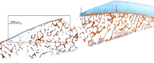
Figure 1.

In vivo preparation (standard debridement) of chondral defects in osteoarthritic knees. Representative section of the debrided surface in 1 of the 5 samples (12.5%) displaying one large area with a missing bone plate and an open bone marrow space (Masson’s trichrome-Goldner stain). Red arrow: tide mark line.
