

Abstract
This report presents anterior segment optical coherence tomography (AS-OCT) images of Kayser-Fleischer ring (KFR) in a child. The AS-OCT images highlight differential reflectivity of the KFR depending on amount of copper deposited in cornea, thus supporting the role of AS-OCT as a follow-up tool. Utility of AS-OCT for diagnosing and documenting the KFR in children otherwise uncooperative for detailed slit lamp examination is discussed.
Keywords: Ophthalmology, Liver disease
Background
Kayser-Fleischer ring (KFR) is pathognomic for Wilson’s disease and represents deposition of copper in the posterior corneal layers.1 The ophthalmologist’s role becomes very important in diagnosis of the disease as the KFR often eludes the observer’s eye on naked eye examination, especially in its early stages. Furthermore, Wilson’s disease usually presents in young children, sometimes even in the preschool age,2 when a detailed magnified focal slit examination in bright light using the slit lamp may not be possible.
In this regard, we present the utility of anterior segment optical coherence tomography (AS-OCT) as a diagnostic and documentation tool for imaging the KFR. The ability of AS-OCT to image corneal copper differentially depending on amount of the metal deposited is also discussed.
Case presentation
A 7-year-old female child was brought to our outpatient services with the suspected diagnosis of Wilson’s disease. The 24-hour urine copper had been noted to be high, while the serum ceruloplasmin levels were reduced. Systemic examination by a paediatrician had revealed hepatomegaly. There was no history of altered behaviour or psychosis. Patient was referred to the ophthalmology department for detection of KFR.
The visual acuity was at least 20/200 in both eyes. The child was uncooperative for detailed slit lamp examination. The anterior segment was examined under diffuse low light conditions (figure 1A), while thorough slit examination was not possible. A 360° circumferential greenish brown ring was noted in the perilimbal area with an accompanying lucid interval in both eyes. However, the precise level of the ring could not be established. The rest of the ocular examination was apparently within normal limits.
Figure 1.
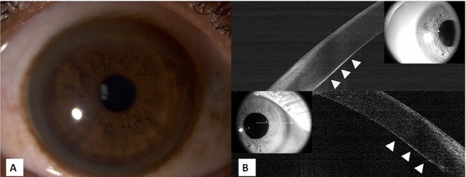
(A) Clinical photograph of the KFR. (B) OCT images of the temporal (upper image) and nasal (lower image) limbus of the right eye depicting the ring as a hyper-reflective band (arrowheads), more evident temporally than nasally as in figure 1A. KFR, Kayser-Fleischer ring; OCT, optical coherence tomography.
With a doubtful clinical diagnosis of KFR, AS-OCT (RTVue, Optovue, Fremont, California, USA) was performed in both eyes. The child was relatively more cooperative for the investigation. A highly reflective band was noted in the posterior cornea involving the area of the Descemet’s membrane and posterior stroma. The band was noted to extend all around the corneal circumference. However, differential reflectivity was easily appreciable with the density being more temporally as compared with nasal limbus (figure 1B).
Hence, the diagnosis of KFR was confirmed.
Investigations
Twenty-four-hour urine copper extraction: 180 mcg/24 hours
Serum ceruloplasmin: 12 mg/dL
Haemoglobin: 11 mg/dL
AS-OCT: optical coherence tomography images revealed KFR as discussed above (figure 1B).
Outcome and follow-up
The child was referred back to the paediatrician with the confirmed ocular diagnosis of KFR, who advised copper chelation therapy. The child is currently on follow-up with the authors as well for sequential monitoring of the KFR both clinically and on AS-OCT.
Discussion
The KFR typically starts without symptoms at the vertical poles of the cornea due to deposition of copper in its deeper layers and then progresses circumferentially. It is known to gradually disappear with normalisation of serum copper levels. Slit lamp is considered the gold standard for diagnosis, while gonioscopy is needed for ruling out KFR in early cases.3 However, in children as well as young adults with neurological deficits due to Wilson’s disease, such intricate microscopic examination may not be possible. Over and above this, clinical examination is highly subjective and dependent on observer skill level. The available clinical scoring methods for judging therapeutic response in Wilson’s disease with KFR are very cumbersome.4
Previously, Scheimpflug imaging has been used for detection of KFR with high specificity.5 In that series of 21 patients, only one patient was found to have a false-positive reading. The study design, however, involved imaging the inferior cornea only. Also, the sensitivity was doubtful, and the authors concluded ‘Scheimpflug does not seem to be more sensitive in detecting KFRs than an experienced ophthalmologist’.
AS-OCT is more easily available than Scheimpflug-based imaging systems. The imaging process involves fixation at an in-built target for a few seconds only, without exposure to bright light, as against a detailed slit-lamp examination. Further availability of autofocusing module makes image acquisition fairly easy. We believe that using ancillary AS-OCT imaging for diagnosis may obviate the need of a skilled cornea specialist to some extent. In a small series of seven patients with KFR, it has been shown that AS-OCT may be useful for serial measurements.6 The authors also discussed the possibility of using the density of the hyper-reflective band as a tool for measurement and gauging response to therapy. We plan to follow-up this patient and perform sequential AS-OCT imaging. Correlation with changes in serum copper and ceruloplasmin levels will also be interesting.
AS-OCT is therefore a useful tool for determining presence of KFR in difficult cases. Given its resolution, reproducibility and repeatability, AS-OCT may also be a useful adjunct for sequentially monitoring therapeutic response and making diagnosis in doubtful cases of Wilson’s disease.
Learning points.
Copper deposits of Kayser-Fleischer ring are seen as a hyper-reflective layer in the posterior cornea on anterior segment optical coherence tomography (AS-OCT).
AS-OCT may be a used as a diagnostic adjunct in difficult and early cases of Wilson’s disease.
Because of its ability to diagnose differential deposition of copper depending on the amount deposited, it is likely to be useful in sequential monitoring of Wilson’s disease. However, further studies are needed to prove its role in management of Wilson’s disease.
Footnotes
Contributors: AR and BT contributed to diagnosis, management, imaging and writing the manuscript. NG contributed to imaging and writing the manuscript. PKM performed crititical review of the manuscript.
Competing interests: None declared.
Patient consent: Consent obtained from guardian.
Provenance and peer review: Not commissioned; externally peer reviewed.
References
- 1. Schrag A, Schott JM. Images in clinical medicine. Kayser-Fleischer rings in Wilson’s disease. N Engl J Med 2012;366:e18 10.1056/NEJMicm1101534 [DOI] [PubMed] [Google Scholar]
- 2. Manolaki N, Nikolopoulou G, Daikos GL, et al. . Wilson disease in children: analysis of 57 cases. J Pediatr Gastroenterol Nutr 2009;48:72–7. 10.1097/MPG.0b013e31817d80b8 [DOI] [PubMed] [Google Scholar]
- 3. Patel AD, Bozdech M. Wilson disease. Arch Ophthalmol 2001;119:1556–7. [DOI] [PubMed] [Google Scholar]
- 4. Esmaeli B, Burnstine MA, Martonyi CL, et al. . Regression of Kayser-Fleischer rings during oral zinc therapy: correlation with systemic manifestations of Wilson’s disease. Cornea 1996;15:582–8. [PubMed] [Google Scholar]
- 5. Telinius N, Ott P, Hjortdal J. Detection of Kayser-Fleischer ring using Scheimpflug imaging. Acta Ophthalmol 2017;95:e248–e249. 10.1111/aos.13271 [DOI] [PubMed] [Google Scholar]
- 6. Sridhar MS. Advantages of anterior segment optical coherence tomography evaluation of the Kayser-Fleischer ring in Wilson disease. Cornea 2017;36:1–346. 10.1097/ICO.0000000000001126 [DOI] [PubMed] [Google Scholar]