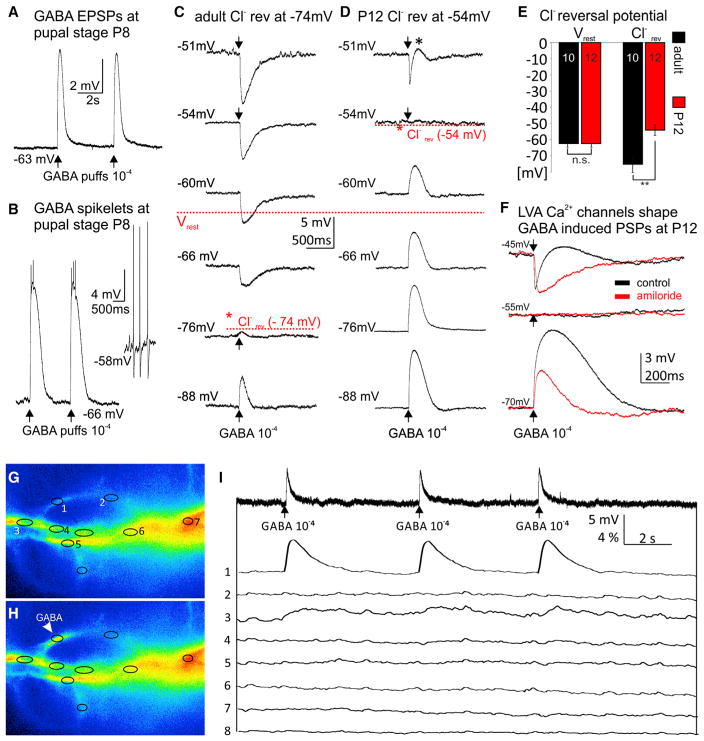
Figure 4. GABA Induces Depolarization, LVA Ca2+ Channel Activation, and Dendritic Ca2+ Signals during Immature Stages.
(A) During pupal life somatic current clamp recordings revealed depolarizations upon brief (5 ms) puffs of GABA (10−4 M) onto dendrites.
(B) For some high order branches within the GABAergic domain GABA puffs caused large amplitude depolarizations with spikelets. GABA-induced spikelets displayed distinctly different amplitudes and shapes as compared to spontaneously occurring action potentials as recorded from the soma (see inset).
(C and D) Sharp electrode recordings were used to determine the chloride reversal potential in adult (C) and pupal (D) MN5. (C) Representative responses to GABA puffs at differentmembranepotentials revealeda chloride reversal potential of −74mV for the adult flight MN5, more than 10m V more hyperpolarized than the average resting membrane potential (−63 mV). (D) By contrast, at P12 the chloride reversal was at −54 mV, about 10 mV more depolarized than resting membrane potential.
(E) Quantification revealed that resting membrane potential remained unchanged through development (−63 ± 2 mV), but chloride reversal was significantly shifted from values more positive than resting during pupal life (−54 ± 3 mV) to values more negative than resting in mature stages (−74 ± 4 mV).
(F) GABA-induced depolarizations are amplified by Ca2+ influx through LVA channels. Sharp electrode recording from a representative pupal stage P12 MN5 held at −70 mV displayed a large depolarizing response to dendritic GABA puffs (lower traces, black). Pharmacological blockade of LVA DmαG Ca2+ channels by amiloride (10−3 M) significantly reduced amplitude and duration of this GABA response (lower traces, red). No response to GABA puff was observed when holding the cell at the chloride reversal potential (middle traces). A biphasic response, hyperpolarization followed by depolarization, was observed when holding membrane potential at −45 mV (upper traces, black). Blockade of LVA channels isolated the GABA-induced hyperpolarizing response (upper traces, red).
(G–I) During pupal life GABA puffs (10−4 M) cause somatic depolarizations but local dendritic Ca2+ signals at the site of GABAAR activation. Ca2+ imaging of parts of MN5 dendrites before (G) and after (H) GABA puff to ROI 1. (I) Simultaneous somatic current clamp recording (upper trace) and Ca2+ imaging from ROIs 1–8 as defined in (G) and (H) in response to three GABA puffs to ROI 1 (for signal spread, see Movie S5).
See also Figures S5D–S5F for pharmacological evidence that GABA-mediated depolarizations required chloride channels. Local dendritic Ca2+ responses to GABA puffs at different dendritic sites are shown in Figure S6 and Movie S6.

