Abstract
Matrix Assisted Laser Desorption/Ionization Time-of-Flight Mass Spectrometry (MALDI-TOF MS) has been demonstrated to be useful for tick identification at the species level. More recently, this tool has been successfully applied for the detection of bacterial pathogens directly in tick vectors. The present work has assessed the detection of Borrelia burgdorferi sensu lato in Ixodes ricinus tick vector by MALDI-TOF MS. To this aim, experimental infection model of I. ricinus ticks by B. afzelii was carried out and specimens collected in the field were also included in the study. Borrelia infectious status of I. ricinus ticks was molecularly controlled using half-idiosome to classify specimens. Among the 39 ticks engorged on infected mice, 14 were confirmed to be infected by B. afzelii. For field collection, 14.8% (n = 12/81) I. ricinus ticks were validated molecularly as infected by B. burgdorferi sl. To determine the body part allowing the detection of MS protein profile changes between non-infected and B. afzelii infected specimens, ticks were dissected in three compartments (i.e. 4 legs, capitulum and half-idiosome) prior to MS analysis. Highly reproducible MS spectra were obtained for I. ricinus ticks according to the compartment tested and their infectious status. However, no MS profile change was found when paired body part comparison between non-infected and B. afzelii infected specimens was made. Statistical analyses did not succeed to discover, per body part, specific MS peaks distinguishing Borrelia-infected from non-infected ticks whatever their origins, laboratory reared or field collected. Despite the unsuccessful of MALDI-TOF MS to classify tick specimens according to their B. afzelii infectious status, this proteomic tool remains a promising method for rapid, economic and accurate identification of tick species. Moreover, the singularity of MS spectra between legs and half-idiosome of I. ricinus could be used to reinforce this proteomic identification by submission of both these compartments to MS.
Introduction
Lyme borreliosis is the most prevalent vector borne disease in the northern hemisphere [1]. This multi-systemic disease presents a large variety of associated clinical signs which hamper clinical diagnosis. Lyme disease is caused by bacteria belonging to the species complex Borrelia burgdorferi sensu lato (sl). B. burgdorferi sl complex includes, up to now, 21 known bacteria species [2]. Among these species 5 are commonly found in human pathology, and B. afzelii is the most prevalent bacterium in Europe [1]. These pathogens are transmitted by blood feeding of infectious ticks belonging to the Ixodes genus. In western Europe, I. ricinus is the most common vector [1].
Until now, there is no human licensed vaccine available, prevention and vector controls remain the main protective measures. To inform populations and to establish efficient protective devices, realization of vector epidemiological studies is required to map tick species and to determine their Borrelia infectious status. Tick species identification could be carried out morphologically and the detection of B. burgdorferi sl. is mainly achieved using molecular biology methods [3]. The expertise required for tick morphological identification and the high costs and time necessary for molecular pathogen detection are limitation factors for rapid characterization of tick species and their Borrelia infectious status.
Recently, Matrix Assisted Laser Desorption/Ionization Time-of-Flight Mass Spectrometry (MALDI-TOF MS) has been successfully applied for the identification of several arthropod families, including ticks [4]. Different tick body parts could be used for their identification by MS, either using whole specimens [5] or tick legs [6,7]. As protein repertory is not equivalent according to tick body part of the same species, the same tick compartment should be used for MS spectra queried against the MS reference database and database creation.
Moreover, this last decade, MALDI-TOF MS has been introduced in clinical microbiology laboratories for the identification and classification of micro-organisms, including bacteria and yeast [8]. The sample preparation simplicity, rapidity and reagent low-costs contributed to the popularity of this tool for microbial routine analyses [9]. Consequently, based on the success of this tool for bacteria and tick identification, MALDI-TOF MS was assessed for the determination of tick bacteria-infectious status. A pioneering study established the proof-of-concept of Borrelia crocidurae detection in soft ticks, Ornithodoros sonrai, by submitting leg protein extracts to MS [10]. More recent works, studying the detection of Rickettsia spp. pathogens in infected ticks using MALDI-TOF MS, reported the dual identification of tick species and Rickettsia spp. infectious status using tick legs [11] or tick hemolymph [12]. Indeed, reproducible changes in MS protein profiles were observed between Rickettsia-free and–infected conspecific specimens.
The aim of the present study was to assess MALDI-TOF MS ability to detect B. afzelii in I. ricinus tick vector which could be useful for entomological diagnosis. I. ricinus ticks collected in the field or infected experimentally by B. afzelii NE4049 were used for this demonstration. The development of a rapid, economic and reliable method for dual identification; the determination of tick species and their Borrelia infectious status, is becoming more and more essential in the framework of Lyme disease vectors monitoring and pathogen circulation.
Material and methods
Borrelia afzelii NE4049 culture
Borrelia afzelii NE4049 [13] was cultured in the BSK-H medium (Sigma, Saint Quentin Fallavier, France) under anaerobic conditions for 8 days. The medium was then centrifuged and the pellet washed three times with PBS. Finally, the pellet was resuspended with 100 μL of PBS and borrelial density (bacteria/μL) was determined using a Petroff-Hausser counting chamber.
Ticks
A total of 149 I. ricinus ticks collected in the field (n = 81) or laboratory-reared (n = 68) were used for this study. Field collection of ticks was done in the Murbach area (GPS: N47.918961 / E007.210436; Alsace, France) in June 2016, by dragging a white flannel flag (1x1 m) over low vegetation. Ticks species were determined by morphological identification under a binocular loupe at a magnification of ×56 (Leica M80, Leica, Nanterre, France) using taxonomic keys [14]. Only I. ricinus ticks were included in this work. The laboratory rearing of I. ricinus ticks was performed as previously described [15,16]. For Borrelia experimental infections of ticks, I. ricinus specimens from the larva stage were fed on C3H/HeN mice infected (n = 39) by B. afzelii strain NE4049 or not infected (n = 29) as previously described [16,17]. At the nymphal stage, tick specimens were frozenly killed until future analyses.
Mice used in tick experiments were three to four-week-old C3H/HeN pathogen-free. They were obtained from our breeding colony and provided food and water ad libitum. At the end of the experiment, mice were killed by isoflurane gas overdose.
Tick dissection
Ticks were processed as previously described [6]. Briefly, each specimen was rinsed 1 minute once with 70% (v/v) ethanol, twice with distilled water and then air-dried. Ticks were individually dissected with a sterile surgical blade under a binocular loupe. Four legs and the capitulum were removed and the idiosome was longitudinally cut in two equal parts. The half-idiosome harboring legs were used for molecular analysis. The three other body parts, the four legs, the capitulum and the half-idiosome without the legs, were prepared individually for MALDI-TOF MS analysis. For ticks collected in the field, only the four legs and the half-idiosome legs less, were submitted to MALDI-TOF MS, the remaining body part was used for molecular analyses.
DNA extraction
DNA of each half-idiosome with legs was individually extracted with ammonium hydroxide (Sigma-Aldrich) as previously described [18,19]. Purified DNA from each tick specimen was either immediately used or stored at -80°C until use.
Molecular identification of ticks
DNA from 14 morphologically identified I. ricinus specimen collected in the field were validated by sequencing a PCR fragment of 480 bp from cytochrome oxidase I (COI) gene as previously described [20]. The sequences were analyzed using the 4Peaks software (version 1.7.1) (Softnic® Corporate, Barcelona, Spain), and were then blasted against GenBank (http://blast.ncbi.nlm.nih.gov).
Molecular detection of B. burgdorferi sl in ticks
The presence of B. burgdorferi sl in ticks was determined by a real-time PCR assay targeting a conserved region of the flagellin gene. Species genotyping was achieved by melting curve analysis as previously described [21]. DNA from B. afzelii, B garinii and B. burgdorferi ss were added or not to the PCR mix as positive and negative controls, respectively.
Sample homogenization and MALDI-TOF MS analysis
Each compartment dissected was homogenized individually using a FastPrep-24 device (MP Biomedicals, Illkirch-Graffenstaden, France) and glass beads (Sigma, Lyon, France) in a mix (50/50) of 15 μL 70% (v/v) formic acid (Sigma) and 15 μL 50% (v/v) acetonitrile (Fluka, Buchs, Switzerland) for protein extraction according to the standardized automated setting described by Nebbak et al. [22]. A quick spin centrifugation at 200 g for 1 min was then performed and 1 μL of the supernatant of each sample was spotted on the MALDI-TOF steel target plate in quadruplicate (Bruker Daltonics, Wissembourg, France). After air-drying, 1 μL of matrix solution composed of saturated α-cyano-4-hydroxycinnamic acid (Sigma, Lyon, France), 50% (v/v) acetonitrile, 2.5% (v/v) trifluoroacetic acid (Aldrich, Dorset, UK) and HPLC-grade water was added. Matrix solution was loaded in duplicate onto each MALDI-TOF plate with and without bacterial control (Pseudomonas aeruginosa ATCC 27853) respectively as a positive or negative control. Spectra were acquired on a Microflex LT MALDI-TOF Mass Spectrometer (Bruker Daltonics) as previously described [23].
Mixing Borrelia with tick protein extracts
Pelleted B. afzelii were suspended in water to obtain 1.107 bacteria/μL. Serial dilution in water were done to add 106, 105 or 104 bacteria to half-idiosome protein extract from Borrelia-free I. ricinus. One microliter of this mix was spotted in quadruplicate on the MALDI-TOF target plate. Half-idiosome protein extract from Borrelia-free I. ricinus and B. afzelii bacteria protein extract were loaded on the MS target plate as controls.
MS spectra analysis
MS spectra profiles were firstly controlled visually with flexAnalysis v3.3 software (Bruker Daltonics). MS spectra were then exported to ClinProTools v2.2 and MALDI-Biotyper v3.0. (Bruker Daltonics) for data processing (smoothing, baseline subtraction, peak picking). MS spectra reproducibility was assessed by the comparison of the average spectral profiles (MSP, Main Spectrum Profile) obtained from the four spots for each specimen according to body part and infectious status with MALDI-Biotyper v3.0 software (Bruker Daltonics). MS spectra reproducibility and specificity taking into account tick body part and Borrelia infectious status were objectified using clustering analyses and Composite Correlation Index (CCI) tool. Cluster analyses (MS dendrogram) were performed based on comparison of the MSP given by MALDI-Biotyper v3.0. software and clustered according to protein mass profile (i.e., their mass signals and intensities). The CCI tool from MALDI-Biotyper v3.0. software was also used, to assess the spectral variations within and between each sample group, according to the body part and Borrelia infectious status, as previously described [23,24]. Higher correlation values (expressed by mean ± standard deviation–SD) reflect higher reproducibility for the MS spectra, and were used to estimate MS spectra distance for each condition (body part and Borrelia-infectious status). To visualize MS spectra distribution from ticks collected in the field, according to body part and/or Borrelia-infectious status, principal component analysis (PCA) from ClinProTools v2.2 software was performed. To list discriminating peaks between compartments and/or infectious status, MS spectra were analysed using the genetic algorithm (GA) model from ClinProTools v 2.2 software. The maximum number of generations was set to 250 and the number of neighbours was three for K nearest neighbour (KNN) classification. A manual inspection and validation of the selected peaks gave recognition capability (RC) value together with the highest cross-validation (CV) value to assess the reliability and accuracy of the model. The discriminating peak masses generated by the model were searched in the peak report created for each compartment and condition.
Database creation and blind tests
The reference MS spectra were created using spectra from legs and half-idiosomes of five I. ricinus ticks at the nymphal stage using MALDI-Biotyper software v3.0. (Bruker Daltonics) [25]. MS spectra were created with an unbiased algorithm using information on the peak position, intensity and frequency. The reproducibility of the MS profiles per body part was evaluated with MALDI-Biotyper software v3.0., which assigns log score values (LSVs) based on the degree of confidence with which the query spectrum identifies to the reference spectrum. LSVs ranged from 0 to 3. According to previous studies [6,23], a LSV of at least 1.8 should be obtained to be considered reliable for species identification. Data were analysed by using GraphPad Prism software version 5.01 (GraphPad, San Diego, CA, USA).
Ethical statement
The protocols to maintain tick colony (N°APAFIS 886–2015062209279407) and to infect ticks on Borrelia infected mice (N°APAFIS 885–2015062209113374) were approved by the Comité Régional d’Ethique en Matière d’Expérimentation Animale de Strasbourg (CREMEAS—Committee on the Ethics of Animal Experiments of the University of Strasbourg). These protocols follow the European directive 2010/63/EU. The authority who issued the permission to collect ticks for each location was the ONF (Office National des forêts, France). The field studies did not involve endangered or protected species.
Results and discussion
Molecular detection of B. burgdorferi sl in ticks
Among the 39 I. ricinus larva, laboratory-reared and experimentally exposed to mice infected by B. afzelii, 36% of the ticks (14/39) were found infected by B. afzelii at the nymphal stage, according to real-time PCR results. The absence of B. afzelii was confirmed by RT-PCR in the 29 I. ricinus nymphs, laboratory-reared and engorged on pathogen-free mice at the larva stage.
A total of 81 ticks at the nymphal stage were collected by flagging in the Murbach area (France). All these specimens were classified as I. ricinus by morphological identification. Among them, the DNA extracted from half-idiosome of 13 specimens, randomly selected, were submitted to tick species identification by molecular tool. BLAST analyses corroborated morphological identification showing 99% sequence similarities with I. ricinus COI sequence from GenBank (Accession number: KF197134.1). Concerning their Borrelia infectious status, 12 ticks (14.8%) were found infected, including six, three and two specimens by B. afzelii, B. burgdorferi ss and B. garinii, respectively, and one co-infected by B. garinii and B. burgdorferi ss. This rate of infection is commonly found in this area [3] and B. afzelii is the most prevalent species in Europe [26].
Assessment of MS spectra specificity between Borrelia-free and–infected ticks according to body part
The research of MS spectra changes according to I. ricinus infectious status was done in three body compartments, the four legs, the capitulum and the half-idiosome legs less. The legs were chosen because all anterior descriptions of distinctive MS spectra in ticks between pathogen-free and bacteria-infected specimens were achieved using legs [10,11] or hemolymph collected at the legs level [12]. The detection of B. crocidurae in the legs of soft ticks and some Rickettsia species in the legs and hemolymph of hard ticks underlined that these bacteria disseminate in the ticks at sufficiently elevated concentration to modify MS spectra. The capitulum was tested to comfort the specificity of leg MS profile changes following Borrelia infection. Indeed, if MS profile changes were detected at the leg level, it appeared essential to submit to MS another body part of the same tick specimen to assess whether MS pattern modifications were also detected in this second compartment and whether some of these MS peak changes were shared between legs and capitulum. Finally, as the presence of B. burgdorferi sl was repeatedly reported in the gut of infected I. ricinus ticks [27,28], the half-idiosomes were also submitted to MS analysis.
Reproducibility of MS spectra according to tick compartment
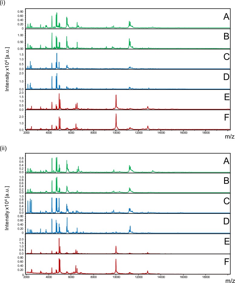
Before comparing MS spectra from the same tick compartment according to the Borrelia-infectious status, reproducible MS spectra had to be obtained for I. ricinus for each compartment and Borrelia-infectious status. The MS spectra comparison from five laboratory reared specimens infected (Fig 1 –ii) or not (Fig 1 –i) by Borrelia visually revealed reproducible protein profiles for each body part. Interestingly, MS spectra from legs (A,B) and capitula (C,D) were closely related. Conversely, MS spectra from half-idiosomes (E,F) appeared more distinct compared to the MS spectra from the two other body parts. Nevertheless, for each body part, no visible MS peak clearly distinguished Borrelia-infected (ii) from pathogen-free specimens (i).
Fig 1. Comparison of MALDI-TOF MS spectra from legs, capitula and half-idiosomes of adult I. ricinus pathogen-free (i) or infected by Borrelia afzelii (ii).
Representative MS spectra of legs (A, B), capitula (C, D) and half-idiosomes (E, F) from laboratory reared I. ricinus homogenized automatically using FastPrep-24 device with glass powder. a.u., arbitrary units; m/z, mass-to-charge ratio.
Specificity of MS spectra according to tick Borrelia-infectious status
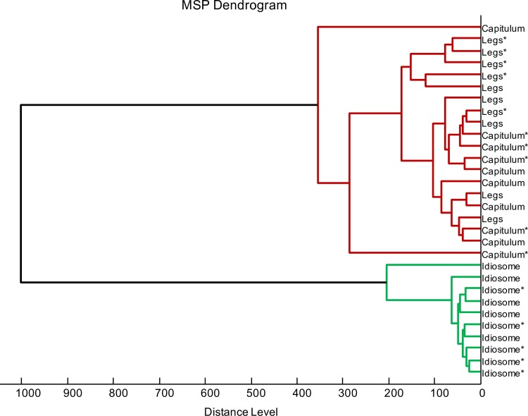
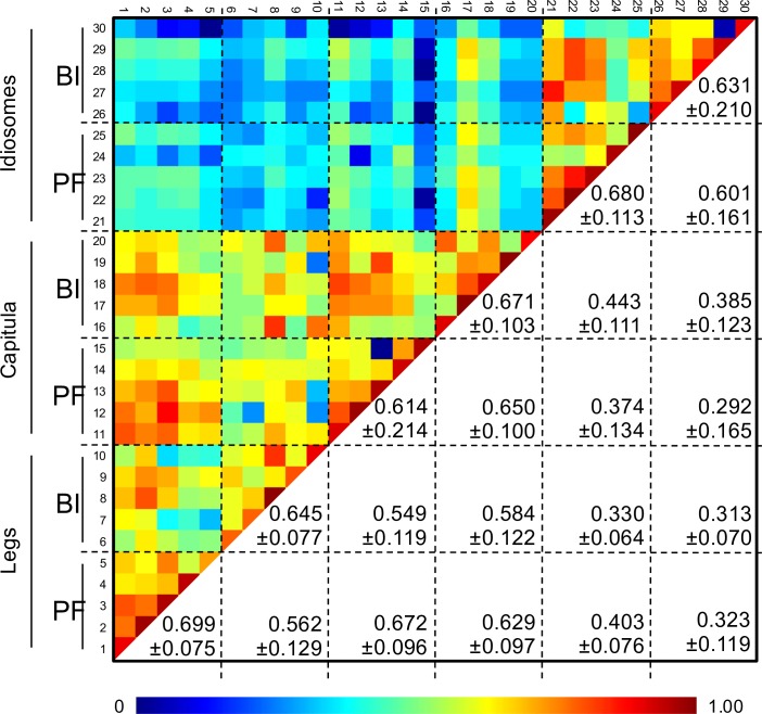
To assess whether specific MS profiles could be associated to tick infectious status per body part, five MS profiles per condition were used to perform clustering and correlation analyses. The dendrogram showed that half-idiosome MS spectra clustered on distinct branches from legs and capitula (Fig 2). Moreover, these last two body parts were found imbricated suggesting the absence of specific MS profiles distinguishing legs and capitula. Additionally, an overlapping of MS spectra from Borrelia-infected and pathogen-free specimens was observed for each compartment on the dendrogram. These results indicated that MS protein profiles were weakly affected by Borrelia infection and comforted the low specificity of MS spectra to distinguish tick legs and capitula. These data were confirmed by CCI matrix highlighting a low correlation of MS spectra between idiosomes and legs or capitula (mean±SD: 0.358 ± 0.121; Fig 3). The high CCI obtained between MS spectra from legs and capitula regardless of their infectious status also strongly suggest the lack of specificity in the MS profile. The superimposition of average MS profiles from idiosomes between Borrelia-infected and pathogen-free specimens using ClinProTool software (Bruker), did not reveal differences in peak position. The detection of few peaks of low intensity modifications could explain the decrease of MS spectra correlation for paired comparison of the idiosome profiles according to Borrelia-infectious status. These intensity MS peak changes could be attributed to tick response to bacterial infection. Indeed, previous works reported transcriptome [29] or protein repertoire [30] changes according to the infection tick status. However, the MS profile changes appeared insufficient to associate a specific protein pattern to I. ricinus infected by Borrelia. The appearance and/or disappearance of at least a few MS peaks following tick infection by a specific Borrelia seems necessary to sustain the detection of a specific MS profile.
Fig 2. MSP dendrogram of MALDI-TOF MS spectra from legs, capitula and half-idiosomes of adult I. ricinus pathogen-free or infected by Borrelia afzelii.
Five specimens per body part and B. afzelii infectious status were used to construct the dendrogram. The dendrogram was created using Biotyper v3.0 software and distance units correspond to the relative similarity of MS spectra. The specimens infected by B. afzelii were indicated by asterisks (*).
Fig 3. Assessment of I. ricinus MS spectra reproducibility according to tick body parts and Borrelia infectious status using composite correlation index (CCI).
MS spectra from five specimens per body part and B. afzelii infectious status were analysed using the CCI tool. Body part and infectious status are indicated on the left side of the heat map. Levels of MS spectra reproducibility are indicated in red and blue revealing relatedness and incongruence between spectra, respectively. CCI matrix was calculated using MALDI-Biotyper v3.0. software with default settings (mass range 3.0–12.0 kDa; resolution 4; 8 intervals; auto-correction off). The values correspond to the mean coefficient correlation and respective standard deviations obtained for paired condition comparisons. CCI were expressed as mean ± standard deviation. BI, Borrelia-infected; PF, pathogen-free.
Comparison of I. ricinus MS profiles between laboratory-reared and field collected specimens
A comparison of MS spectra distribution from specimens collected in the field taking into account diversity of Borrelia species identified in I. ricinus ticks was performed. To visualize their distribution according to their Borrelia-infectious status, PCA was performed. Twelve I. ricinus ticks (co)-infected by a B. burgdorferi sl species, plus five Borrelia-free specimens and five laboratory reared I. ricinus ticks were included in this analysis. An intertwining of the dot-reflecting MS spectra distribution from half-idiosomes (Fig 4A) and legs (Fig 4B) was observed independently of their infectious status. No clustering was found for MS spectra from ticks infected neither by the same Borrelia species, nor between Borrelia-free and–infected specimens in each compartment (Fig 4A and 4B). Then, these results strengthened the uselessness of MALDI-TOF MS to distinct I. ricinus specimens according to their Borrelia-infectious status in both these body parts. Interestingly, the dots corresponding to MS spectra from Borrelia-free specimen, laboratory reared and collected in the field, overlapped, underlining the specificity of the protein profiles to each I. ricinus body part independently of their origins (i.e., laboratory or field).
Fig 4. Principal component analysis (PCA) from MS spectra of idiosomes and legs of I. ricinus infected or not by Borrelia sp.
PCA dimensional image from MS spectra of I. ricinus idiosomes (A) and legs (B) Borrelia-free (red dots, n = 10), infected by B. afzelii (green dots, n = 6), B. burgdorferi (blue dots, n = 3), B. garinii (yellow dots, n = 2), co-infected by B. garinii and B. burgdorferi (purple dots, n = 1). (C) PCA dimensional image from the same MS spectra of I. ricinus idiosomes (red dots, n = 22) and legs (green dots, n = 22). The contributions of PC1, PC2 and PC3 were 38.4%, 15.5% and 7.2%, respectively. Among the Borrelia-free specimens, five were laboratory reared and the other five came from field collection.
Conversely, the submission of these same last 22 samples per body part to PCA highlighted a clear separation of the dots from the legs and idiosomes (Fig 4C), confirming a specificity of MS spectra between these two compartments. Thirteen discriminating MS peaks were exhibited using the Genetic Algorithm model (ClinProTools software) between idiosomes and legs from five laboratory reared and pathogen-free I. ricinus specimens (Table 1). Recognition capability (RC) and a cross validation (CV) 100% values were obtained which confirms their accuracy for tick identification. Indeed, MS profiles are poorly affected by the environmental conditions [31] and, even specific, there is a cross recognition between the Rickettsia-free and infected tick [11].
Table 1. Mass peak list distinguishing legs and idiosomes from laboratory reared and pathogen-free I. ricinus specimens.
| Mass (m/z) | Start Mass | End Mass | Legs | Idiosome |
|---|---|---|---|---|
| 3329.42 | 3321.25 | 3335.84 | - | + |
| 3724.17 | 3716.85 | 3728.83 | + | - |
| 3751.09 | 3742.66 | 3755.75 | + | - |
| 4453.72 | 4446.31 | 4463.82 | + | - |
| 4721.00 | 4717.60 | 4727.42 | + | - |
| 4996.13 | 4981.44 | 5005.29 | - | + |
| 5496.18 | 5483.74 | 5503.89 | - | + |
| 5701.28 | 5695.40 | 5716.65 | + | - |
| 6203.19 | 6188.93 | 6213.30 | - | + |
| 6311.90 | 6293.38 | 6321.68 | - | + |
| 6411.56 | 6395.70 | 6421.23 | - | + |
| 9991.35 | 9975.31 | 10009.66 | - | + |
| 12821.56 | 12769.80 | 12859.35 | - | + |
| Total | 5 | 8 |
Da. Daltons; m/z. mass to charge.
The inefficiency of MS spectra to distinguish Borrelia-infected from pathogen-free I. ricinus ticks regardless of the body part tested could be attributed to different factors. Firstly, at a distance of blood feeding, B. burdorgferi bacteria are mainly confined to tick gut [27]. The sequestration of the Borrelia in the gut could explain the lack of probative MS spectra changes in the legs and capitula of B. afzelii infected ticks. Moreover, the bacterial load seems to be decreasing over time especially for laboratory-reared ticks [32]. On the contrary, R. slovaca, R. conorii and R. massilliae can be observed directly by tick hemolymph drop staining [33]. The dissemination of bacteria to the entire tick body vectors explains their detection in legs [11] and hemolymph of ticks [12].
The inability to distinguish Borrelia-infected from pathogen-free I. ricinus ticks at the idiosome level could be attributed, on the one hand, to the low gut Borrelia inoculum in unfed ticks (approximately 2000 spirochetes [27]) and, on the other hand, to the variety of other resident bacteria species hiding their detection [34]. The blood meal was reported to ensure the gut multiplication and the dissemination of the Borrelia to the tick salivary glands [27]. Then, the research of MS profiles changes in ticks recently blood-engorged could be an alternative. Nevertheless, the blood contained in the idiosomes from freshly engorged ticks was already reported to strongly modify MS spectra, impairing tick identification [5].
An alternative proteomic strategy for elucidation tick B. burgdorferi sl-infectious status could be to analyze peptide MS profiles instead of intact proteins. This approach was successfully applied for Culicoides species identification [35]. With this method, it would be possible to identify unambiguously differential MS peaks attributed to B. burgdorferi sl-infection by peptide sequencing using tandem MS (MS/MS). However, the resort to more sophisticated mass spectrometry apparatus is required such as MALDI-TOF MS/MS or liquid chromatography electrospray ionization (LC/ESI) MS/MS machines [35]. The MS window range was also change (i.e., 400 to 4kDa), hampering the use of resulting MS spectra to query the home-made MS reference database for tick species identification done on MALDI-TOF MS. Moreover, an additional step consisting in sample enzymatic digestion (e.g. Trypsin) prior to MS submission is necessary. The combination of the sample preparation steps plus tandem MS analysis not only increases dramatically the processing time for determination of tick infectious status from few minutes by MALDI-TOF MS [4,10] to several hours, but also the cost per analyze. Taken together, tandem MS could appear less attractive compared to molecular biology for tick Borrelia burgdorferi sl-infectious status. Nevertheless, proteomic methods present the advantage to detect pathogenic agent protein products which is more convincing to claim that bacteria are alive than DNA amplification which could correspond to trace of death bacteria. It also participates in the demonstration of vector competence of an arthropod vector [36].
MS profile change following the addition of B. afzelii to half-idiosome
The ratio of bacteria/tick protein abundances seems then too low for tick species and Borrelia detection by MS. To evaluate sensitivity of MALDI-TOF MS for this dual detection, serial dilutions of B. afzelii were added to half-idiosome I. ricinus Borrelia-free protein extract. The comparison of the resulting MS profiles of these mix samples with unmixed ones, revealed that MS peaks were shared with B. afzelii MS spectra, only for the tick sample added with the highest bacteria concentration (Fig 5). Interestingly, MS peaks were neither commonly found between sample mix of tick and half-idiosome with Borrelia-infected I. ricinus specimens. The MS spirochetes detection limit appeared to be around 106 Borrelia per half-idiosome, which is 500 fold upper than the borrelial inoculum found in a Borrelia-infected unengorged nymph [27]. Conversely, our molecular assay detects B. burgdorferi sl DNA to a concentration of 2 bacteria/μL [37].
Fig 5. Sensitivity of MALDI-TOF MS for Borrelia detection in mix protein extract.
Representative MS spectra from half-‡idiosome I. ricinus protein extract, without (A) or with the addition of 104 (B), 105 (C) or 106 (D) Borrelia afzelii bacteria. MS profiles from 106 Borrelia afzelii alone (E) and half-idiosome protein extract from I. ricinus infected by Borrelia afzelii (F) were shown. MS peaks commonly found between B. afzelii and half-idiosome I. ricinus protein extract with the addition of 106 were indicated by dashed lines.
MS reference spectra database creation and validation step
The MS spectra from legs, capitula and idiosomes of the 5 specimens, laboratory-reared and non-exposed, used for clustering analysis, were loaded into MALDI-Biotyper v.3.0. (Bruker Daltonics) to create a reference MS database. Then, the remaining 144 legs, 144 idiosomes and 63 capitula of I. ricinus laboratory reared or collected in the field, infected or not by Borrelia, were subjected to MALDI-TOF MS analysis. Overall, 98.9% (347/351) of the MS spectra queried against the database, obtained LSVs over 1.8, the threshold established for relevant identification [4]. The four samples which did not reach the LSV threshold were attributed to low quality of MS spectra. The implementation of a preprocessing step of quality control should be developed to remove low quality MS spectra which may induce irrelevant identification [38].
Among MS spectra with LSVs over 1.8, 100%, 97.2% and 73.0% of the idiosome, leg and capitula MS spectra, respectively, yielded correct identification of I. ricinus body part. For legs and capitula, cross-recognition occurred due to the proximity of the MS spectra between these two body parts, as reported above. However, for laboratory reared specimens, lower heterogeneity of LSVs from legs and idiosomes were obtained in comparison to capitula (Fig 6).
Fig 6. Comparison of LSVs from MS spectra of I. ricinus ticks according to body part, origin and Borrelia-infectious status.
Dashed line represent the threshold value for relevant identification (LSVs>1.8). LSV, log score value; NE, non-exposed; BF; Borrelia-free; BI; Borrelia-infeted.
Interestingly, LSV ranges were equivalent between Borrelia-infected and–free, for each body part, comforting the absence of MS profile changes according to Borrelia-infectious status (Fig 5).
The MS spectra specificity of legs and idiosomes from I. ricinus allows to submit these two body parts independently to MS for specimen identification. The corroboration of the species determination using two distinct body parts from the same specimen should reinforce identification by this proteomic tool. In the future, the creation of MS spectra reference database using both these compartments could improve specimen identification relevance by MALDI-TOF MS. It is interesting to note that lower LSVs were obtained for body parts from specimens collected in the field compared to the laboratory reared ones, and this phenomenon was less pronounced for the idiosomes than for the legs. Solely MS spectra from laboratory reared specimens were included in the database, which could explain higher matching level for this group (Fig 6). Nevertheless, correct relevant identifications were also obtained for field specimens of both body parts. Despite the proximity of MS spectra between the legs and capitula of I. ricinus, for specimen identification by MS spectra query against the MS database, the use of the same body part, homogenized in the conditions as those included in the MS database, is recommended to improve matching level and then the reliability of identification.
Conclusion
The present study failed to attribute specific MS profiles distinguishing Borrelia-infected and pathogen-free specimens for each body part tested. The low Borrelia inoculum and the absence of bacteria dissemination in the tick, at a distance from blood feeding, is likely explain this failure. Then, MALDI-TOF MS did not appear sufficiently noticeable for dual detection of Borrelia and tick species. The selection of a more specific body part such as gut and/or the use of other mass spectrometry strategies (i.e. targeted proteomics like Selected Reaction Monitoring in tandem with mass spectrometry [39]) could improve concomitant tick identification and/or pathogen detection [40]. However, the great reproducibility of the MS spectra generated from the three compartments would allow an identification using each one of these compartments. Moreover, the tick MS spectra specificity, from legs and idiosomes, opens new opportunities for the arthropod identification validation. Finally, the MALDI-TOF MS remains a rapid, economic, relevant and now worth validating tool for ticks and probably for arthropod identification.
Acknowledgments
We thank URMITE laboratory for it warms welcome for the two weeks training course at the benefit of PB, co-author of this work. This manuscript was revised by Marie-Christine MICHELLET an English teacher. We are grateful to Professor L. Sabatier for the critical reading of the manuscript.
Abbreviations
- sl
sensu lato
- ss
sensu stricto
- MALDI-TOF MS
Matrix Assisted Laser Desorption/Ionization Time-of-Flight Mass Spectrometry
- BSK
Barbour-Stoenner-Kelly-based media
- PCR
Polymerase Chain Reaction
- CCI
Composite Correlation Index
- PCA
Principal Component Analysis
- GA
Genetic algorithm
- KNN
K Nearest Neighbourg
- RC
recognition capability
- CV
Cross Validation
- LSV
Log Score Value
Data Availability
All relevant data are within the paper.
Funding Statement
This work has been carried out thanks to the support of the French National Reference Center for Borrelia.
References
- 1.Stanek G, Wormser GP, Gray J, Strle F. Lyme borreliosis. Lancet Lond Engl. 2012;379: 461–473. [DOI] [PubMed] [Google Scholar]
- 2.Cutler SJ, Ruzic-Sabljic E, Potkonjak A. Emerging borreliae—Expanding beyond Lyme borreliosis. Mol Cell Probes. 2016; 22–27. doi: 10.1016/j.mcp.2016.08.003 [DOI] [PubMed] [Google Scholar]
- 3.Ferquel E, Garnier M, Marie J, Bernede-Bauduin C, Baranton G, Perez-Eid C, et al. Prevalence of Borrelia burgdorferi sensu lato and Anaplasmataceae members in Ixodes ricinus ticks in Alsace, a focus of Lyme Borreliosis endemicity in France. Appl Environ Microbiol. 2006;72: 3074–3078. doi: 10.1128/AEM.72.4.3074-3078.2006 [DOI] [PMC free article] [PubMed] [Google Scholar]
- 4.Yssouf A, Almeras L, Raoult D, Parola P. Emerging tools for identification of arthropod vectors. Future Microbiol. 2016;11: 549–566. doi: 10.2217/fmb.16.5 [DOI] [PubMed] [Google Scholar]
- 5.Karger A, Kampen H, Bettin B, Dautel H, Ziller M, Hoffmann B, et al. Species determination and characterization of developmental stages of ticks by whole-animal matrix-assisted laser desorption/ionization mass spectrometry. Ticks Tick-Borne Dis. 2012;3: 78–89. doi: 10.1016/j.ttbdis.2011.11.002 [DOI] [PubMed] [Google Scholar]
- 6.Yssouf A, Flaudrops C, Drali R, Kernif T, Socolovschi C, Berenger J-M, et al. Matrix-assisted laser desorption ionization-time of flight mass spectrometry for rapid identification of tick vectors. J Clin Microbiol. 2013;51: 522–8. doi: 10.1128/JCM.02665-12 [DOI] [PMC free article] [PubMed] [Google Scholar]
- 7.Kumsa B, Laroche M, Almeras L, Mediannikov O, Raoult D, Parola P. Morphological, molecular and MALDI-TOF mass spectrometry identification of ixodid tick species collected in Oromia, Ethiopia. Parasitol Res. 2016;115: 4199–4210. doi: 10.1007/s00436-016-5197-9 [DOI] [PubMed] [Google Scholar]
- 8.Croxatto A, Prod’hom G, Greub G. Applications of MALDI-TOF mass spectrometry in clinical diagnostic microbiology. FEMS Microbiol Rev. 2012;36: 380–407. doi: 10.1111/j.1574-6976.2011.00298.x [DOI] [PubMed] [Google Scholar]
- 9.Seng P, Drancourt M, Gouriet F, Scola BL, Fournier P-E, Rolain JM, et al. Ongoing Revolution in Bacteriology: Routine Identification of Bacteria by Matrix-Assisted Laser Desorption Ionization Time-of-Flight Mass Spectrometry. Clin Infect Dis. 2009;49: 543–551. doi: 10.1086/600885 [DOI] [PubMed] [Google Scholar]
- 10.Fotso Fotso A, Mediannikov O, Diatta G, Almeras L, Flaudrops C, Parola P, et al. MALDI-TOF mass spectrometry detection of pathogens in vectors: the Borrelia crocidurae/Ornithodoros sonrai paradigm. PLoS Negl Trop Dis. 2014;8: e2984 doi: 10.1371/journal.pntd.0002984 [DOI] [PMC free article] [PubMed] [Google Scholar]
- 11.Yssouf A, Almeras L, Terras J, Socolovschi C, Raoult D, Parola P. Detection of Rickettsia spp in ticks by MALDI-TOF MS. PLoS Negl Trop Dis. 2015;9: e0003473 doi: 10.1371/journal.pntd.0003473 [DOI] [PMC free article] [PubMed] [Google Scholar]
- 12.Yssouf A, Almeras L, Berenger J-M, Laroche M, Raoult D, Parola P. Identification of tick species and disseminate pathogen using hemolymph by MALDI-TOF MS. Ticks Tick-Borne Dis. 2015;6: 579–586. doi: 10.1016/j.ttbdis.2015.04.013 [DOI] [PubMed] [Google Scholar]
- 13.Tonetti N, Voordouw MJ, Durand J, Monnier S, Gern L. Genetic variation in transmission success of the Lyme borreliosis pathogen Borrelia afzelii. Ticks Tick-Borne Dis. 2015;6: 334–343. doi: 10.1016/j.ttbdis.2015.02.007 [DOI] [PubMed] [Google Scholar]
- 14.Pérez-Eid C. Les tiques. Identification, biologie, importance médicale et vétérinaire Lavoisier; 2007. [Google Scholar]
- 15.Kern A, Collin E, Barthel C, Michel C, Jaulhac B, Boulanger N. Tick saliva represses innate immunity and cutaneous inflammation in a murine model of lyme disease. Vector Borne Zoonotic Dis. 2011;11: 1343–50. doi: 10.1089/vbz.2010.0197 [DOI] [PubMed] [Google Scholar]
- 16.Mbow ML, Rutti B, Brossard M. Infiltration of CD4+ CD8+ T cells, and expression of ICAM-1, Ia antigens, IL-1 alpha and TNF-alpha in the skin lesion of BALB/c mice undergoing repeated infestations with nymphal Ixodes ricinus ticks. Immunology. 1994;82: 596–602. [PMC free article] [PubMed] [Google Scholar]
- 17.Kern A, Collin E, Barthel C, Michel C, Jaulhac B, Boulanger N. Tick saliva represses innate immunity and cutaneous inflammation in a murine model of Lyme disease. Vector Borne Zoonotic Dis Larchmt N. 2011;11: 1343–1350. doi: 10.1089/vbz.2010.0197 [DOI] [PubMed] [Google Scholar]
- 18.Guy EC, Stanek G. Detection of Borrelia burgdorferi in patients with Lyme disease by the polymerase chain reaction. J Clin Pathol. 1991;44: 610–611. [DOI] [PMC free article] [PubMed] [Google Scholar]
- 19.Rijpkema S, Golubić D, Molkenboer M, Verbeek-De Kruif N, Schellekens J. Identification of four genomic groups of Borrelia burgdorferi sensu lato in Ixodes ricinus ticks collected in a Lyme borreliosis endemic region of northern Croatia. Exp Appl Acarol. 1996;20: 23–30. [DOI] [PubMed] [Google Scholar]
- 20.Duron O, Noël V, McCoy KD, Bonazzi M, Sidi-Boumedine K, Morel O, et al. The Recent Evolution of a Maternally-Inherited Endosymbiont of Ticks Led to the Emergence of the Q Fever Pathogen, Coxiella burnetii. PLoS Pathog. 2015;11: e1004892 doi: 10.1371/journal.ppat.1004892 [DOI] [PMC free article] [PubMed] [Google Scholar]
- 21.Hidri N, Barraud O, de Martino S, Garnier F, Paraf F, Martin C, et al. Lyme endocarditis. Clin Microbiol Infect. 2012;18: E531–2. doi: 10.1111/1469-0691.12016 [DOI] [PubMed] [Google Scholar]
- 22.Nebbak A, El Hamzaoui B, Berenger J-M, Bitam I, Raoult D, Almeras L, et al. Comparative analysis of storage conditions and homogenization methods for tick and flea species for identification by MALDI-TOF MS. Med Vet Entomol. 2017; doi: 10.1111/mve.12250 [DOI] [PubMed] [Google Scholar]
- 23.Nebbak A, Willcox AC, Bitam I, Raoult D, Parola P, Almeras L. Standardization of sample homogenization for mosquito identification using an innovative proteomic tool based on protein profiling. Proteomics. 2016; doi: 10.1002/pmic.201600287 [DOI] [PubMed] [Google Scholar]
- 24.Diarra AZ, Almeras L, Laroche M, Berenger J-M, Koné AK, Bocoum Z, et al. Molecular and MALDI-TOF identification of ticks and tick-associated bacteria in Mali. PLoS Negl Trop Dis. 2017;11: e0005762 doi: 10.1371/journal.pntd.0005762 [DOI] [PMC free article] [PubMed] [Google Scholar]
- 25.Lafri I, Almeras L, Bitam I, Caputo A, Yssouf A, Forestier C-L, et al. Identification of Algerian Field-Caught Phlebotomine Sand Fly Vectors by MALDI-TOF MS. PLoS Negl Trop Dis. 2016;10: e0004351 doi: 10.1371/journal.pntd.0004351 [DOI] [PMC free article] [PubMed] [Google Scholar]
- 26.Stanek G, Wormser G, Gray J, Strle F. Lyme borreliosis. Lancet. Elsevier Ltd; 2012;379: 461–73. doi: 10.1016/S0140-6736(11)60103-7 [DOI] [PubMed] [Google Scholar]
- 27.Piesman J, Schneider BS, Zeidner NS. Use of quantitative PCR to measure density of Borrelia burgdorferi in the midgut and salivary glands of feeding tick vectors. J Clin Microbiol. 2001;39: 4145–4148. doi: 10.1128/JCM.39.11.4145-4148.2001 [DOI] [PMC free article] [PubMed] [Google Scholar]
- 28.Dunham-Ems SM, Caimano MJ, Pal U, Wolgemuth CW, Eggers CH, Balic A, et al. Live imaging reveals a biphasic mode of dissemination of Borrelia burgdorferi within ticks. J Clin Invest. 2009;119: 3652–3665. doi: 10.1172/JCI39401 [DOI] [PMC free article] [PubMed] [Google Scholar]
- 29.Rudenko N, Golovchenko M, Edwards MJ, Grubhoffer L. Differential expression of Ixodes ricinus tick genes induced by blood feeding or Borrelia burgdorferi infection. J Med Entomol. 2005;42: 36–41. [DOI] [PubMed] [Google Scholar]
- 30.Rudenko N, Golovchenko M, Grubhoffer L. Gene organization of a novel defensin of Ixodes ricinus: first annotation of an intron/exon structure in a hard tick defensin gene and first evidence of the occurrence of two isoforms of one member of the arthropod defensin family. Insect Mol Biol. 2007;16: 501–507. doi: 10.1111/j.1365-2583.2007.00745.x [DOI] [PubMed] [Google Scholar]
- 31.Dieme C, Yssouf A, Vega-Rúa A, Berenger J-M, Failloux A-B, Raoult D, et al. Accurate identification of Culicidae at aquatic developmental stages by MALDI-TOF MS profiling. Parasit Vectors. 2014;7: 544 doi: 10.1186/s13071-014-0544-0 [DOI] [PMC free article] [PubMed] [Google Scholar]
- 32.Jacquet M, Genné D, Belli A, Maluenda E, Sarr A, Voordouw MJ. The abundance of the Lyme disease pathogen Borrelia afzeliideclines over time in the tick vector Ixodes ricinus. Parasit Vectors. 2017;10: 257 doi: 10.1186/s13071-017-2187-4 [DOI] [PMC free article] [PubMed] [Google Scholar]
- 33.Beati L, Humair PF, Aeschlimann A, Raoult D. Identification of spotted fever group rickettsiae isolated from Dermacentor marginatus and Ixodes ricinus ticks collected in Switzerland. Am J Trop Med Hyg. 1994;51: 138–148. [DOI] [PubMed] [Google Scholar]
- 34.Van Treuren W, Ponnusamy L, Brinkerhoff RJ, Gonzalez A, Parobek CM, Juliano JJ, et al. Variation in the Microbiota of Ixodes Ticks with Regard to Geography, Species, and Sex. Appl Environ Microbiol. 2015;81: 6200–6209. doi: 10.1128/AEM.01562-15 [DOI] [PMC free article] [PubMed] [Google Scholar]
- 35.Uhlmann KR, Gibb S, Kalkhof S, Arroyo-Abad U, Schulz C, Hoffmann B, et al. Species determination of Culicoides biting midges via peptide profiling using matrix-assisted laser desorption ionization mass spectrometry. Parasit Vectors. 2014;7: 392 doi: 10.1186/1756-3305-7-392 [DOI] [PMC free article] [PubMed] [Google Scholar]
- 36.Kahl O, Gern L, Eisen L, Lane RS. Ecological research on Borrelia burgdorferi sensu lato: terminology and some methodological pitfalls Lyme borreliosis: biology, epidemiology and control. CABI; First edition; 2002. pp. 29–46. [Google Scholar]
- 37.Boyer PH, De Martino SJ, Hansmann Y, Zilliox L, Boulanger N, Jaulhac B. No evidence of Borrelia mayonii in an endemic area for Lyme borreliosis in France. Parasit Vectors. 2017;10: 282 doi: 10.1186/s13071-017-2212-7 [DOI] [PMC free article] [PubMed] [Google Scholar]
- 38.Yssouf A, Parola P, Lindström A, Lilja T, L’Ambert G, Bondesson U, et al. Identification of European mosquito species by MALDI-TOF MS. Parasitol Res. 2014;113: 2375–2378. doi: 10.1007/s00436-014-3876-y [DOI] [PubMed] [Google Scholar]
- 39.Schnell G, Boeuf A, Westermann B, Jaulhac B, Lipsker D, Carapito C, et al. Discovery and targeted proteomics on cutaneous biopsies infected by borrelia to investigate lyme disease. Mol Cell Proteomics MCP. 2015;14: 1254–1264. doi: 10.1074/mcp.M114.046540 [DOI] [PMC free article] [PubMed] [Google Scholar]
- 40.Schnell G, Boeuf A, Westermann B, Jaulhac B, Carapito C, Boulanger N, et al. Discovery and targeted proteomics on cutaneous biopsies: a promising work toward an early diagnosis of Lyme disease. Mol Cell Proteomics. 2015;14: 1254–64. doi: 10.1074/mcp.M114.046540 [DOI] [PMC free article] [PubMed] [Google Scholar]
Associated Data
This section collects any data citations, data availability statements, or supplementary materials included in this article.
Data Availability Statement
All relevant data are within the paper.


