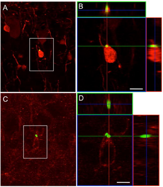
Figure 5.

SVZ neuroblasts that take up the MPIO become interneurons in the OB. (A) Confocal image of a coronal section through the olfactory bulb of a rat that received an MPIO injection in the SVZ 4 weeks prior to tissue processing. The boxed region shows a calretinin-expressing (A) and 5T4-expressing (C) granule cells that contain MPIO particles. (B and D) Confocal 3-D reconstruction of orthogonal projections in the x–z (top) and y–z (right) planes confirms the presence of the MPIO within the cell body. Scale bar = 5 μm.
