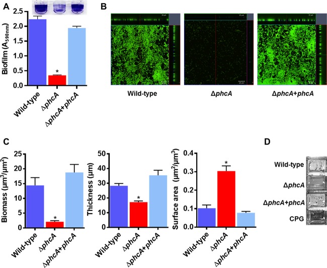
FIG 5 .
PhcA is required for normal R. solanacearum attachment to abiotic surfaces. (A) Biofilm formation by R. solanacearum strains was quantified using the PVC plate-crystal violet staining method; data shown reflect results from two biological replicates, each performed with 12 technical replicates (n = 24); the asterisk indicates P < 0.0001 (ANOVA). (B) Confocal laser microscopy images showing orthogonal views of biofilm formed by R. solanacearum cultures after 24 h of static incubation in a glass chamber slide and staining performed with a BacLight Live/Dead kit. Live cells are green, and dead cells are red. Images are representative of results from two biological replicates with four technical replicates each (scale bar in inset, 20 µm). (C) R. solanacearum ΔphcA mutant cells produce in vitro biofilms on glass that are thinner and more diffuse than those of wild-type cells. Comstat analysis was used to calculate the biomass, thickness, and surface area of 24-h biofilms (n = 8). Asterisks indicate P < 0.05 (ANOVA). (D) Pellicles formed by R. solanacearum strains on the surface of rich broth after 48 h of static incubation; images are representative of results from three biological replicates, each performed with two technical replicates.

