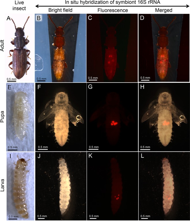
FIG 3 .
Localization of bacteriomes in adults, pupae, and larvae of O. surinamensis. (A to D) Adults. (E to H) Pupae. (I to L) Larvae. (A, E, and I) Live insect images. (B, F, and J) Bright-field images of the insects subjected to whole-mount in situ hybridization targeting 16S rRNA of the symbiont. (C, G, and K) Epifluorescence dissection microscopic images of the same insects, in which four bacteriomes in the abdomen are visualized in red. (D, H, and L) Merged images. In panels B to D, F to H, and J to L, the insect bodies were gently pressed by a coverslip to locate the four bacteriomes to the same focal plane. In panels B to D, thin silicon rubber plates for stabilizing the position of the insect body are seen on both sides.

