



Abstract
Arteriovenous malformations (AVMs) are high-flow vascular anomalies that have demonstrated a very high recurrence rate after endovascular treatment, surgical treatment, or a combination of both. Surgical treatments have shown good response when they are small and well localized but a poor response when diffuse. A better understanding of the nature of the lesion has led to a better response rate and a safer treatment for these patients. This has been accomplished through a detailed understanding of the angioarchitecture of the lesion, enabling a tailored approach in reaching and targeting the nidus of the AVM with different liquid embolic agents, more specifically ethanol. Flow reduction techniques help in exposing the nidus to sclerosant agents. A clinical classification, the Schobinger classification, will help determine the appropriate time to start or to pursue therapy.
Keywords: arteriovenous malformation, vascular anomalies, nidus destruction, embolization, ethanol injection
Objectives : Upon completion of this article, the reader will be able to identify the indications for treatment of these patients as well as the specific approach to be used depending on the architecture of the arteriovenous malformation.
Accreditation : This activity has been planned and implemented in accordance with the Essential Areas and Policies of the Accreditation Council for Continuing Medical Education (ACCME) through the joint providership of Tufts University School of Medicine (TUSM) and Thieme Medical Publishers, New York. TUSM is accredited by the ACCME to provide continuing medical education for physicians.
Credit : Tufts University School of Medicine designates this journal-based CME activity for a maximum of 1 AMA PRA Category 1 Credit ™. Physicians should claim only the credit commensurate with the extent of their participation in the activity.
Arteriovenous malformations (AVMs) are high-flow, low-resistant lesions that are the result of a failure in orderly resorption of the primitive blood vessels during the early gestational period resulting in a direct communication between the arterial and the venous systems due to absent normal capillary network. 1 2 AVMs are usually present at birth but may not be clinically evident. They commonly become evident during childhood and are often exacerbated at puberty or with pregnancies. 3 4 They can be combined with syndromes. Clinically, a purple or red discoloration of the skin can be seen and be confused with a cutaneous port wine stain. Closer examination reveals increased temperature and dilated veins, most of the time associated with a palpable thrill. 5 These lesions can cause significant morbidity and even mortality at advanced stages of their evolution. Cutaneous ischemia with ulceration or infection and hemorrhage are the most common local complications. If the malformation is extensive and centralized in the body, high output cardiac failure can occur. 6 The clinical staging classification will ultimately decide if and when the patient will undergo endovascular/percutaneous or surgical treatments. The aim of interventional therapy is to target the nidus of the vascular malformations in the vast majority of cases knowing that proximal embolization will fail and can often times exacerbate patient symptomatology. 7 The means to reach the nidus and ultimately treat it will largely depend on the architecture of the lesion and its location. To have good assessment of this architecture, a good imaging workup is necessary. Magnetic resonance imaging (MRI), Doppler ultrasound, and in certain circumstances computed tomography will help in this matter before a more invasive angiogram is performed to determine treatment approach. Different embolic agents can be used depending on the presented situation.
Classification
Three classification systems exist for determining proper treatment of patients affected by AVMs. The first is a clinical classification: Schobinger's clinical classification of AVMs symptomatology ( Table 1 ). 8 In this classification, four stages are described. Stage 1 is quiescent and no treatment is warranted. Depending on symptomatology and after weighing risks and benefits, treatment could start at stage 2 for certain patients and is definitely pertinent at stages 3 and 4.
Table 1. Clinical classification of Schobinger.
| Stage I | Quiescence: may or may not have vascular skin stain, warmth of the affected tissues, and arteriovenous shunts can be detected by Doppler ultrasound. The arteriovenous malformation is present but causes no clinical symptoms |
| Stage II | Expansion of arteriovenous malformation lesion: Stage I plus enlargement, pulsations, palpable thrill, audible bruit, and enlarged arterialized tortuous/tense veins |
| Stage III | Destructive tissue changes: Stage II plus dystrophic skin changes, skin ulcerations that can be nonhealing, bleeding from ulcerated skin or mucosal surfaces, overt tissue necrosis, and lytic lesions of bone may occur |
| Stage IV | Decompensation: Stage III plus congestive cardiac failure with increased cardiac output, abnormally lowered peripheral vascular resistance, and venous hypertension secondary to tissue and skin changes |
The other two classifications describe the angioarchitecture of the AVM to help determine the best possible approach to eradicate the nidus of the AVM. The first classification was described in 2006 by Cho et al, 9 and more recently, one classification was proposed by Yakes and Baumgartner ( Figs. 1 and 2 ). 10
Fig. 1.

Angiographic classification according to Cho et al. 9
Fig. 2.

Angiographic classification according to Yakes and Baumgartner. 10
Patient Workup
Clinical Examination
The first step for a successful outcome is patient assessment. A complete physical examination should be undertaken at this stage. As said previously, for soft-tissue AVMs, a purple or red discoloration of the skin can be seen. Closer examination reveals increased temperature and dilated veins, most of the time associated with a palpable thrill.
If in a peripheral location, limb length/size discrepancy can be observed in associated limb overgrowth syndrome such as Parkes–Weber syndrome which is associated with a RASA-1 mutation. 11 The presence of macrocephalia and or hamartoma in patients with AVM is often associated with a phosphatase N tensin homolog (PTEN) mutation. 12 This mutation is typically seen in the Bannayan–Riley–Ruvalcaba (BRRS) and Cowden syndromes but can also be seen in a more general disorder called PTEN hamartoma tumor syndrome (PHTS). 12 These patients present typically multiple intramuscular AVMs with ectopic fat overgrowth, hamartomas, and intracranial developmental anomalies. 13 Typically, these patients do not respond well to embolotherapy and have a propensity to recur after embolization and develop malignant tumors.
To stage properly, signs of tissue destruction should be assessed: dystrophic skin changes, nonhealing ulcerative lesion, bleeding, and necrosis. Local pain is a symptom that is also assessed. Its presence can be secondary to venous hypertension. More systemic symptoms like increased shortness of breath can also be an ominous sign especially in central lesions. Their presence can be an early sign of heart failure. A chest X-ray, electrocardiography, and cardiac ultrasound can complete the workup along with routine laboratory work for any patient going for an interventional procedure under general anesthesia.
Imaging Workup
Noninvasive imaging is also mandatory. It allows proper confirmation of diagnosis, sets a baseline for the patient, and helps define the architecture of the malformation.
A critical imaging modality is color Doppler ultrasound, especially for soft-tissue AVMs. The lesion shows multiple feeding arteries with increased diastolic flow and an increased systolic–diastolic flow venous return. 14 The gray scale component of the ultrasound allows to confirm the absence of a soft-tissue mass, which, if present, could orient to an alternative diagnosis, mostly a hypervascular tumor. Increased velocities on spectral Doppler within the main feeding arteries on follow-up visits could indicate evolution of the lesion and can facilitate the decisional process to start treatment. Also, a thorough examination with the color Doppler ultrasound can help target the nidus for direct punctures by identifying the portions of the AVM that have the most turbulence and high flow, reflected by the sites of most prominent aliasing.
MRI is the best examination to evaluate the extension of the malformation in adjacent structures especially for bone involvement. MRI findings include dilated feeding and draining vessels with little tissue matrix and no venous lakes. 15 Signal voids are typically observed in these vessels on both T1- and T2-weighted spin echo sequence, whereas a hypersignal is observed on gradient echo and contrast-enhanced angiographic sequences indicating a high-flow lesion. 16 Gadolinium-enhanced MR angiography is helpful to evaluate feeding arteries and draining veins. The presence of an early venous filling is typically seen in AVMs. Using time-resolved MR angiography sequences, it is now possible to evaluate the dynamic enhancement of AVMs. 17 18 19 20 21 Since these sequences have a high temporal resolution, there is a compromise on spatial resolution which is lower than conventional three-dimensional MR angiography using parallel imaging techniques.
Computed tomography angiography is rarely used as a routine modality. It will mainly be used in AVMs that have bony involvement and lesions that have dilated veins or venous aneurysms. 22 This will facilitate guidance and planning for a venous approach.
Catheter angiography is mandatory before any therapeutic interventions. It allows to evaluate precisely the feeding arteries and draining veins of the malformation and the feasibility of embolization. The angiographic characteristics of AVMs are dilatation and lengthening of afferent arteries, with early opacification of enlarged vein. Selective and superselective catheterizations are necessary to demonstrate the full extent of these high-flow malformations and to allow precise mapping of feeding and draining vessels.
Treatment
Treatment of AVMs is complex and should be reserved for symptomatic cases. Stage I lesions are usually left for observation and close follow-up. Stage II malformation can be treated if they are well localized. Stage III and IV lesions should be treated because of the risk of progression, serious hemorrhage, and terminal cardiac failure. 4 23 Extensive stage I lesions should be managed conservatively with compressive stockings because extensive resection and complex reconstruction of the lesion can be worse than the lesion itself and recurrence is likely. 4 24 Depending on the anatomy and extent of the malformation, the treatment can be palliative or curative. It has been shown in the past that the vast majority of these lesions recur over time and that resection (with or without embolization) has a lower recurrence rate and longer time to recurrence. 25 This statement holds true only for localized lesions and not diffuse forms which are usually treated endovascularly through embolization with a palliative goal. Also, endovascular techniques and approaches have been refined, which leads to a better treatment of these patients. Optimal embolization can allow curative surgical resection. 26
The type of signs and symptoms will also define the endovascular approach. Bleeding and pain will mainly be due to venous hypertension and decreasing the inflow more aggressively will be the primary goal. If sign of skin destruction through ischemia is dominating the clinical picture, treatment will be aimed at decreasing this steal phenomenon first by reducing the arteriovenous shunting.
Treatments of these patients are not without risks and the operator must be aware of the complications which should be thoroughly explained to the patient. Most of the complications are related to ethanol injection. The most feared complication remains cardiovascular collapse secondary to pulmonary hypertension caused by ethanol injection, especially in large doses. This has been reported in 0.2% of cases. Skin necrosis (15.5%), nerve injury (4.8%), muscular fibrosis (1.5%), and hemoglobinuria (0.95%) have also been reported. 27 With ethanol embolization alone, Do et al have reported a 68% success rate (cure and improvement). 28 When aggressive embolization can be combined with resection surgery, low recurrence rates are observed if the AVM is completely resected (18%) or in AVMs with precise limits with surrounding tissues (16%). 26
Liquid Embolic Agents
Different embolic agents can be used to treat AVM. The three liquid embolics used are ethanol, ethylene vinyl alcohol (EVOH) copolymer, and glue.
Ethanol
Ethanol is the only agent that can achieve nidal destruction because of its fundamental properties. This agent can be potentially curative. It destroys the endothelium of the vascular walls causing fracture up to the internal elastica lamina. This fracture will promote platelet adhesion and thrombosis and will inhibit further secretion of angiogenesis factors causing permanent occlusion of the exposed vessels and prohibiting further neovascularization. 29 However, for this to happen, the vessel wall has to be sufficiently exposed to high concentrations of ethanol for a certain amount of time. This can be optimized by techniques for controlling the inflow and the outflow of the lesion. 30 Pure ethanol can cause spasm in the pulmonary arteries thereby creating right ventricular straining that, if significant enough, can lead to systemic vascular collapse. Appropriate measures have to be taken to ensure the safety of the procedure. For example, monitoring of pulmonary artery pressures may be done especially if use of large doses of ethanol is considered. This will allow baseline mean and systolic pulmonary arterial pressure measurements. Vasodilator therapy, primarily milrinone, is administered if pulmonary arterial pressures increase to greater than 25 mm Hg compared with baseline values. 31 32 It is noteworthy that pulmonary systolic pressures will rise after ethanol injection secondary to pain. At the same time, systemic pressures will also rise accordingly. Both will diminish after a short period of time. If at any time the pulmonary pressures rise and systemic pressures decrease, it might signify that a threshold has been reached and treatment discontinued. To avoid this complication, injections should be given with small boluses (up to 5 mL) and a wait time of at least 5 minutes should be kept in between injections. Some advocate that limiting the injection to 0.14 mL ethanol/kg every 10 minutes should be sufficient enough to avoid complications from acute pulmonary hypertension. 33 The maximum dose should be 1 mL/kg.
Glue
Glue has no sclerosant effect and therefore does not cause nidal destruction. It is, however, very useful for vessel occlusion and can help slow down the flow in certain situations. It is mixed with lipiodol and will polymerize after its contact to blood or other ionic solutions. The rate of polymerization will depend on the lipiodol concentration. The greater the dilution (more lipiodol), the longer it will take to polymerize allowing for deeper penetration. Glue can, however, be very unpredictable and its learning curve longer for proper use. It is adhesive and can recanalize after some time. 34 We typically use glue lipiodol dilution ratio ranging between 1/2(33%) and 1/4 (20%) depending of the velocity and size of fistulas. We recommend to avoid the use of glue in oral mucosa and superficial location because of skin extrusion.
Ethylene Vinyl Alcohol Copolymer
EVOH (Onyx) also has weak sclerosant properties and acts through polymerization. It is an elastic copolymer (EVOH copolymer), dissolved in dimethyl-sulfoxide (DMSO). It has cohesive properties which prevents fragmentation of the injected cast and lessen the chances of having a catheter stick. It hardens from the outside to inside, allowing deep penetration from a gentle push of the syringe, like lava. It will travel to the path of least resistance allowing a much better control. Injection rate has to be slow and controlled as vasospasm can ensue if injected too vigorously. The rate of injection should be between 0.1 and 0.3 mL/min and a compatible microcatheter should be used. There are different techniques of Onyx injection. When the agent is going in the right direction, it is recommended to continue the slow injection without interruption. If there is a reflux of onyx around the catheter, it is recommended to stop the injection for 1 to 2 minutes to create a solid plug around the catheter and resume the injection to push the new onyx forward (stop-and-go technique). When the onyx is going in a wrong direction, the same principles apply, allowing time to the polymer to solidify and redirect the new injected onyx to lower resistance areas. Occlusive balloon or coils can be used to help Onyx penetration while preventing reflux. 35 Recanalization has also been described over time. 36
General Approach to Achieve Nidus Occlusion
As mentioned previously, the focus of the treatment will be targeting and destruction of the nidus and avoiding proximal occlusion. Proximal occlusion or even partial resection of the nidus will result in a treatment failure and possible enlargement of the AVM by neovascularization. 7 Patients' symptoms could increase accordingly. Every effort should be made to avoid vascular compromise in normal vascular territories and a stepwise approach with multiple sessions at regular intervals is highly recommended.
In general, we rely on an endovascular arterial approach first to reach the nidus. This will allow inflow to decrease. Once this approach is no longer possible due to inadequate feeding artery size, the nidal destruction will be completed by direct percutaneous punctures or a venous approach.
In reality, architecture as well as location and symptomatology have to be taken in consideration and each patient has to have a tailored treatment accordingly. Here is our approach to target the nidus for the best results and keeping the risks at a minimal level.
Yakes Type I
AVMs of this nature will have a direct fistula between the artery and the vein. The treatment will be straightforward: blocking the communication with a mechanical agent such as coils or a vascular plug. The approach can either be from the venous side or the arterial side depending on the ease to reach the target area. Coils can be loaded on a 0.035- or 0.018-inch system. Usage of these devices needs a relatively small vascular network for the coils to remain in place. However, if the arteriovenous communication is wide, a sizable Amplatzer plug may be needed, therefore requiring a bigger introducer sheath making venous access more desirable and allowing a safer and easier occlusion. Amplatzer plugs can take time to occlude and may sometimes remain patent despite proper sizing and placement. In these specific circumstances, occlusion can be completed with a liquid embolic such as glue or Onyx ( Fig. 3 ).
Fig. 3.

( a ) CTA showing venous aneurysm from direct fistula. (b) Selective renal artery angiography with dilated vein and early enhancement of renal vein from at least two fistulas. ( c ) Coil embolization of the fistulas. ( d ) Follow-up angiography. ( e ) Follow-up CTA at 6 months confirming treatment of direct fistulas.
Yakes Type II/Cho Type IIIb
These lesions are characterized by multiple feeding arteries and arterioles that communicate through a nidus, exiting in multiple draining veins. The location of the lesion is important, as it will have an impact on the nature of the embolic agent chosen. For soft-tissue lesions affecting the limbs or the superficial aspect of the body (chest, back), an endovascular arterial approach will first be performed. The size of the feeding arteries has to be adequate enough for very distal catheterization up to the nidus to allow direct delivery of pure ethanol in the microfistulas.
These procedures are performed under general anesthesia mainly due to the severe pain induced by ethanol embolization and the need for close monitoring of pulmonary pressures, as there is a significant risk of cardiopulmonary complications. 37
Endovascular Approach
Arterial access and nonselective and selective angiograms are first performed to map the malformation and confirm the angioarchitecture after which superselective catheterization is performed. Small microcatheters used for neurovascular work are often times necessary. Each pedicle leading to the nidus will be superselectively catheterized and embolized with pure ethanol. The volume and injection rate of ethanol will be based on a previous contrast injection with a digital subtraction angiography (DSA) acquisition performed with the same syringe size. The optimal injection rate should be slow enough to prevent contrast/ethanol reflux but fast and long enough to provide an optimal filling of the nidus and enough contact time of ethanol in the nidus. Because ethanol is radiolucent, injections will be done under double roadmap technique, also called negative roadmap or progressive roadmap ( Fig. 4 ). The first milliliters injected will be the residual contrast in the catheter. This will be seen under fluoroscopy and will allow assessment of the injection rate to avoid reflux. The remaining portion of the injection is pure ethanol. At this point, fluoroscopy can be stopped but care has to be taken to maintain the same injection rate for the first half of the bolus and a lower injection for the remaining ethanol volume. Care should be taken to flush the microcatheter from ethanol very slowly. If any resistance is felt, injections should be stopped. We usually use small boluses of 3 to 5 mL to minimize the risk of nontarget embolization that can occur if spasm ensues during the injection. In more dangerous territories (distal extremities, external carotid branches) or AVM with small feeders and slower flow, one can even use smaller boluses (0.2–1 mL).
Fig. 4.

( a, b ) A 50-year-old woman with foot AVM. ( c ). endovascular approach with distal microcatheter and ethanol injection. ( d ) Direct puncture of nidus. ( e ) Outflow compression to get better exposure of nidus to ethanol. ( f ) Final angiography with satisfactory result.
To ensure proper dwell time between ethanol and vessel wall, decreasing the inflow or the outflow may sometimes be necessary. To achieve this, balloon occlusion catheters can be helpful and can be positioned in the inflow or the outflow vessels. If the lesion is superficial, surgical clamps or tourniquets can be applied on an outflow vein or an inflow artery. If temporary material is used in the outflow vessel, care must be taken as not to release the occlusion too quickly. Large amounts of ethanol or thrombus could be suddenly released into the systemic vein and induce rapid pulmonary hypertension or pulmonary embolism with subsequent cardiopulmonary collapse. Large coils can also be positioned in a collateral or main venous drainage ( Fig. 5 ).
Fig. 5.

( a, b ) Prominent pelvic AVM mainly from uterine artery and important dilated venous draining vein. ( c–e ) Endovascular approach and ethanol injection to decrease inflow. ( f ) Embolization with glue up to the venous sac with a proximal flow control with an occlusion balloon. ( g, h ) Final angiography shows complete resolution of AVM.
Another way to help decrease inflow and prevent recanalization in the presence of persisting fistula is combining a sclerosing agent followed by a mechanical occlusion with a liquid embolic such as Onyx or glue. We use ethanol injection first to ensure proper sclerosing effect of the vessels before vessel occlusion. Other teams propose to combine glue injection followed by ethanolamine oleate injection for type II and III AVMs. 38
Multiple sessions may be needed to embolize the nidus optimally. Frequently, small portions of the nidus remain open, but their access has been maximized through the arterial approach. The remaining feeding arteries are at this point too small or too tortuous to allow proper distal catheterization for safe and efficient ethanol injection. In these circumstances, a venous approach, if retrograde venous catheterization, is possible or direct punctures within the nidus can be performed. Decreasing the inflow via the first few arterial sessions makes these two options possible, safer, and easier to carry out.
Direct Puncture
To achieve direct puncture, ultrasound guidance is used. Doppler wave form analysis helps target the proper vessel. Due to its location, at the boundary between artery and vein, these vessels show the most turbulence. Surgical clamps often will be used as a flow reduction technique.
For lesions that involve distal limbs such as fingers and toes, tourniquets will be used to protect the tip of the digit if it is not affected and also the other nonaffected digits if in proximity. The tourniquet will increase the arterial pressure in these segments and will be protected if reflux occurs from an aggressive injection ( Fig. 6 ).
Fig. 6.
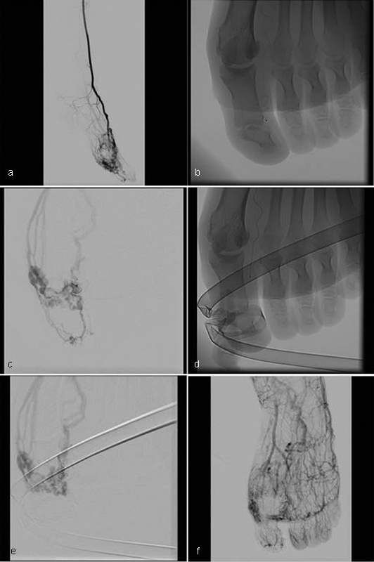
( a ) Foot angiography with AVM involving first toe. (b, c) Distal selective angiography showing normal arteries at the tip of toe. ( d, e ) Protection of distal aspect of toe with tourniquet and ethanol injection through microcatheter and direct puncture. ( f ) Final run with normal aspect of distal toe and treatment of fistulas.
Yakes Type II/Cho Type IIIB, Abdominal and Pelvic AVMS
AVMs that are located deep and more particularly in the abdomen have to be considered differently. Flow reduction techniques can be harder to accomplish and nontarget embolization can cause another set of problems ( Fig. 7 ). These lesions are less likely to be embolized with ethanol and will more than likely necessitate curative surgical resection after embolization. For this reason, the embolic agent preferred will be a liquid embolic, glue or Onyx, that will be followed by surgical resection ( Fig. 8 ). 39
Fig. 7.

( a–e ) A 40-year-old male patient presenting with priapism and nonhealing ulcer of penis from pelvic AVM draining in venous aneurysm and refluxing all the way to penile veins causing venous congestion. ( f ) Endovascular approach and filling of nidus of to the venous aneurysm with Onyx. ( g, h ) Patient asymptomatic at 2-month follow-up with absence of significant shunting on angiography.
Fig. 8.

( a–d ) CTA of a 55-year-old male patient with intermittent bleeding that developed intractable abdominal pain for the past 3 weeks. AVM nidus in mesosigmoid with early draining veins and inferior mesenteric vein increased in size. Wall of sigmoid colon thickened with fat straining confirming ischemic changes. ( e–h ) Embolization with Onyx before surgical resection.
Yakes Type II/Cho Type IIIB, External Carotid AVMs
Malformations of the soft tissues of the head and neck are vascularized by the external carotid artery circuit. For treatment of these lesions, knowledge of communications between the internal and external carotid artery is mandatory. Blockage of some draining veins can be prohibited (cavernous sinus) as opposed to most veins in the peripheral circulation that have no consequences, for the most part. These lesions are treated in the same way. Care must be taken if Onyx is used and one has to ensure of the absence of peripheral vessels close to the skin. This superficial skin can be permanently tattooed by the tantalum powder of the onyx. Good results have been reported after ethanol embolization of Yakes type II/Cho type III extracranial AVMs involving the face or cervical area with or without surgery. 40
Yakes Types IIIA and IIIb/Cho Type II
This architectural construct of these AVMs is characterized by multiple inflow arterioles draining into an aneurysmal venous sac that has either a single outflow or multiple outflow veins. It is believed that the AV fistulas are within the wall of the aneurysmal vein and one of the key components for successful treatment is embolization of this vein if possible. This type of malformation is most often seen in the pelvis ( Fig. 9 ).
Fig. 9.
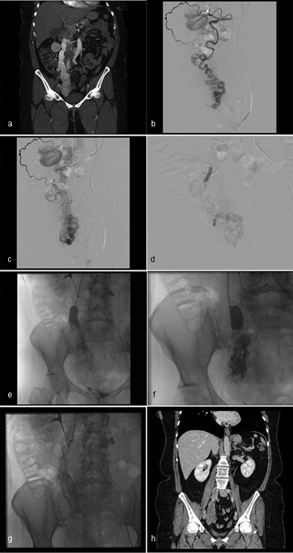
( a ) Pelvic AVM draining into a prominent gonadal vein. (b, c) Feeding artery coming primarily from ovarian branch originating from renal artery. (d) Arterial endovascular approach and ethanol embolization to decrease the inflow. (e, f) Balloon occlusion to decrease outflow and further embolization with ethanol from microcatheter inserted through the balloon catheter. ( g ) Deployment of an Amplatzer plug to avoid thrombus migration. ( h ) Follow-up CTA confirming occlusion of AVM.
The first step remains decreasing the inflow by an arterial endovascular approach with ethanol embolization that can be followed up with glue or Onyx embolization. Coil packing of the aneurysmal venous pouch can be done to complete the embolization in the case of a single draining outflow vein. This venous pouch can be reached either by a retrograde venous approach or direct puncture if accessible. Mechanical occlusion of the venous drainage with coil or Amplatzer plug can be combined with liquid embolic or sclerosant agent injection in the nidus (the push-through method). 41 Retrograde injection of ethanol through an occlusive balloon is effective but can be dangerous if large volume of ethanol is released following balloon deflation. 42 We prefer using on the venous side a permanent mechanical occlusion combined with sclerosant agents.
In case where there are many draining veins, the process can be more time consuming, as a multitude of these veins may need to be occluded with coils or liquid embolics.
Cho Type IIIA
This type of AVM can be difficult to treat. It is characterized by a mesh-like network of arterioles draining into a nidus with multiple outflow venules. The difficulty lies on the fact that the size of these feeding and outflowing vessels is too small to be selectively catheterized and ethanol injection would be too proximal with increased risk of proximal nontarget embolization.
The strategy for this type of lesion remains identical to the one described earlier with an arterial endovascular approach, but the embolization will be performed with Onyx. 43 In this circumstance, the properties of Onyx will allow deep penetration, often penetrating the nidus. Direct puncture under ultrasound guidance can be done as a second step, targeting vessels that have the most turbulence on color Doppler ( Fig. 10 ). Ethanol can be injected after direct puncture of the nidus.
Fig. 10.

( a ) MIP MRA showing a small but very symptomatic AVM in right labial region. (b, c) Angiography shows multiple feeding arteries that are very small in size. ( d, e ) Microcatheterization as deep as possible and injection with Onyx. ( f ) Follow-up angiography demonstrated a residual nidus at superior aspect too small for microcatheterization. ( g ) Direct puncture under ultrasound guidance and ethanol injection. ( h, i )Follow-up angiography demonstrates complete embolization.
Yakes Type IV
Another difficult-to-treat architectural lesion is Yakes type IV. It has the characteristics of an innumerable amount of fistulas infiltrating tissue interspersed with normal capillaries that maintain the viability of the tissue. A 50:50 mixture of ethanol and nonionic contrast can apparently be curative. 10
Patient Followup
All patients undergoing embolization should be followed up closely by clinical results and Doppler ultrasound examinations. Usually, the embolization sessions should be performed every 6 to 8 weeks until a significant devascularization and clinical improvement are obtained. This interval can be lengthened if the patient experiences skin necrosis or nerve injury. If possible, it is preferable to allow for complete healing of necrosis before resuming embolization session. Patient with stage 1 Schobinger AVM can be followed up every 5 years if there is no evidence of AVM growth. We are following stage 2 AVM every year, whereas stage 3 and 4 AVMs will be followed closely between embolization procedures. The management should involve a multidisciplinary team consisting of interventional radiologists, plastic surgeons, dermatologists, and internal medicine specialists.
Following ethanol embolization, a diffuse capillary hypervascularity without significant residual shunting can be observed on Doppler ultrasound or DSA in the embolized area.
Conclusion
The treatment of AVMs has evolved over the past few years. Better understanding of this type of vascular malformation and its hemodynamic specificities has led to a classification system that allows a more tailored treatment depending on the architecture of the malformation. Ethanol injection to obtain nidus destruction remains the key component of the treatment. Use of flow restricting techniques is critical for optimal results.
Patient safety has to be taken into consideration at all times. Meticulous embolization techniques and proper monitoring will decrease the risk of complications. Experienced multidisciplinary teams are necessary to optimally care for these challenging patients.
References
- 1.Mulliken J B, Glowacki J. Classification of pediatric vascular lesions. Plast Reconstr Surg. 1982;70(01):120–121. [PubMed] [Google Scholar]
- 2.Yakes W F, Rossi P, Odink H. How I do it. Arteriovenous malformation management. Cardiovasc Intervent Radiol. 1996;19(02):65–71. doi: 10.1007/BF02563895. [DOI] [PubMed] [Google Scholar]
- 3.Burrows P E, Laor T, Paltiel H, Robertson R L. Diagnostic imaging in the evaluation of vascular birthmarks. Dermatol Clin. 1998;16(03):455–488. doi: 10.1016/s0733-8635(05)70246-1. [DOI] [PubMed] [Google Scholar]
- 4.Kohout M P, Hansen M, Pribaz J J, Mulliken J B. Arteriovenous malformations of the head and neck: natural history and management. Plast Reconstr Surg. 1998;102(03):643–654. doi: 10.1097/00006534-199809030-00006. [DOI] [PubMed] [Google Scholar]
- 5.Vander Kam V, Achauer B M. Arteriovenous malformations: a team approach to management. Plast Surg Nurs. 1995;15(01):53–57. [PubMed] [Google Scholar]
- 6.Enjolras O. Classification and management of the various superficial vascular anomalies: hemangiomas and vascular malformations. J Dermatol. 1997;24(11):701–710. doi: 10.1111/j.1346-8138.1997.tb02522.x. [DOI] [PubMed] [Google Scholar]
- 7.Lee B B, Baumgartner I, Berlien H P et al. Consensus document of the International Union of Angiology (IUA)-2013. Current concept on the management of arterio-venous management. Int Angiol. 2013;32(01):9–36. [PubMed] [Google Scholar]
- 8.Finn M C, Glowacki J, Mulliken J B. Congenital vascular lesions: clinical application of a new classification. J Pediatr Surg. 1983;18(06):894–900. doi: 10.1016/s0022-3468(83)80043-8. [DOI] [PubMed] [Google Scholar]
- 9.Cho S K, Do Y S, Shin S W et al. Arteriovenous malformations of the body and extremities: analysis of therapeutic outcomes and approaches according to a modified angiographic classification. J Endovasc Ther. 2006;13(04):527–538. doi: 10.1583/05-1769.1. [DOI] [PubMed] [Google Scholar]
- 10.Yakes W, Baumgartner I. Interventional treatment of arterio-venous malformations. Gefasschirurgie. 2014;19(04):325–330. [Google Scholar]
- 11.Revencu N, Boon L M, Mulliken J B et al. Parkes Weber syndrome, vein of Galen aneurysmal malformation, and other fast-flow vascular anomalies are caused by RASA1 mutations. Hum Mutat. 2008;29(07):959–965. doi: 10.1002/humu.20746. [DOI] [PubMed] [Google Scholar]
- 12.Kurek K C, Howard E, Tennant L B et al. PTEN hamartoma of soft tissue: a distinctive lesion in PTEN syndromes. Am J Surg Pathol. 2012;36(05):671–687. doi: 10.1097/PAS.0b013e31824dd86c. [DOI] [PMC free article] [PubMed] [Google Scholar]
- 13.Tan W H, Baris H N, Burrows P E et al. The spectrum of vascular anomalies in patients with PTEN mutations: implications for diagnosis and management. J Med Genet. 2007;44(09):594–602. doi: 10.1136/jmg.2007.048934. [DOI] [PMC free article] [PubMed] [Google Scholar]
- 14.Dubois J, Soulez G, Oliva V L, Berthiaume M J, Lapierre C, Therasse E. Soft-tissue venous malformations in adult patients: imaging and therapeutic issues. Radiographics. 2001;21(06):1519–1531. doi: 10.1148/radiographics.21.6.g01nv031519. [DOI] [PubMed] [Google Scholar]
- 15.Hovius S E, Borg D H, Paans P R, Pieterman H. The diagnostic value of magnetic resonance imaging in combination with angiography in patients with vascular malformations: a prospective study. Ann Plast Surg. 1996;37(03):278–285. doi: 10.1097/00000637-199609000-00008. [DOI] [PubMed] [Google Scholar]
- 16.Siegel M J. Magnetic resonance imaging of musculoskeletal soft tissue masses. Radiol Clin North Am. 2001;39(04):701–720. doi: 10.1016/s0033-8389(05)70306-7. [DOI] [PubMed] [Google Scholar]
- 17.Reinacher P C, Stracke P, Reinges M H, Hans F J, Krings T. Contrast-enhanced time-resolved 3-D MRA: applications in neurosurgery and interventional neuroradiology. Neuroradiology. 2007;49 01:S3–S13. doi: 10.1007/s00234-007-1468-6. [DOI] [PubMed] [Google Scholar]
- 18.Saleh R S, Lohan D G, Villablanca J P, Duckwiler G, Kee S T, Finn J P. Assessment of craniospinal arteriovenous malformations at 3T with highly temporally and highly spatially resolved contrast-enhanced MR angiography. AJNR Am J Neuroradiol. 2008;29(05):1024–1031. doi: 10.3174/ajnr.A0947. [DOI] [PMC free article] [PubMed] [Google Scholar]
- 19.Taschner C A, Gieseke J, Le Thuc V et al. Intracranial arteriovenous malformation: time-resolved contrast-enhanced MR angiography with combination of parallel imaging, keyhole acquisition, and k-space sampling techniques at 1.5 T. Radiology. 2008;246(03):871–879. doi: 10.1148/radiol.2463070293. [DOI] [PubMed] [Google Scholar]
- 20.Wu Y, Kim N, Korosec F R et al. 3D time-resolved contrast-enhanced cerebrovascular MR angiography with subsecond frame update times using radial k-space trajectories and highly constrained projection reconstruction. AJNR Am J Neuroradiol. 2007;28(10):2001–2004. doi: 10.3174/ajnr.A0772. [DOI] [PMC free article] [PubMed] [Google Scholar]
- 21.Ziyeh S, Strecker R, Berlis A, Weber J, Klisch J, Mader I. Dynamic 3D MR angiography of intra- and extracranial vascular malformations at 3T: a technical note. AJNR Am J Neuroradiol. 2005;26(03):630–634. [PMC free article] [PubMed] [Google Scholar]
- 22.Tao Q, Lv B, Bhatia K S, Qiao B, Zheng C Q, Chen Z F. Three-dimensional CT angiography for the diagnosis and assessment of arteriovenous malformations in the oral and maxillofacial region. J Craniomaxillofac Surg. 2010;38(01):32–37. doi: 10.1016/j.jcms.2009.10.021. [DOI] [PubMed] [Google Scholar]
- 23.Mulliken J B, Fishman S J, Burrows P E. Vascular anomalies. Curr Probl Surg. 2000;37(08):517–584. doi: 10.1016/s0011-3840(00)80013-1. [DOI] [PubMed] [Google Scholar]
- 24.Upton J, Coombs C J, Mulliken J B, Burrows P E, Pap S. Vascular malformations of the upper limb: a review of 270 patients. J Hand Surg Am. 1999;24(05):1019–1035. doi: 10.1053/jhsu.1999.1019. [DOI] [PubMed] [Google Scholar]
- 25.Liu A S, Mulliken J B, Zurakowski D, Fishman S J, Greene A K. Extracranial arteriovenous malformations: natural progression and recurrence after treatment. Plast Reconstr Surg. 2010;125(04):1185–1194. doi: 10.1097/PRS.0b013e3181d18070. [DOI] [PubMed] [Google Scholar]
- 26.Goldenberg D C, Hiraki P Y, Caldas J G, Puglia P, Marques T M, Gemperli R. Surgical treatment of extracranial arteriovenous malformations after multiple embolizations: outcomes in a series of 31 patients. Plast Reconstr Surg. 2015;135(02):543–552. doi: 10.1097/PRS.0000000000000890. [DOI] [PubMed] [Google Scholar]
- 27.Qiu Y, Chen H, Lin X, Hu X, Jin Y, Ma G. Outcomes and complications of sclerotherapy for venous malformations. Vasc Endovascular Surg. 2013;47(06):454–461. doi: 10.1177/1538574413492390. [DOI] [PubMed] [Google Scholar]
- 28.Do Y S, Yakes W F, Shin S W et al. Ethanol embolization of arteriovenous malformations: interim results. Radiology. 2005;235(02):674–682. doi: 10.1148/radiol.2352040449. [DOI] [PubMed] [Google Scholar]
- 29.Prasetyono T O, Kreshanti P. Efficacy of intra-lesional alcohol injection as alternative and/or complementary treatment of vascular malformations: a systematic review. J Plast Reconstr Aesthet Surg. 2010;63(07):1071–1079. doi: 10.1016/j.bjps.2009.04.020. [DOI] [PubMed] [Google Scholar]
- 30.Jackson J E, Mansfield A O, Allison D J. Treatment of high-flow vascular malformations by venous embolization aided by flow occlusion techniques. Cardiovasc Intervent Radiol. 1996;19(05):323–328. doi: 10.1007/BF02570183. [DOI] [PubMed] [Google Scholar]
- 31.Legiehn G M, Heran M K.Classification, diagnosis, and interventional radiologic management of vascular malformations Orthop Clin North Am 20063703435–474., vii–viii [DOI] [PubMed] [Google Scholar]
- 32.Kim J S, Nam M H, Do Y S et al. Efficacy of milrinone versus nitroglycerin in controlling pulmonary arterial hypertension induced by intravenous injections of absolute ethanol in anesthetized dogs. J Vasc Interv Radiol. 2010;21(06):882–887. doi: 10.1016/j.jvir.2010.02.011. [DOI] [PubMed] [Google Scholar]
- 33.Shin B S, Do Y S, Cho H S et al. Effects of repeat bolus ethanol injections on cardiopulmonary hemodynamic changes during embolotherapy of arteriovenous malformations of the extremities. J Vasc Interv Radiol. 2010;21(01):81–89. doi: 10.1016/j.jvir.2009.09.026. [DOI] [PubMed] [Google Scholar]
- 34.Rao V R, Mandalam K R, Gupta A K, Kumar S, Joseph S. Dissolution of isobutyl 2-cyanoacrylate on long-term follow-up. AJNR Am J Neuroradiol. 1989;10(01):135–141. [PMC free article] [PubMed] [Google Scholar]
- 35.Chiu A H, Aw G, Wenderoth J D. Double-lumen arterial balloon catheter technique for Onyx embolization of dural arteriovenous fistulas: initial experience. J Neurointerv Surg. 2014;6(05):400–403. doi: 10.1136/neurintsurg-2013-010768. [DOI] [PMC free article] [PubMed] [Google Scholar]
- 36.Bauer A M, Bain M D, Rasmussen P A. Onyx resorbtion with AVM recanalization after complete AVM obliteration. Interv Neuroradiol. 2015;21(03):351–356. doi: 10.1177/1591019915581985. [DOI] [PMC free article] [PubMed] [Google Scholar]
- 37.Shin B S, Do Y S, Cho H S et al. Cardiovascular effects and predictability of cardiovascular collapse after repeated intravenous bolus injections of absolute ethanol in anesthetized pigs. J Vasc Interv Radiol. 2010;21(12):1867–1872. doi: 10.1016/j.jvir.2010.08.007. [DOI] [PubMed] [Google Scholar]
- 38.Kitagawa A, Izumi Y, Hagihara M et al. Ethanolamine oleate sclerotherapy combined with transarterial embolization using n-butyl cyanoacrylate for extracranial arteriovenous malformations. Cardiovasc Intervent Radiol. 2014;37(02):371–380. doi: 10.1007/s00270-013-0653-8. [DOI] [PubMed] [Google Scholar]
- 39.Cassinotto C, Lapuyade B. Pancreatic arteriovenous malformation embolization with onyx. J Vasc Interv Radiol. 2015;26(03):442–444. doi: 10.1016/j.jvir.2014.11.036. [DOI] [PubMed] [Google Scholar]
- 40.Su L, Wang D, Han Y, Wang Z, Zheng L, Fan X. Absolute ethanol embolization of infiltrating-diffuse extracranial arteriovenous malformations in the head and neck. Eur J Vasc Endovasc Surg. 2015;50(01):114–121. doi: 10.1016/j.ejvs.2015.02.020. [DOI] [PubMed] [Google Scholar]
- 41.Wohlgemuth W A, Müller-Wille R, Teusch V I et al. The retrograde transvenous push-through method: a novel treatment of peripheral arteriovenous malformations with dominant venous outflow. Cardiovasc Intervent Radiol. 2015;38(03):623–631. doi: 10.1007/s00270-015-1063-x. [DOI] [PubMed] [Google Scholar]
- 42.van der Linden E, van Baalen J M, Pattynama P M. Retrograde transvenous ethanol embolization of high-flow peripheral arteriovenous malformations. Cardiovasc Intervent Radiol. 2012;35(04):820–825. doi: 10.1007/s00270-011-0265-0. [DOI] [PubMed] [Google Scholar]
- 43.Numan F, Omeroglu A, Kara B, Cantaşdemir M, Adaletli I, Kantarci F. Embolization of peripheral vascular malformations with ethylene vinyl alcohol copolymer (Onyx) J Vasc Interv Radiol. 2004;15(09):939–946. doi: 10.1097/01.RVI.0000130862.23109.52. [DOI] [PubMed] [Google Scholar]